
Retina
Encyclopedia
The vertebrate retina is a light-sensitive tissue lining the inner surface of the eye
. The optics of the eye create an image of the visual world on the retina, which serves much the same function as the film in a camera. Light striking the retina initiates a cascade of chemical and electrical events that ultimately trigger nerve impulses. These are sent to various visual centers of the brain
through the fibers of the optic nerve
.
In vertebrate embryonic development, the retina and the optic nerve
originate as outgrowths of the developing brain
, so the retina is considered part of the central nervous system
(CNS). It is the only part of the CNS that can be visualized non-invasively.
The retina is a layered structure with several layers of neurons interconnected by synapses
. The only neurons that are directly sensitive to light are the photoreceptor cells. These are mainly of two types: the rods
and cones
. Rods function mainly in dim light and provide black-and-white vision, while cones support daytime vision and the perception of colour. A third, much rarer type of photoreceptor, the photosensitive ganglion cell
, is important for reflexive responses to bright daylight.
Neural signals from the rods and cones undergo processing by other neuron
s of the retina. The output takes the form of action potential
s in retinal ganglion cells whose axon
s form the optic nerve
. Several important features of visual perception
can be traced to the retinal encoding and processing of light.
's main eyes, set in the front-and-centre of the cephalothorax and forming tubes extending well into the cephalothorax, are more acute in daylight than a cat's and 10 times more acute than a dragonfly's.
 The vertebrate retina has ten distinct layers. From closest to farthest from the vitreous body - that is, from closest to the front exterior of the head towards the interior and back of the head:
These can be simplified into 4 main processing stages: photoreception, transmission to bipolar cells, transmission to ganglion cells which also contain photoreceptors, the photosensitive ganglion cell
s, and transmission along the optic nerve. At each synaptic stage there are also laterally connecting horizontal and amacrine cells.
The optic nerve is a central tract of many axons of ganglion cells connecting primarily to the lateral geniculate body, a visual relay station in the diencephalon (the rear of the forebrain). It also projects to the superior colliculus
, the suprachiasmatic nucleus
, and the nucleus of the optic tract. It passes through the other layers creating the Optic disc
in primates.
Additional structures, not directly associated with vision, are found as outgrowths of the retina in some vertebrate groups. In bird
s, the pecten
is a vascular structure of complex shape that projects from the retina into the vitreous humour
; it supplies oxygen and nutrients to the eye, and may also aid in vision. Reptile
s have a similar, but much simpler, structure, referred to as the papillary cone.
, a part of the retina sometimes called "the blind spot" because it lacks photoreceptors, is located at the optic papilla, a nasal zone where the optic-nerve fibers leave the eye. It appears as an oval white area of 3mm². Temporal (in the direction of the temples) to this disc is the macula
. At its center is the fovea
, a pit that is responsible for our sharp central vision but is actually less sensitive to light because of its lack of rods. Human and non-human primate
s possess one fovea as opposed to certain bird species such as hawks who actually are bifoviate and dogs and cats who possess no fovea but a central band known as the visual streak. Around the fovea extends the central retina for about 6 mm and then the peripheral retina. The edge of the retina is defined by the ora serrata
. The length from one ora to the other (or macula), the most sensitive area along the horizontal meridian is about 3.2 mm.
 In section the retina is no more than 0.5 mm thick. It has three layers of nerve
cells and two of synapse
s, including the unique ribbon synapse
s. The optic nerve carries the ganglion cell
axon
s to the brain and the blood vessels that open into the retina. The ganglion cells lie innermost in the retina while the photoreceptive cells lie outermost. Because of this counter-intuitive arrangement, light must first pass through and around the ganglion cells and through the thickness of the retina, (including its capillary vessels, not shown) before reaching the rods and cones. However it does not pass through the epithelium
or the choroid
(both of which are opaque).
The white blood cell
s in the capillaries in front of the photoreceptors can be perceived as tiny bright moving dots when looking into blue light. This is known as the blue field entoptic phenomenon
(or Scheerer's phenomenon).
Between the ganglion cell layer and the rods and cones there are two layers of neuropil
s where synaptic contacts are made. The neuropil layers are the outer plexiform layer
and the inner plexiform layer
. In the outer the rods and cones connect to the vertically running bipolar cells, and the horizontally oriented horizontal cells connect to ganglion cells.
The central retina is cone-dominated and the peripheral retina is rod-dominated. In total there are about seven million cones and a hundred million rods. At the centre of the macula is the foveal pit where the cones are smallest and in a hexagonal mosaic, the most efficient and highest density. Below the pit the other retina layers are displaced, before building up along the foveal slope until the rim of the fovea or parafovea which is the thickest portion of the retina. The macula has a yellow pigmentation from screening pigments and is known as the macula lutea. The area directly surrounding the fovea has the highest density of rods converging on single bipolars. Since the cones have a much lesser power of merging signals, the fovea allows for the sharpest vision the eye can attain.
Though the rod and cones are a mosaic of sorts, transmission from receptors to bipolars to ganglion cells is not the case, Since there are about 150 million receptors and only 1 million optic nerve fibers, there must be convergence and thus mixing of signals. Moreover, the horizontal action of the horizontal and amacrine cells can allow one area of the retina to control another (e.g., one stimulus inhibiting another). This inhibition is key to the sum of messages sent to the higher regions of the brain. In some lower vertebrates, (e.g., the pigeon) there is a "centrifugal" control of messages, that is, one layer can control another, or higher regions of the brain can drive the retinal nerve cells, but in primates this does not occur.
retina has the photoreceptors at the front side of the retina, with processing neurons and capillaries behind them. Because of this, cephalopods do not have a blind spot
.
The cephalopod retina does not originate as an outgrowth of the brain, as the vertebrate one does. It was originally argued that this difference shows that vertebrate and cephalopod eyes are not homologous
but have evolved separately. The evolutionary biologist Richard Dawkins
cites the imperfect structure of the human retina as confounding claims by creationists or intelligent design
theorists that the human eye is so perfect it must have a designer.
In 2009 Kröger anatomically showed in Zebrafish that though the inverted arrangement is nonadaptive in that it creates avoidable scattering of light (and thus loss of light and image blur), it has space-saving advantages for small-eyed animals in which there is a minimal vitreous body, as the space between the lens and the photoreceptors’ light-sensitive outer segments is completely filled with retinal cells.
The difference between vertebrate and cephalopod retinas presents an interesting puzzle of evolutionary path which is not yet fully settled. From an evolutionary perspective, a convoluted structure such as the inverted retina can generally come about as a consequence of two alternative processes; (a) an advantageous "good" compromise between competing functional limitations, or (b) as a historical maladaptive relic of the convoluted path of organ evolution and transformation. Vision is an important adaptation in higher vertebrates. Therefore, if the retina is indeed “wired wrongly” or “badly designed” (from an optical engineering point of view) then it sensible to look for it to possibly have some very significant physiological advantage. One such suggestion is based on the argument that the mammalian photoreceptor amplification process requires vast quantities of metabolic energy, and consequently, it requires massive and homogeneous supply of blood. Indeed, a unique network of blood vessels gives impression of being well adapted to provide the photoreceptor layer with copious quantities of blood. This lead to a proposition that the inverted retina is an adaptation to deliver abundant quantities of oxygen to the photoreceptor cells commensurate with their high energy demands.
Yet, the cephalopods have a non-inverted retina which is comparable in resolving power to the eyes of many vertebrates. Hence, at least for cold-blooded vertebrates, the inverted retinal structure is almost certainly not an adaptive necessity. All together, the inverted retina structure remains a mystery. Why should such an unlikely arrangement have appeared in the first place some 600 million years ago in the earliest of vertebrates who had presumably no need for high acuity vision and in all probability possessed photoreceptors with lower metabolic rates than those of higher warm-blooded vertebrates today? If the non-inverted retina works so well for the cold-blooded cephalopods, why did evolution go down the path of having inverted retina in most cold-blooded vertebrates (e.g., fish)? We are likely to be still missing a satisfactory scientific understanding of either (a) some hidden optimal physiological advantages of the inverted retina structure, or (b) a well-reasoned evolutionary path of how the retina has evolved in early stages from some other organ or structures so that the inverted retina is a sub-optimal relic of the evolutionary path. E.g., early in the Evolution of the eye
, there was a cup-shaped hollow, with the vertebrate retina represented by a single cell layer that lined the eye's cavity. This layer then became increasingly photosensitive, evolving into rods and cones. Later, this single layer would be supplemented by additional neurons, creating cross-connections for logic processing and encoding of signals. In the primitive eye, there was no lens or vitreous body. Added layers then likely came to be positioned "on top" of older structures in the bottom of the cup, finally ending up between the rods and cones and the vitreous body.
The cones respond to bright light and mediate high-resolution colour vision during daylight illumination (also called photopic vision). The rods are saturated at daylight levels and don't contribute to pattern vision. However, rods do respond to dim light and mediate lower-resolution, monochromatic vision under very low levels of illumination (called scotopic vision). The illumination in most office settings falls between these two levels and is called mesopic vision. At these light levels, both the rods and cones are actively contributing pattern information to that exiting the eye. What contribution the rod information makes to pattern vision under these circumstances is unclear.
The response of cones to various wavelengths of light is called their spectral sensitivity. In normal human vision, the spectral sensitivity of a cone falls into one of three subgroups. These are often called blue, green, and red cones but more accurately are short, medium, and long wavelength sensitive cone subgroups. It is a lack of one or more of the cone subtypes that causes individuals to have deficiencies in colour vision or various kinds of colour blindness
. These individuals are not blind to objects of a particular colour but experience the inability to distinguish between two groups of colours that can be distinguished by people with normal vision. Humans have three different types of cones (trichromatic vision) while most other mammals lack cones with red sensitive pigment and therefore have poorer (dichromatic) colour vision. However, some animals have four spectral subgroups, e.g., the trout adds an ultraviolet subgroup to short, medium and long subgroups that are similar to humans. Some fish are sensitive to the polarization of light as well.
When light falls on a receptor it sends a proportional response synaptically to bipolar cell
s which in turn signal the retinal ganglion cells. The receptors are also 'cross-linked' by horizontal cell
s and amacrine cell
s, which modify the synaptic signal before the ganglion cells. Rod and cone signals are intermixed and combine, although rods are mostly active in very poorly lit conditions and saturate in broad daylight, while cones function in brighter lighting because they are not sensitive enough to work at very low light levels.
Despite the fact that all are nerve cells, only the retinal ganglion cells and few amacrine cells create action potentials. In the photoreceptors, exposure to light hyperpolarizes the membrane in a series of graded shifts. The outer cell segment contains a photopigment
. Inside the cell the normal levels of cyclic guanosine monophosphate
(cGMP) keep the Na+ channel open and thus in the resting state the cell is depolarised. The photon
causes the retinal
bound to the receptor protein to isomerise to trans-retinal
. This causes receptor to activate multiple G-proteins. This in turn causes the Ga-subunit of the protein to activate a phosphodiesterase (PDE6), which degrades cGMP, resulting in the closing of Na+ cyclic nucleotide-gated ion channel
s (CNGs). Thus the cell is hyperpolarised. The amount of neurotransmitter released is reduced in bright light and increases as light levels fall. The actual photopigment is bleached away in bright light and only replaced as a chemical process, so in a transition from bright light to darkness the eye can take up to thirty minutes to reach full sensitivity (see Adaptation (eye)
).
In the retinal ganglion cells there are two types of response, depending on the receptive field
of the cell. The receptive fields of retinal ganglion cells comprise a central approximately circular area, where light has one effect on the firing of the cell, and an annular surround, where light has the opposite effect on the firing of the cell. In ON cells, an increment in light intensity in the centre of the receptive field causes the firing rate to increase. In OFF cells, it makes it decrease. In a linear model, this response profile is well described by a Difference of Gaussians
and is the basis for edge detection
algorithms. Beyond this simple difference ganglion cells are also differentiated by chromatic sensitivity and the type of spatial summation. Cells showing linear spatial summation are termed X cells (also called parvocellular, P, or midget ganglion cells), and those showing non-linear summation are Y cells (also called magnocellular, M, or parasol retinal ganglion cells), although the correspondence between X and Y cells (in the cat retina) and P and M cells (in the primate retina) is not as simple as it once seemed.
In the transfer of visual signals to the brain, the visual pathway, the retina is vertically divided in two, a temporal (nearer to the temple) half and a nasal (nearer to the nose) half. The axons from the nasal half cross the brain at the optic chiasma to join with axons from the temporal half of the other eye before passing into the lateral geniculate body.
Although there are more than 130 million retinal receptors, there are only approximately 1.2 million fibres (axons) in the optic nerve; a large amount of pre-processing is performed within the retina. The fovea produces the most accurate information. Despite occupying about 0.01% of the visual field (less than 2° of visual angle
), about 10% of axons in the optic nerve are devoted to the fovea. The resolution limit of the fovea has been determined at around 10,000 points. The information capacity is estimated at 500,000 bits per second (for more information on bits, see information theory
) without colour or around 600,000 bits per second including colour .
 The retina, unlike a camera, does not simply send a picture to the brain. The retina spatially encodes (compresses) the image to fit the limited capacity of the optic nerve. Compression is necessary because there are 100 times more Photoreceptor cells than ganglion cells as mentioned above. The retina does so by "decorrelating"
the incoming images in a manner to be described below. These operations are carried out by the center surround structures as implemented by the bipolar and ganglion cells.
There are two types of center surround structures in the retina—on-centers and off-centers. On-centers have a positively weighted center and a negatively weighted surround. Off-centers are just the opposite. Positive weighting is more commonly known as excitatory and negative weighting is more commonly known as inhibitory.
These center surround structures are not physical in the sense that one cannot see them by staining samples of tissue and examining the retina's anatomy. The center surround structures are logical (i.e., mathematically abstract) in the sense that they depend on the connection strengths between ganglion and bipolar cells. It is believed that the connection strengths between cells is caused by the number and types of ion channel
s embedded in the synapse
s between the ganglion and bipolar cells. Stephen Kuffler
in the 1950s was the first person to begin to understand these center surround structures in the retina of cats. See Receptive field
for figures and more information on center surround structures. See chapter 3 of David Hubel's on-line book (listed below) for an excellent introduction.
The center surround structures are mathematically equivalent to the edge detection
algorithms used by computer programmers to extract or enhance the edges in a digital photograph. Thus the retina performs operations on the image to enhance the edges of objects within its visual field. For example, in a picture of a dog, a cat and a car, it is the edges of these objects that contain the most information. In order for higher functions in the brain (or in a computer for that matter) to extract and classify objects such as a dog and a cat, the retina is the first step to separating out the various objects within the scene.
As an example, the following matrix
is at the heart of the computer algorithm
that implements edge detection. This matrix is the computer equivalent to the center surround structure. In this example, each box (element) within this matrix would be connected to one photoreceptor. The photoreceptor in the center is the current receptor being processed. The center photoreceptor is multiplied by the +1 weight factor. The surrounding photoreceptors are the "nearest neighbors" to the center and are multiplied by the -1/8 value. The sum of all nine of these elements is finally calculated. This summation is repeated for every photoreceptor in the image by shifting left to the end of a row and then down to the next line.
The total sum of this matrix is zero if all the inputs from the nine photoreceptors are the same value. The zero result indicates the image was uniform (non-changing) within this small patch. Negative or positive sums mean something was varying (changing) within this small patch of nine photoreceptors.
The above matrix is only an approximation to what really happens inside the retina. The differences are:
Here is an example of an input image and how edge detection would modify it.

Once the image is spatially encoded by the center surround structures, the signal is sent out the optical nerve (via the axons of the ganglion cells) through the optic chiasm
to the LGN (lateral geniculate nucleus
). The exact function of the LGN is unknown at this time. The output of the LGN is then sent to the back of the brain. Specifically the output of the LGN "radiates" out to the V1 Primary visual cortex.
Simplified Signal Flow: Photoreceptors → Bipolar → Ganglion → Chiasm → LGN → V1 cortex

has been used to image individual rods and cones in the living human retina and a company based in Scotland have engineered technology that allows physicians to observe the complete retina without any discomfort to patients.
The electroretinogram
is used to measure non-invasively the retina's electrical activity, which is affected by certain diseases. A relatively new technology, now becoming widely available, is optical coherence tomography
(OCT). This non-invasive technique allows one to obtain a 3D
volumetric or high resolution cross-sectional tomogram of the retinal fine structure with histologic-quality.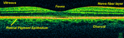 Treatment depends upon the nature of the disease or disorder. Transplantation
of retinas has been attempted, but without much success. At MIT
, The University of Southern California, and the University of New South Wales
, an "artificial retina" is under development: an implant which will bypass the photoreceptors of the retina and stimulate the attached nerve cells directly, with signals from a digital camera.
 There are two circulations, both supplied by the ophthalmic artery. The uveal circulation consists of arteries entering the globe outside the optic nerve, these supply the uvea and outer and middle layers of the retina. The retinal circulation, on the other hand, supplies the inner layer of the retina and passes with the optic nerve as a branch of the ophthalmic artery called the central artery of the retina. The unique structure of the blood vessel
s in the retina has been used for biometric identification
.
The vascular topographical geometry in the retina is known to conform to structural
principles that are related to certain physical properties . The analysis of the geometrical structure is very important as deviations from the optimal principles may indicate some cardiovascular diseases, such as hypertension and atherosclerosis ; a comprehensive analysis is given by Patton et al (2006) . The identification of vascular bifurcations is one of the basic steps in this analysis. Azzopardi and Petkov (2011) propose a computer vision
algorithm that automatically detects these retinal features. Their results are evaluated against the ground truth data of vascular bifurcations of retinal fundus images that are obtained from the DRIVE data set.
, Haldan Keffer Hartline
and Ragnar Granit
won the 1967 Nobel Prize in Physiology or Medicine
for their scientific research on the retina.
A recent University of Pennsylvania
study calculated the approximate bandwidth
of human retinas is 8.75 megabits per second, whereas guinea pig
retinas transfer at 875 kilobits.
MacLaren & Pearson and colleagues at University College London
and Moorfields Eye Hospital
in London showed in 2006 that photoreceptor cells could be transplanted successfully in the mouse retina if donor cells were at a critical developmental stage. Recently Ader and colleagues in Dublin showed using the electron microscope that transplanted photoreceptors formed synaptic connections.
holds promise as a potential avenue to cure a wide range of retinal diseases. This involves using a non-infectious virus to shuttle a gene into a part of the retina. Recombinant adeno-associated virus
(rAAV) vectors possess a number of features that render them ideally suited for retinal gene therapy, including a lack of pathogenicity, minimal immunogenicity, and the ability to transduce postmitotic cells in a stable and efficient manner. rAAV vectors are increasingly utilized for their ability to mediate efficient transduction of retinal pigment epithelium (RPE), photoreceptor cells and retinal ganglion cells. Each cell type can be specifically targeted by choosing the appropriate combination of AAV serotype
, promoter, and intraocular injection site.
The unique architecture of the retina and its relatively immune-privileged environment help this process. Tight junctions that form the blood retinal barrier separate the subretinal space from the blood supply, thus protecting it from microbes and most immune-mediated damage, and enhancing its potential to respond to vector-mediated therapies. The highly compartmentalized anatomy of the eye facilitates accurate delivery of therapeutic vector suspensions to specific tissues under direct visualization using microsurgical techniques. In the sheltered environment of the retina, AAV vectors are able to maintain high levels of transgene
expression in the retinal pigmented epithelium (RPE), photoreceptors, or ganglion cells for long periods of time after a single treatment. In addition, the eye and the visual system can be routinely and easily monitored for visual function and retinal structural changes after injections with noninvasive advanced technology, such as visual acuities, contrast sensitivity, fundus
auto-fluorescence (FAF), dark-adapted visual thresholds, vascular diameters, pupillometry, electroretinography
(ERG), multifocal ERG and optical coherence tomography
(OCT).
This strategy is effective against retinal diseases that have been studied including neovascular diseases that are features of age-related macular degeneration, diabetic retinopathy
and retinopathy of prematurity
. Since the regulation of vascularization in the mature retina involves a balance between endogenous positive growth factors, such as vascular endothelial growth factor
(VEGF) and inhibitors of angiogenesis
, such as pigment epithelium-derived factor (PEDF), rAAV-mediated expression of PEDF, angiostatin, and the soluble VEGF receptor sFlt-1, which are all antiangiogenic proteins, have been shown to reduce aberrant vessel formation in animal models. Since specific gene therapies cannot readily be used to treat a significant fraction of patients with retinal dystrophy, there is a major interest in developing a more generally applicable survival factor therapy. Neurotrophic factors
have the ability to modulate neuronal growth during development to maintain existing cells and to allow recovery of injured neuronal populations in the eye. AAV encoding neurotrophic factors such as fibroblast growth factor (FGF) family members and GDNF either protected photoreceptors from apoptosis or slowed down cell death.
However, treatment of inherited retinal degenerative diseases such as retinitis pigmentosa
and Leber congenital amaurosis (LCA) via gene replacement therapy constitutes the most straightforward and therefore the most promising approach for treating the autosomal recessive retinal disease. Leber Congenital Amaurosis (LCA2) is a defect of the RPE65
gene, which is responsible for the synthesis of 11-cis retinal, an important molecule in the visual phototransduction
, and gene replacement therapy studies utilizing rpe65-encoding AAV have yielded hopeful results in animal models. Based on several encouraging reports from animal models, at least three clinical trials are currently underway for the treatment of LCA using modified AAV vectors carrying the RPE65 cDNA and have reported positive preliminary results.
Eye
Eyes are organs that detect light and convert it into electro-chemical impulses in neurons. The simplest photoreceptors in conscious vision connect light to movement...
. The optics of the eye create an image of the visual world on the retina, which serves much the same function as the film in a camera. Light striking the retina initiates a cascade of chemical and electrical events that ultimately trigger nerve impulses. These are sent to various visual centers of the brain
Brain
The brain is the center of the nervous system in all vertebrate and most invertebrate animals—only a few primitive invertebrates such as sponges, jellyfish, sea squirts and starfishes do not have one. It is located in the head, usually close to primary sensory apparatus such as vision, hearing,...
through the fibers of the optic nerve
Optic nerve
The optic nerve, also called cranial nerve 2, transmits visual information from the retina to the brain. Derived from the embryonic retinal ganglion cell, a diverticulum located in the diencephalon, the optic nerve doesn't regenerate after transection.-Anatomy:The optic nerve is the second of...
.
In vertebrate embryonic development, the retina and the optic nerve
Optic nerve
The optic nerve, also called cranial nerve 2, transmits visual information from the retina to the brain. Derived from the embryonic retinal ganglion cell, a diverticulum located in the diencephalon, the optic nerve doesn't regenerate after transection.-Anatomy:The optic nerve is the second of...
originate as outgrowths of the developing brain
Brain
The brain is the center of the nervous system in all vertebrate and most invertebrate animals—only a few primitive invertebrates such as sponges, jellyfish, sea squirts and starfishes do not have one. It is located in the head, usually close to primary sensory apparatus such as vision, hearing,...
, so the retina is considered part of the central nervous system
Central nervous system
The central nervous system is the part of the nervous system that integrates the information that it receives from, and coordinates the activity of, all parts of the bodies of bilaterian animals—that is, all multicellular animals except sponges and radially symmetric animals such as jellyfish...
(CNS). It is the only part of the CNS that can be visualized non-invasively.
The retina is a layered structure with several layers of neurons interconnected by synapses
Chemical synapse
Chemical synapses are specialized junctions through which neurons signal to each other and to non-neuronal cells such as those in muscles or glands. Chemical synapses allow neurons to form circuits within the central nervous system. They are crucial to the biological computations that underlie...
. The only neurons that are directly sensitive to light are the photoreceptor cells. These are mainly of two types: the rods
Rod cell
Rod cells, or rods, are photoreceptor cells in the retina of the eye that can function in less intense light than can the other type of visual photoreceptor, cone cells. Named for their cylindrical shape, rods are concentrated at the outer edges of the retina and are used in peripheral vision. On...
and cones
Cone cell
Cone cells, or cones, are photoreceptor cells in the retina of the eye that are responsible for color vision; they function best in relatively bright light, as opposed to rod cells that work better in dim light. If the retina is exposed to an intense visual stimulus, a negative afterimage will be...
. Rods function mainly in dim light and provide black-and-white vision, while cones support daytime vision and the perception of colour. A third, much rarer type of photoreceptor, the photosensitive ganglion cell
Photosensitive ganglion cell
Photosensitive ganglion cells, also called photosensitive Retinal Ganglion Cells , intrinsically photosensitive Retinal Ganglion Cells or melanopsin-containing ganglion cells, are a type of neuron in the retina of the mammalian eye.They were discovered in the early 1990sand are, unlike other...
, is important for reflexive responses to bright daylight.
Neural signals from the rods and cones undergo processing by other neuron
Neuron
A neuron is an electrically excitable cell that processes and transmits information by electrical and chemical signaling. Chemical signaling occurs via synapses, specialized connections with other cells. Neurons connect to each other to form networks. Neurons are the core components of the nervous...
s of the retina. The output takes the form of action potential
Action potential
In physiology, an action potential is a short-lasting event in which the electrical membrane potential of a cell rapidly rises and falls, following a consistent trajectory. Action potentials occur in several types of animal cells, called excitable cells, which include neurons, muscle cells, and...
s in retinal ganglion cells whose axon
Axon
An axon is a long, slender projection of a nerve cell, or neuron, that conducts electrical impulses away from the neuron's cell body or soma....
s form the optic nerve
Optic nerve
The optic nerve, also called cranial nerve 2, transmits visual information from the retina to the brain. Derived from the embryonic retinal ganglion cell, a diverticulum located in the diencephalon, the optic nerve doesn't regenerate after transection.-Anatomy:The optic nerve is the second of...
. Several important features of visual perception
Visual perception
Visual perception is the ability to interpret information and surroundings from the effects of visible light reaching the eye. The resulting perception is also known as eyesight, sight, or vision...
can be traced to the retinal encoding and processing of light.
Invertebrate retinae
The retinae of a jumping spiderJumping spider
The jumping spider family contains more than 500 described genera and about 5,000 described species, making it the largest family of spiders with about 13% of all species. Jumping spiders have some of the best vision among invertebrates and use it in courtship, hunting and navigation...
's main eyes, set in the front-and-centre of the cephalothorax and forming tubes extending well into the cephalothorax, are more acute in daylight than a cat's and 10 times more acute than a dragonfly's.
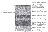
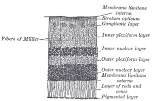
Anatomy of vertebrate retina
- Inner limiting membraneInner limiting membraneThe inner limiting membrane is the boundary between the retina and the vitreous body, formed by astrocytes and the end feet of Müller cells. It is separated from the vitreous humor by a basal lamina....
– Müller cellMuller gliaMüller glia, or Müller cells, are glial cells found in the vertebrate retina, which normally serve the functions of any normal glial cells. However, following injury to the retina, it has been seen that Müller glia undergo dedifferentiation into multipotent progenitor cells...
footplates - Nerve fiber layerNerve fiber layerThe retinal nerve fiber layer is formed by the expansion of the fibers of the optic nerve; it is thickest near the porus opticus, gradually diminishing toward the ora serrata....
– axons of the ganglion cell nuclei - Ganglion cell layerGanglion cell layerThe ganglion cell layer is a layer of the retina that consists of retinal ganglion cells.In the macula lutea, the layer forms several strata....
– contains nuclei of ganglion cells, the axons of which become the optic nerve fibers for messages. - Inner plexiform layerInner plexiform layerThe inner plexiform layer is an area of the retina that is made up of a dense reticulum of fibrils formed by interlaced dendrites of retinal ganglion cells and cells of the inner nuclear layer. Within this reticulum a few branched spongioblasts are sometimes embedded....
– contains the synapse between the bipolar cell axons and the dendrites of the ganglion and amacrine cells. - Inner nuclear layerInner nuclear layerThe inner nuclear layer or layer of inner granules is made up of a number of closely packed cells, of which there are three varieties, viz.: bipolar cells, horizontal cells, and amacrine cells.-Bipolar cells:...
– contains the nuclei and surrounding cell bodies (perikarya) of the bipolar cells. - Outer plexiform layerOuter plexiform layerThe outer plexiform layer is a layer of neuronal synapses in the retina of the eye. It consists of a dense network of synapses between dendrites of horizontal cells from the inner nuclear layer, and photoreceptor cell inner segments from the outer nuclear layer...
– projections of rods and cones ending in the rod spherule and cone pedicle, respectively. These make synapses with dendrites of bipolar In the macular regionMaculaThe macula or macula lutea is an oval-shaped highly pigmented yellow spot near the center of the retina of the human eye. It has a diameter of around 5 mm and is often histologically defined as having two or more layers of ganglion cells...
, this is known as the Fiber layer of Henle. - Outer nuclear layerOuter nuclear layerThe outer nuclear layer , is one of the layers of the vertebrate retina, the light-detecting portion of the eye...
– cell bodies of rods and cones - External limiting membraneExternal limiting membraneThe external limiting membrane is one of the ten distinct layers of the retina of the eye. It has a network-like structure and is situated at the bases of the rods and cones.-Additional images:-External links:...
– layer that separates the inner segment portions of the photoreceptors from their cell nucleus - Photoreceptor layer – rodsRod cellRod cells, or rods, are photoreceptor cells in the retina of the eye that can function in less intense light than can the other type of visual photoreceptor, cone cells. Named for their cylindrical shape, rods are concentrated at the outer edges of the retina and are used in peripheral vision. On...
/conesCone cellCone cells, or cones, are photoreceptor cells in the retina of the eye that are responsible for color vision; they function best in relatively bright light, as opposed to rod cells that work better in dim light. If the retina is exposed to an intense visual stimulus, a negative afterimage will be... - Retinal pigment epithelium - single layer of cuboidal cells
These can be simplified into 4 main processing stages: photoreception, transmission to bipolar cells, transmission to ganglion cells which also contain photoreceptors, the photosensitive ganglion cell
Photosensitive ganglion cell
Photosensitive ganglion cells, also called photosensitive Retinal Ganglion Cells , intrinsically photosensitive Retinal Ganglion Cells or melanopsin-containing ganglion cells, are a type of neuron in the retina of the mammalian eye.They were discovered in the early 1990sand are, unlike other...
s, and transmission along the optic nerve. At each synaptic stage there are also laterally connecting horizontal and amacrine cells.
The optic nerve is a central tract of many axons of ganglion cells connecting primarily to the lateral geniculate body, a visual relay station in the diencephalon (the rear of the forebrain). It also projects to the superior colliculus
Superior colliculus
The optic tectum or simply tectum is a paired structure that forms a major component of the vertebrate midbrain. In mammals this structure is more commonly called the superior colliculus , but, even in mammals, the adjective tectal is commonly used. The tectum is a layered structure, with a...
, the suprachiasmatic nucleus
Suprachiasmatic nucleus
The suprachiasmatic nucleus or nuclei, abbreviated SCN, is a tiny region on the brain's midline, situated directly above the optic chiasm. It is responsible for controlling circadian rhythms...
, and the nucleus of the optic tract. It passes through the other layers creating the Optic disc
Optic disc
The optic disc or optic nerve head is the location where ganglion cell axons exit the eye to form the optic nerve. There are no light sensitive rods or cones to respond to a light stimulus at this point. This causes a break in the visual field called "the blind spot" or the "physiological blind spot"...
in primates.
Additional structures, not directly associated with vision, are found as outgrowths of the retina in some vertebrate groups. In bird
Bird
Birds are feathered, winged, bipedal, endothermic , egg-laying, vertebrate animals. Around 10,000 living species and 188 families makes them the most speciose class of tetrapod vertebrates. They inhabit ecosystems across the globe, from the Arctic to the Antarctic. Extant birds range in size from...
s, the pecten
Pecten oculi
The pecten or pecten oculi is a comb-like structure of blood vessels belonging to the choroid in the eye of a bird. It is non-sensory and is a pigmented structure that projects into the vitreous body from the point where the optic nerve enters the eyeball. The pecten is believed to both nourish the...
is a vascular structure of complex shape that projects from the retina into the vitreous humour
Vitreous humour
The vitreous humour or vitreous humor is the clear gel that fills the space between the lens and the retina of the eyeball of humans and other vertebrates...
; it supplies oxygen and nutrients to the eye, and may also aid in vision. Reptile
Reptile
Reptiles are members of a class of air-breathing, ectothermic vertebrates which are characterized by laying shelled eggs , and having skin covered in scales and/or scutes. They are tetrapods, either having four limbs or being descended from four-limbed ancestors...
s have a similar, but much simpler, structure, referred to as the papillary cone.
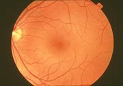
Physical structure of human retina
In adult humans, the entire retina is approximately 72% of a sphere about 22 mm in diameter. The entire retina contains about 7 million cones and 75 to 150 million rods. The optic discOptic disc
The optic disc or optic nerve head is the location where ganglion cell axons exit the eye to form the optic nerve. There are no light sensitive rods or cones to respond to a light stimulus at this point. This causes a break in the visual field called "the blind spot" or the "physiological blind spot"...
, a part of the retina sometimes called "the blind spot" because it lacks photoreceptors, is located at the optic papilla, a nasal zone where the optic-nerve fibers leave the eye. It appears as an oval white area of 3mm². Temporal (in the direction of the temples) to this disc is the macula
Macula
The macula or macula lutea is an oval-shaped highly pigmented yellow spot near the center of the retina of the human eye. It has a diameter of around 5 mm and is often histologically defined as having two or more layers of ganglion cells...
. At its center is the fovea
Fovea
The fovea centralis, also generally known as the fovea , is a part of the eye, located in the center of the macula region of the retina....
, a pit that is responsible for our sharp central vision but is actually less sensitive to light because of its lack of rods. Human and non-human primate
Primate
A primate is a mammal of the order Primates , which contains prosimians and simians. Primates arose from ancestors that lived in the trees of tropical forests; many primate characteristics represent adaptations to life in this challenging three-dimensional environment...
s possess one fovea as opposed to certain bird species such as hawks who actually are bifoviate and dogs and cats who possess no fovea but a central band known as the visual streak. Around the fovea extends the central retina for about 6 mm and then the peripheral retina. The edge of the retina is defined by the ora serrata
Ora serrata
The ora serrata is the serrated junction between the retina and the ciliary body. This junction marks the transition from the simple non-photosensitive area of the retina to the complex, multi-layered photosensitive region. In animals in which the region does not have a serrated appearance, it is...
. The length from one ora to the other (or macula), the most sensitive area along the horizontal meridian is about 3.2 mm.
Nerve
A peripheral nerve, or simply nerve, is an enclosed, cable-like bundle of peripheral axons . A nerve provides a common pathway for the electrochemical nerve impulses that are transmitted along each of the axons. Nerves are found only in the peripheral nervous system...
cells and two of synapse
Synapse
In the nervous system, a synapse is a structure that permits a neuron to pass an electrical or chemical signal to another cell...
s, including the unique ribbon synapse
Ribbon synapse
Ribbon synapse is a type of synapse linking some particular neuronal cells, which have unique features, such as their morphology, mechanisms of multivesicular release and calcium channel positioning which promote high speed of neurotransmitter release and an ongoing cycle of exocytosis and...
s. The optic nerve carries the ganglion cell
Ganglion cell
A retinal ganglion cell is a type of neuron located near the inner surface of the retina of the eye. It receives visual information from photoreceptors via two intermediate neuron types: bipolar cells and amacrine cells...
axon
Axon
An axon is a long, slender projection of a nerve cell, or neuron, that conducts electrical impulses away from the neuron's cell body or soma....
s to the brain and the blood vessels that open into the retina. The ganglion cells lie innermost in the retina while the photoreceptive cells lie outermost. Because of this counter-intuitive arrangement, light must first pass through and around the ganglion cells and through the thickness of the retina, (including its capillary vessels, not shown) before reaching the rods and cones. However it does not pass through the epithelium
Epithelium
Epithelium is one of the four basic types of animal tissue, along with connective tissue, muscle tissue and nervous tissue. Epithelial tissues line the cavities and surfaces of structures throughout the body, and also form many glands. Functions of epithelial cells include secretion, selective...
or the choroid
Choroid
The choroid, also known as the choroidea or choroid coat, is the vascular layer of the eye, containing connective tissue, and lying between the retina and the sclera. The human choroid is thickest at the far extreme rear of the eye , while in the outlying areas it narrows to 0.1 mm...
(both of which are opaque).
The white blood cell
White blood cell
White blood cells, or leukocytes , are cells of the immune system involved in defending the body against both infectious disease and foreign materials. Five different and diverse types of leukocytes exist, but they are all produced and derived from a multipotent cell in the bone marrow known as a...
s in the capillaries in front of the photoreceptors can be perceived as tiny bright moving dots when looking into blue light. This is known as the blue field entoptic phenomenon
Blue field entoptic phenomenon
The blue field entoptic phenomenon or Scheerer's phenomenon is the appearance of tiny bright dots moving quickly along squiggly lines in the visual field, especially when looking into bright blue light...
(or Scheerer's phenomenon).
Between the ganglion cell layer and the rods and cones there are two layers of neuropil
Neuropil
In neuroanatomy, a neuropil, which is sometimes referred to as a neuropile, is a region between neuronal cell bodies in the gray matter of the brain and blood-brain barrier . It consists of a dense tangle of axon terminals, dendrites and glial cell processes...
s where synaptic contacts are made. The neuropil layers are the outer plexiform layer
Outer plexiform layer
The outer plexiform layer is a layer of neuronal synapses in the retina of the eye. It consists of a dense network of synapses between dendrites of horizontal cells from the inner nuclear layer, and photoreceptor cell inner segments from the outer nuclear layer...
and the inner plexiform layer
Inner plexiform layer
The inner plexiform layer is an area of the retina that is made up of a dense reticulum of fibrils formed by interlaced dendrites of retinal ganglion cells and cells of the inner nuclear layer. Within this reticulum a few branched spongioblasts are sometimes embedded....
. In the outer the rods and cones connect to the vertically running bipolar cells, and the horizontally oriented horizontal cells connect to ganglion cells.
The central retina is cone-dominated and the peripheral retina is rod-dominated. In total there are about seven million cones and a hundred million rods. At the centre of the macula is the foveal pit where the cones are smallest and in a hexagonal mosaic, the most efficient and highest density. Below the pit the other retina layers are displaced, before building up along the foveal slope until the rim of the fovea or parafovea which is the thickest portion of the retina. The macula has a yellow pigmentation from screening pigments and is known as the macula lutea. The area directly surrounding the fovea has the highest density of rods converging on single bipolars. Since the cones have a much lesser power of merging signals, the fovea allows for the sharpest vision the eye can attain.
Though the rod and cones are a mosaic of sorts, transmission from receptors to bipolars to ganglion cells is not the case, Since there are about 150 million receptors and only 1 million optic nerve fibers, there must be convergence and thus mixing of signals. Moreover, the horizontal action of the horizontal and amacrine cells can allow one area of the retina to control another (e.g., one stimulus inhibiting another). This inhibition is key to the sum of messages sent to the higher regions of the brain. In some lower vertebrates, (e.g., the pigeon) there is a "centrifugal" control of messages, that is, one layer can control another, or higher regions of the brain can drive the retinal nerve cells, but in primates this does not occur.
Vertebrate and cephalopod retina differences
The vertebrate retina is inverted in the sense that the light sensing cells sit at the back side of the retina, so that light has to pass through layers of neurons and capillaries before it reaches the rods and cones. By contrast, the cephalopodCephalopod
A cephalopod is any member of the molluscan class Cephalopoda . These exclusively marine animals are characterized by bilateral body symmetry, a prominent head, and a set of arms or tentacles modified from the primitive molluscan foot...
retina has the photoreceptors at the front side of the retina, with processing neurons and capillaries behind them. Because of this, cephalopods do not have a blind spot
Blind spot
Blind spot may refer to:In ophthalmology:*Scotoma, an obscuration of the visual field*Optic disc, also known as the anatomical blind spot, the specific region of the retina where the optic nerve and blood vessels pass through to connect to the back of the eye*Blind spot , also known as the...
.
The cephalopod retina does not originate as an outgrowth of the brain, as the vertebrate one does. It was originally argued that this difference shows that vertebrate and cephalopod eyes are not homologous
Homology (biology)
Homology forms the basis of organization for comparative biology. In 1843, Richard Owen defined homology as "the same organ in different animals under every variety of form and function". Organs as different as a bat's wing, a seal's flipper, a cat's paw and a human hand have a common underlying...
but have evolved separately. The evolutionary biologist Richard Dawkins
Richard Dawkins
Clinton Richard Dawkins, FRS, FRSL , known as Richard Dawkins, is a British ethologist, evolutionary biologist and author...
cites the imperfect structure of the human retina as confounding claims by creationists or intelligent design
Intelligent design
Intelligent design is the proposition that "certain features of the universe and of living things are best explained by an intelligent cause, not an undirected process such as natural selection." It is a form of creationism and a contemporary adaptation of the traditional teleological argument for...
theorists that the human eye is so perfect it must have a designer.
In 2009 Kröger anatomically showed in Zebrafish that though the inverted arrangement is nonadaptive in that it creates avoidable scattering of light (and thus loss of light and image blur), it has space-saving advantages for small-eyed animals in which there is a minimal vitreous body, as the space between the lens and the photoreceptors’ light-sensitive outer segments is completely filled with retinal cells.
The difference between vertebrate and cephalopod retinas presents an interesting puzzle of evolutionary path which is not yet fully settled. From an evolutionary perspective, a convoluted structure such as the inverted retina can generally come about as a consequence of two alternative processes; (a) an advantageous "good" compromise between competing functional limitations, or (b) as a historical maladaptive relic of the convoluted path of organ evolution and transformation. Vision is an important adaptation in higher vertebrates. Therefore, if the retina is indeed “wired wrongly” or “badly designed” (from an optical engineering point of view) then it sensible to look for it to possibly have some very significant physiological advantage. One such suggestion is based on the argument that the mammalian photoreceptor amplification process requires vast quantities of metabolic energy, and consequently, it requires massive and homogeneous supply of blood. Indeed, a unique network of blood vessels gives impression of being well adapted to provide the photoreceptor layer with copious quantities of blood. This lead to a proposition that the inverted retina is an adaptation to deliver abundant quantities of oxygen to the photoreceptor cells commensurate with their high energy demands.
Yet, the cephalopods have a non-inverted retina which is comparable in resolving power to the eyes of many vertebrates. Hence, at least for cold-blooded vertebrates, the inverted retinal structure is almost certainly not an adaptive necessity. All together, the inverted retina structure remains a mystery. Why should such an unlikely arrangement have appeared in the first place some 600 million years ago in the earliest of vertebrates who had presumably no need for high acuity vision and in all probability possessed photoreceptors with lower metabolic rates than those of higher warm-blooded vertebrates today? If the non-inverted retina works so well for the cold-blooded cephalopods, why did evolution go down the path of having inverted retina in most cold-blooded vertebrates (e.g., fish)? We are likely to be still missing a satisfactory scientific understanding of either (a) some hidden optimal physiological advantages of the inverted retina structure, or (b) a well-reasoned evolutionary path of how the retina has evolved in early stages from some other organ or structures so that the inverted retina is a sub-optimal relic of the evolutionary path. E.g., early in the Evolution of the eye
Evolution of the eye
The evolution of the eye has been a subject of significant study, as a distinctive example of a homologous organ present in a wide variety of taxa. Certain components of the eye, such as the visual pigments, appear to have a common ancestry – that is, they evolved once, before the animals radiated...
, there was a cup-shaped hollow, with the vertebrate retina represented by a single cell layer that lined the eye's cavity. This layer then became increasingly photosensitive, evolving into rods and cones. Later, this single layer would be supplemented by additional neurons, creating cross-connections for logic processing and encoding of signals. In the primitive eye, there was no lens or vitreous body. Added layers then likely came to be positioned "on top" of older structures in the bottom of the cup, finally ending up between the rods and cones and the vitreous body.
Physiology
An image is produced by the patterned excitation of the cones and rods in the retina. The excitation is processed by the neuronal system and various parts of the brain working in parallel to form a representation of the external environment in the brain.The cones respond to bright light and mediate high-resolution colour vision during daylight illumination (also called photopic vision). The rods are saturated at daylight levels and don't contribute to pattern vision. However, rods do respond to dim light and mediate lower-resolution, monochromatic vision under very low levels of illumination (called scotopic vision). The illumination in most office settings falls between these two levels and is called mesopic vision. At these light levels, both the rods and cones are actively contributing pattern information to that exiting the eye. What contribution the rod information makes to pattern vision under these circumstances is unclear.
The response of cones to various wavelengths of light is called their spectral sensitivity. In normal human vision, the spectral sensitivity of a cone falls into one of three subgroups. These are often called blue, green, and red cones but more accurately are short, medium, and long wavelength sensitive cone subgroups. It is a lack of one or more of the cone subtypes that causes individuals to have deficiencies in colour vision or various kinds of colour blindness
Color blindness
Color blindness or color vision deficiency is the inability or decreased ability to see color, or perceive color differences, under lighting conditions when color vision is not normally impaired...
. These individuals are not blind to objects of a particular colour but experience the inability to distinguish between two groups of colours that can be distinguished by people with normal vision. Humans have three different types of cones (trichromatic vision) while most other mammals lack cones with red sensitive pigment and therefore have poorer (dichromatic) colour vision. However, some animals have four spectral subgroups, e.g., the trout adds an ultraviolet subgroup to short, medium and long subgroups that are similar to humans. Some fish are sensitive to the polarization of light as well.
When light falls on a receptor it sends a proportional response synaptically to bipolar cell
Bipolar cell
As a part of the retina, the bipolar cell exists between photoreceptors and ganglion cells. They act, directly or indirectly, to transmit signals from the photoreceptors to the ganglion cells.-Overview:...
s which in turn signal the retinal ganglion cells. The receptors are also 'cross-linked' by horizontal cell
Horizontal cell
Horizontal cells are the laterally interconnecting neurons in the outer plexiform layer of the retina of mammalian eyes. They help integrate and regulate the input from multiple photoreceptor cells...
s and amacrine cell
Amacrine cell
Amacrine cells are interneurons in the retina. Amacrine cells are responsible for 70% of input to retinal ganglion cells. Bipolar cells, which are responsible for the other 30% of input to retinal ganglia, are regulated by amacrine cells.-Overview:...
s, which modify the synaptic signal before the ganglion cells. Rod and cone signals are intermixed and combine, although rods are mostly active in very poorly lit conditions and saturate in broad daylight, while cones function in brighter lighting because they are not sensitive enough to work at very low light levels.
Despite the fact that all are nerve cells, only the retinal ganglion cells and few amacrine cells create action potentials. In the photoreceptors, exposure to light hyperpolarizes the membrane in a series of graded shifts. The outer cell segment contains a photopigment
Photopigment
Photopigments are unstable pigments that undergo a chemical change when they absorb light. The term is generally applied to the non-protein chromophore moiety of photosensitive chromoproteins, such as the pigments involved in photosynthesis and photoreception...
. Inside the cell the normal levels of cyclic guanosine monophosphate
Cyclic guanosine monophosphate
Cyclic guanosine monophosphate is a cyclic nucleotide derived from guanosine triphosphate . cGMP acts as a second messenger much like cyclic AMP...
(cGMP) keep the Na+ channel open and thus in the resting state the cell is depolarised. The photon
Photon
In physics, a photon is an elementary particle, the quantum of the electromagnetic interaction and the basic unit of light and all other forms of electromagnetic radiation. It is also the force carrier for the electromagnetic force...
causes the retinal
Retinal
Retinal, also called retinaldehyde or vitamin A aldehyde, is one of the many forms of vitamin A . Retinal is a polyene chromophore, and bound to proteins called opsins, is the chemical basis of animal vision...
bound to the receptor protein to isomerise to trans-retinal
Retinal
Retinal, also called retinaldehyde or vitamin A aldehyde, is one of the many forms of vitamin A . Retinal is a polyene chromophore, and bound to proteins called opsins, is the chemical basis of animal vision...
. This causes receptor to activate multiple G-proteins. This in turn causes the Ga-subunit of the protein to activate a phosphodiesterase (PDE6), which degrades cGMP, resulting in the closing of Na+ cyclic nucleotide-gated ion channel
Cyclic nucleotide-gated ion channel
Cyclic nucleotide-gated ion channels are ion channels that function in response to the binding of cyclic nucleotides. CNG channels are nonselective cation channels that are found in the membranes of various types of cells.- Discovery :...
s (CNGs). Thus the cell is hyperpolarised. The amount of neurotransmitter released is reduced in bright light and increases as light levels fall. The actual photopigment is bleached away in bright light and only replaced as a chemical process, so in a transition from bright light to darkness the eye can take up to thirty minutes to reach full sensitivity (see Adaptation (eye)
Adaptation (eye)
In ocular physiology, adaptation is the ability of the eye to adjust to various levels of darkness and light.-Efficacy:The human eye can function from very dark to very bright levels of light; its sensing capabilities reach across nine orders of magnitude. This means that the brightest and the...
).
In the retinal ganglion cells there are two types of response, depending on the receptive field
Receptive field
The receptive field of a sensory neuron is a region of space in which the presence of a stimulus will alter the firing of that neuron. Receptive fields have been identified for neurons of the auditory system, the somatosensory system, and the visual system....
of the cell. The receptive fields of retinal ganglion cells comprise a central approximately circular area, where light has one effect on the firing of the cell, and an annular surround, where light has the opposite effect on the firing of the cell. In ON cells, an increment in light intensity in the centre of the receptive field causes the firing rate to increase. In OFF cells, it makes it decrease. In a linear model, this response profile is well described by a Difference of Gaussians
Difference of Gaussians
In computer vision, Difference of Gaussians is a grayscale image enhancement algorithm that involves the subtraction of one blurred version of an original grayscale image from another, less blurred version of the original. The blurred images are obtained by convolving the original grayscale image...
and is the basis for edge detection
Edge detection
Edge detection is a fundamental tool in image processing and computer vision, particularly in the areas of feature detection and feature extraction, which aim at identifying points in a digital image at which the image brightness changes sharply or, more formally, has discontinuities...
algorithms. Beyond this simple difference ganglion cells are also differentiated by chromatic sensitivity and the type of spatial summation. Cells showing linear spatial summation are termed X cells (also called parvocellular, P, or midget ganglion cells), and those showing non-linear summation are Y cells (also called magnocellular, M, or parasol retinal ganglion cells), although the correspondence between X and Y cells (in the cat retina) and P and M cells (in the primate retina) is not as simple as it once seemed.
In the transfer of visual signals to the brain, the visual pathway, the retina is vertically divided in two, a temporal (nearer to the temple) half and a nasal (nearer to the nose) half. The axons from the nasal half cross the brain at the optic chiasma to join with axons from the temporal half of the other eye before passing into the lateral geniculate body.
Although there are more than 130 million retinal receptors, there are only approximately 1.2 million fibres (axons) in the optic nerve; a large amount of pre-processing is performed within the retina. The fovea produces the most accurate information. Despite occupying about 0.01% of the visual field (less than 2° of visual angle
Visual angle
The visual angle is the angle a viewed object subtends at the eye, usually stated in degrees of arc.It also is called the object's angular size....
), about 10% of axons in the optic nerve are devoted to the fovea. The resolution limit of the fovea has been determined at around 10,000 points. The information capacity is estimated at 500,000 bits per second (for more information on bits, see information theory
Information theory
Information theory is a branch of applied mathematics and electrical engineering involving the quantification of information. Information theory was developed by Claude E. Shannon to find fundamental limits on signal processing operations such as compressing data and on reliably storing and...
) without colour or around 600,000 bits per second including colour .
Spatial encoding
Decorrelation
Decorrelation is a general term for any process that is used to reduce autocorrelation within a signal, or cross-correlation within a set of signals, while preserving other aspects of the signal. A frequently used method of decorrelation is the use of a matched linear filter to reduce the...
the incoming images in a manner to be described below. These operations are carried out by the center surround structures as implemented by the bipolar and ganglion cells.
There are two types of center surround structures in the retina—on-centers and off-centers. On-centers have a positively weighted center and a negatively weighted surround. Off-centers are just the opposite. Positive weighting is more commonly known as excitatory and negative weighting is more commonly known as inhibitory.
These center surround structures are not physical in the sense that one cannot see them by staining samples of tissue and examining the retina's anatomy. The center surround structures are logical (i.e., mathematically abstract) in the sense that they depend on the connection strengths between ganglion and bipolar cells. It is believed that the connection strengths between cells is caused by the number and types of ion channel
Ion channel
Ion channels are pore-forming proteins that help establish and control the small voltage gradient across the plasma membrane of cells by allowing the flow of ions down their electrochemical gradient. They are present in the membranes that surround all biological cells...
s embedded in the synapse
Synapse
In the nervous system, a synapse is a structure that permits a neuron to pass an electrical or chemical signal to another cell...
s between the ganglion and bipolar cells. Stephen Kuffler
Stephen Kuffler
Stephen William Kuffler was a pre-eminent Hungarian-American neurophysiologist. Often, he's been referred to as the "Father of Modern Neuroscience". He founded the Harvard Neurobiology department in 1966, and made numerous seminal contributions to our understanding of vision, neural coding, and...
in the 1950s was the first person to begin to understand these center surround structures in the retina of cats. See Receptive field
Receptive field
The receptive field of a sensory neuron is a region of space in which the presence of a stimulus will alter the firing of that neuron. Receptive fields have been identified for neurons of the auditory system, the somatosensory system, and the visual system....
for figures and more information on center surround structures. See chapter 3 of David Hubel's on-line book (listed below) for an excellent introduction.
The center surround structures are mathematically equivalent to the edge detection
Edge detection
Edge detection is a fundamental tool in image processing and computer vision, particularly in the areas of feature detection and feature extraction, which aim at identifying points in a digital image at which the image brightness changes sharply or, more formally, has discontinuities...
algorithms used by computer programmers to extract or enhance the edges in a digital photograph. Thus the retina performs operations on the image to enhance the edges of objects within its visual field. For example, in a picture of a dog, a cat and a car, it is the edges of these objects that contain the most information. In order for higher functions in the brain (or in a computer for that matter) to extract and classify objects such as a dog and a cat, the retina is the first step to separating out the various objects within the scene.
As an example, the following matrix
Matrix (mathematics)
In mathematics, a matrix is a rectangular array of numbers, symbols, or expressions. The individual items in a matrix are called its elements or entries. An example of a matrix with six elements isMatrices of the same size can be added or subtracted element by element...
is at the heart of the computer algorithm
Algorithm
In mathematics and computer science, an algorithm is an effective method expressed as a finite list of well-defined instructions for calculating a function. Algorithms are used for calculation, data processing, and automated reasoning...
that implements edge detection. This matrix is the computer equivalent to the center surround structure. In this example, each box (element) within this matrix would be connected to one photoreceptor. The photoreceptor in the center is the current receptor being processed. The center photoreceptor is multiplied by the +1 weight factor. The surrounding photoreceptors are the "nearest neighbors" to the center and are multiplied by the -1/8 value. The sum of all nine of these elements is finally calculated. This summation is repeated for every photoreceptor in the image by shifting left to the end of a row and then down to the next line.
| -1/8 | -1/8 | -1/8 |
| -1/8 | +1 | -1/8 |
| -1/8 | -1/8 | -1/8 |
The total sum of this matrix is zero if all the inputs from the nine photoreceptors are the same value. The zero result indicates the image was uniform (non-changing) within this small patch. Negative or positive sums mean something was varying (changing) within this small patch of nine photoreceptors.
The above matrix is only an approximation to what really happens inside the retina. The differences are:
- The above example is called "balanced". The term balanced means that the sum of the negative weights is equal to the sum of the positive weights so that they cancel out perfectly. Retinal ganglion cells are almost never perfectly balanced.
- The table is square while the center surround structures in the retina are circular.
- Neurons operate on spike trains traveling down nerve cell axons. Computers operate on a single Floating pointFloating pointIn computing, floating point describes a method of representing real numbers in a way that can support a wide range of values. Numbers are, in general, represented approximately to a fixed number of significant digits and scaled using an exponent. The base for the scaling is normally 2, 10 or 16...
number that is essentially constant from each input pixelPixelIn digital imaging, a pixel, or pel, is a single point in a raster image, or the smallest addressable screen element in a display device; it is the smallest unit of picture that can be represented or controlled....
. (The computer pixel is basically the equivalent of a biological photoreceptor.) - The retina performs all these calculations in parallel while the computer operates on each pixel one at a time. There are no repeated summations and shifting as there would be in a computer.
- Finally, the horizontalHorizontal cellHorizontal cells are the laterally interconnecting neurons in the outer plexiform layer of the retina of mammalian eyes. They help integrate and regulate the input from multiple photoreceptor cells...
and amacrine cellAmacrine cellAmacrine cells are interneurons in the retina. Amacrine cells are responsible for 70% of input to retinal ganglion cells. Bipolar cells, which are responsible for the other 30% of input to retinal ganglia, are regulated by amacrine cells.-Overview:...
s play a significant role in this process but that is not represented here.
Here is an example of an input image and how edge detection would modify it.
Once the image is spatially encoded by the center surround structures, the signal is sent out the optical nerve (via the axons of the ganglion cells) through the optic chiasm
Optic chiasm
The optic chiasm or optic chiasma is the part of the brain where the optic nerves partially cross...
to the LGN (lateral geniculate nucleus
Lateral geniculate nucleus
The lateral geniculate nucleus is the primary relay center for visual information received from the retina of the eye. The LGN is found inside the thalamus of the brain....
). The exact function of the LGN is unknown at this time. The output of the LGN is then sent to the back of the brain. Specifically the output of the LGN "radiates" out to the V1 Primary visual cortex.
Simplified Signal Flow: Photoreceptors → Bipolar → Ganglion → Chiasm → LGN → V1 cortex
Diseases and disorders
There are many inherited and acquired diseases or disorders that may affect the retina. Some of them include:- Retinitis pigmentosaRetinitis pigmentosaRetinitis pigmentosa is a group of genetic eye conditions that leads to incurable blindness. In the progression of symptoms for RP, night blindness generally precedes tunnel vision by years or even decades. Many people with RP do not become legally blind until their 40s or 50s and retain some...
is a group of genetic diseases that affect the retina and causes the loss of night vision and peripheral vision. - Macular degenerationMacular degenerationAge-related macular degeneration is a medical condition which usually affects older adults and results in a loss of vision in the center of the visual field because of damage to the retina. It occurs in “dry” and “wet” forms. It is a major cause of blindness and visual impairment in older adults...
describes a group of diseases characterized by loss of central vision because of death or impairment of the cells in the maculaMaculaThe macula or macula lutea is an oval-shaped highly pigmented yellow spot near the center of the retina of the human eye. It has a diameter of around 5 mm and is often histologically defined as having two or more layers of ganglion cells...
. - Cone-rod dystrophy (CORD) describes a number of diseases where vision loss is caused by deterioration of the conesCone cellCone cells, or cones, are photoreceptor cells in the retina of the eye that are responsible for color vision; they function best in relatively bright light, as opposed to rod cells that work better in dim light. If the retina is exposed to an intense visual stimulus, a negative afterimage will be...
and/or rodsRod cellRod cells, or rods, are photoreceptor cells in the retina of the eye that can function in less intense light than can the other type of visual photoreceptor, cone cells. Named for their cylindrical shape, rods are concentrated at the outer edges of the retina and are used in peripheral vision. On...
in the retina. - In retinal separation, the retina detaches from the back of the eyeball. IgnipunctureIgnipunctureIgnipuncture is the original procedure of closing a retina break in retinal separation by transfixation of the break with cautery. The procedure was pioneered and named by Jules Gonin in the early 1900s...
is an outdated treatment method. The term retinal detachmentRetinal detachmentRetinal detachment is a disorder of the eye in which the retina peels away from its underlying layer of support tissue. Initial detachment may be localized, but without rapid treatment the entire retina may detach, leading to vision loss and blindness. It is a medical emergency.The retina is a...
is used to describe a separation of the neurosensory retina from the retinal pigment epithelium. There are several modern treatment methods for fixing a retinal detachmentRetinal detachmentRetinal detachment is a disorder of the eye in which the retina peels away from its underlying layer of support tissue. Initial detachment may be localized, but without rapid treatment the entire retina may detach, leading to vision loss and blindness. It is a medical emergency.The retina is a...
: pneumatic retinopexy, scleral buckleScleral buckleA scleral buckle is one of several ophthalmologic procedures that can be used to repair a retinal detachment. Retinal detachments are usually caused by retinal tears, and a scleral buckle can be used to close the retinal break....
, cryotherapyCryotherapyCryotherapy is the local or general use of low temperatures in medical therapy or the removal of heat from a body part. The term "cryotherapy" comes from the Greek cryo meaning cold and the word therapy meaning cure...
, laser photocoagulationLaser photocoagulationLaser coagulation or laser photocoagulation surgery is used to treat a number of eye diseases and has become widely used in recent decades...
and pars plana vitrectomyVitrectomyVitrectomy is a surgery to remove some or all of the vitreous humor from the eye. Anterior vitrectomy entails removing small portions of the vitreous from the front structures of the eye—often because these are tangled in an intraocular lens or other structures...
. - Both hypertensionHypertensionHypertension or high blood pressure is a cardiac chronic medical condition in which the systemic arterial blood pressure is elevated. What that means is that the heart is having to work harder than it should to pump the blood around the body. Blood pressure involves two measurements, systolic and...
and diabetes mellitusDiabetes mellitusDiabetes mellitus, often simply referred to as diabetes, is a group of metabolic diseases in which a person has high blood sugar, either because the body does not produce enough insulin, or because cells do not respond to the insulin that is produced...
can cause damage to the tiny blood vessels that supply the retina, leading to hypertensive retinopathyHypertensive retinopathyHypertensive retinopathy is damage to the retina due to high blood pressure .-Pathophysiology:The retina is one of the "target organs" that are damaged by sustained hypertension....
and diabetic retinopathyDiabetic retinopathyDiabetic retinopathy is retinopathy caused by complications of diabetes mellitus, which can eventually lead to blindness....
. - RetinoblastomaRetinoblastomaRetinoblastoma is a rapidly developing cancer that develops in the cells of retina, the light-detecting tissue of the eye. In the developed world, Rb has one of the best cure rates of all childhood cancers , with more than nine out of every ten sufferers surviving into...
is a cancerCancerCancer , known medically as a malignant neoplasm, is a large group of different diseases, all involving unregulated cell growth. In cancer, cells divide and grow uncontrollably, forming malignant tumors, and invade nearby parts of the body. The cancer may also spread to more distant parts of the...
of the retina. - Retinal diseases in dogDogThe domestic dog is a domesticated form of the gray wolf, a member of the Canidae family of the order Carnivora. The term is used for both feral and pet varieties. The dog may have been the first animal to be domesticated, and has been the most widely kept working, hunting, and companion animal in...
s include retinal dysplasiaRetinal dysplasiaRetinal dysplasia is an eye disease affecting the retina of animals and, less commonly, humans. It is usually a nonprogressive disease and can be caused by viral infections, drugs, vitamin A deficiency, or genetic defects...
, progressive retinal atrophyProgressive retinal atrophyProgressive retinal atrophy is a group of genetic diseases seen in certain breeds of dogs and, more rarely, cats. Similar to retinitis pigmentosa in humans, it is characterized by the bilateral degeneration of the retina, causing progressive vision loss culminating in blindness...
, and sudden acquired retinal degenerationSudden acquired retinal degenerationSudden acquired retinal degeneration syndrome is a disease in dogs causing sudden blindness. It can occur in any breed, but female dogs may be predisposed...
. - Lipemia retinalis is a white appearance of the retina, and can occur by lipid deposition in lipoprotein lipase deficiencyLipoprotein lipase deficiencyLipoprotein lipase deficiency is caused by a mutation in the gene which codes lipoprotein lipase....
.
Diagnosis and treatment
A number of different instruments are available for the diagnosis of diseases and disorders affecting the retina. An ophthalmoscope is used to examine the retina. Recently, adaptive opticsAdaptive optics
Adaptive optics is a technology used to improve the performance of optical systems by reducing the effect of wavefront distortions. It is used in astronomical telescopes and laser communication systems to remove the effects of atmospheric distortion, and in retinal imaging systems to reduce the...
has been used to image individual rods and cones in the living human retina and a company based in Scotland have engineered technology that allows physicians to observe the complete retina without any discomfort to patients.
The electroretinogram
Electroretinography
Electroretinography measures the electrical responses of various cell types in the retina, including the photoreceptors , inner retinal cells , and the ganglion cells. Electrodes are usually placed on the cornea and the skin near the eye, although it is possible to record the ERG from skin electrodes...
is used to measure non-invasively the retina's electrical activity, which is affected by certain diseases. A relatively new technology, now becoming widely available, is optical coherence tomography
Optical coherence tomography
Optical coherence tomography is an optical signal acquisition and processing method. It captures micrometer-resolution, three-dimensional images from within optical scattering media . Optical coherence tomography is an interferometric technique, typically employing near-infrared light...
(OCT). This non-invasive technique allows one to obtain a 3D
Dimension
In physics and mathematics, the dimension of a space or object is informally defined as the minimum number of coordinates needed to specify any point within it. Thus a line has a dimension of one because only one coordinate is needed to specify a point on it...
volumetric or high resolution cross-sectional tomogram of the retinal fine structure with histologic-quality.
Organ transplant
Organ transplantation is the moving of an organ from one body to another or from a donor site on the patient's own body, for the purpose of replacing the recipient's damaged or absent organ. The emerging field of regenerative medicine is allowing scientists and engineers to create organs to be...
of retinas has been attempted, but without much success. At MIT
Massachusetts Institute of Technology
The Massachusetts Institute of Technology is a private research university located in Cambridge, Massachusetts. MIT has five schools and one college, containing a total of 32 academic departments, with a strong emphasis on scientific and technological education and research.Founded in 1861 in...
, The University of Southern California, and the University of New South Wales
University of New South Wales
The University of New South Wales , is a research-focused university based in Kensington, a suburb in Sydney, New South Wales, Australia...
, an "artificial retina" is under development: an implant which will bypass the photoreceptors of the retina and stimulate the attached nerve cells directly, with signals from a digital camera.
Retinal blood supply
Blood vessel
The blood vessels are the part of the circulatory system that transports blood throughout the body. There are three major types of blood vessels: the arteries, which carry the blood away from the heart; the capillaries, which enable the actual exchange of water and chemicals between the blood and...
s in the retina has been used for biometric identification
Retinal scan
A retinal scan is a biometric technique that uses the unique patterns on a person's retina to identify them. It is not to be confused with another ocular-based technology, iris recognition.-Introduction:...
.
The vascular topographical geometry in the retina is known to conform to structural
principles that are related to certain physical properties . The analysis of the geometrical structure is very important as deviations from the optimal principles may indicate some cardiovascular diseases, such as hypertension and atherosclerosis ; a comprehensive analysis is given by Patton et al (2006) . The identification of vascular bifurcations is one of the basic steps in this analysis. Azzopardi and Petkov (2011) propose a computer vision
Computer vision
Computer vision is a field that includes methods for acquiring, processing, analysing, and understanding images and, in general, high-dimensional data from the real world in order to produce numerical or symbolic information, e.g., in the forms of decisions...
algorithm that automatically detects these retinal features. Their results are evaluated against the ground truth data of vascular bifurcations of retinal fundus images that are obtained from the DRIVE data set.
Research
George WaldGeorge Wald
George Wald was an American scientist who is best known for his work with pigments in the retina. He won a share of the 1967 Nobel Prize in Physiology or Medicine with Haldan Keffer Hartline and Ragnar Granit.- Research :...
, Haldan Keffer Hartline
Haldan Keffer Hartline
Haldan Keffer Hartline was an American physiologist who was a co-winner of the 1967 Nobel Prize in Physiology or Medicine for his work in analyzing the neurophysiological mechanisms of vision.Hartline began his study of retinal electrophysiology as a National Research Council Fellow at Johns...
and Ragnar Granit
Ragnar Granit
Ragnar Arthur Granit was a Finnish/Swedish scientist who was awarded the Nobel Prize in Physiology or Medicine in 1967 along with Haldan Keffer Hartline and George Wald....
won the 1967 Nobel Prize in Physiology or Medicine
Nobel Prize in Physiology or Medicine
The Nobel Prize in Physiology or Medicine administered by the Nobel Foundation, is awarded once a year for outstanding discoveries in the field of life science and medicine. It is one of five Nobel Prizes established in 1895 by Swedish chemist Alfred Nobel, the inventor of dynamite, in his will...
for their scientific research on the retina.
A recent University of Pennsylvania
University of Pennsylvania
The University of Pennsylvania is a private, Ivy League university located in Philadelphia, Pennsylvania, United States. Penn is the fourth-oldest institution of higher education in the United States,Penn is the fourth-oldest using the founding dates claimed by each institution...
study calculated the approximate bandwidth
Bandwidth (computing)
In computer networking and computer science, bandwidth, network bandwidth, data bandwidth, or digital bandwidth is a measure of available or consumed data communication resources expressed in bits/second or multiples of it .Note that in textbooks on wireless communications, modem data transmission,...
of human retinas is 8.75 megabits per second, whereas guinea pig
Guinea pig
The guinea pig , also called the cavy, is a species of rodent belonging to the family Caviidae and the genus Cavia. Despite their common name, these animals are not in the pig family, nor are they from Guinea...
retinas transfer at 875 kilobits.
MacLaren & Pearson and colleagues at University College London
University College London
University College London is a public research university located in London, United Kingdom and the oldest and largest constituent college of the federal University of London...
and Moorfields Eye Hospital
Moorfields Eye Hospital
Moorfields Eye Hospital NHS Foundation Trust is an NHS eye hospital located in London, United Kingdom. It is the oldest and largest eye hospital in the world and is internationally renowned for its comprehensive clinical and research activities...
in London showed in 2006 that photoreceptor cells could be transplanted successfully in the mouse retina if donor cells were at a critical developmental stage. Recently Ader and colleagues in Dublin showed using the electron microscope that transplanted photoreceptors formed synaptic connections.
Retinal gene therapy
Gene therapyGene therapy
Gene therapy is the insertion, alteration, or removal of genes within an individual's cells and biological tissues to treat disease. It is a technique for correcting defective genes that are responsible for disease development...
holds promise as a potential avenue to cure a wide range of retinal diseases. This involves using a non-infectious virus to shuttle a gene into a part of the retina. Recombinant adeno-associated virus
Adeno-associated virus
Adeno-associated virus is a small virus which infects humans and some other primate species. AAV is not currently known to cause disease and consequently the virus causes a very mild immune response. AAV can infect both dividing and non-dividing cells and may incorporate its genome into that of...
(rAAV) vectors possess a number of features that render them ideally suited for retinal gene therapy, including a lack of pathogenicity, minimal immunogenicity, and the ability to transduce postmitotic cells in a stable and efficient manner. rAAV vectors are increasingly utilized for their ability to mediate efficient transduction of retinal pigment epithelium (RPE), photoreceptor cells and retinal ganglion cells. Each cell type can be specifically targeted by choosing the appropriate combination of AAV serotype
Serotype
Serotype or serovar refers to distinct variations within a subspecies of bacteria or viruses. These microorganisms, viruses, or cells are classified together based on their cell surface antigens...
, promoter, and intraocular injection site.
The unique architecture of the retina and its relatively immune-privileged environment help this process. Tight junctions that form the blood retinal barrier separate the subretinal space from the blood supply, thus protecting it from microbes and most immune-mediated damage, and enhancing its potential to respond to vector-mediated therapies. The highly compartmentalized anatomy of the eye facilitates accurate delivery of therapeutic vector suspensions to specific tissues under direct visualization using microsurgical techniques. In the sheltered environment of the retina, AAV vectors are able to maintain high levels of transgene
Transgene
A transgene is a gene or genetic material that has been transferred naturally or by any of a number of genetic engineering techniques from one organism to another....
expression in the retinal pigmented epithelium (RPE), photoreceptors, or ganglion cells for long periods of time after a single treatment. In addition, the eye and the visual system can be routinely and easily monitored for visual function and retinal structural changes after injections with noninvasive advanced technology, such as visual acuities, contrast sensitivity, fundus
Fundus (eye)
The fundus of the eye is the interior surface of the eye, opposite the lens, and includes the retina, optic disc, macula and fovea, and posterior pole. The fundus can be viewed with an ophthalmoscope. The term may also be inclusive of Bruch's membrane and the choroid.The color of the fundus varies...
auto-fluorescence (FAF), dark-adapted visual thresholds, vascular diameters, pupillometry, electroretinography
Electroretinography
Electroretinography measures the electrical responses of various cell types in the retina, including the photoreceptors , inner retinal cells , and the ganglion cells. Electrodes are usually placed on the cornea and the skin near the eye, although it is possible to record the ERG from skin electrodes...
(ERG), multifocal ERG and optical coherence tomography
Optical coherence tomography
Optical coherence tomography is an optical signal acquisition and processing method. It captures micrometer-resolution, three-dimensional images from within optical scattering media . Optical coherence tomography is an interferometric technique, typically employing near-infrared light...
(OCT).
This strategy is effective against retinal diseases that have been studied including neovascular diseases that are features of age-related macular degeneration, diabetic retinopathy
Diabetic retinopathy
Diabetic retinopathy is retinopathy caused by complications of diabetes mellitus, which can eventually lead to blindness....
and retinopathy of prematurity
Retinopathy of prematurity
Retinopathy of prematurity , previously known as retrolental fibroplasia , is an eye disease that affects prematurely-born babies. It is thought to be caused by disorganized growth of retinal blood vessels which may result in scarring and retinal detachment. ROP can be mild and may resolve...
. Since the regulation of vascularization in the mature retina involves a balance between endogenous positive growth factors, such as vascular endothelial growth factor
Vascular endothelial growth factor
Vascular endothelial growth factor is a signal protein produced by cells that stimulates vasculogenesis and angiogenesis. It is part of the system that restores the oxygen supply to tissues when blood circulation is inadequate....
(VEGF) and inhibitors of angiogenesis
Angiogenesis
Angiogenesis is the physiological process involving the growth of new blood vessels from pre-existing vessels. Though there has been some debate over terminology, vasculogenesis is the term used for spontaneous blood-vessel formation, and intussusception is the term for the formation of new blood...
, such as pigment epithelium-derived factor (PEDF), rAAV-mediated expression of PEDF, angiostatin, and the soluble VEGF receptor sFlt-1, which are all antiangiogenic proteins, have been shown to reduce aberrant vessel formation in animal models. Since specific gene therapies cannot readily be used to treat a significant fraction of patients with retinal dystrophy, there is a major interest in developing a more generally applicable survival factor therapy. Neurotrophic factors
Neurotrophic factors
Neurotrophic factors are a family of proteins that are responsible for the growth and survival of developing neurons and the maintenance of mature neurons...
have the ability to modulate neuronal growth during development to maintain existing cells and to allow recovery of injured neuronal populations in the eye. AAV encoding neurotrophic factors such as fibroblast growth factor (FGF) family members and GDNF either protected photoreceptors from apoptosis or slowed down cell death.
However, treatment of inherited retinal degenerative diseases such as retinitis pigmentosa
Retinitis pigmentosa
Retinitis pigmentosa is a group of genetic eye conditions that leads to incurable blindness. In the progression of symptoms for RP, night blindness generally precedes tunnel vision by years or even decades. Many people with RP do not become legally blind until their 40s or 50s and retain some...
and Leber congenital amaurosis (LCA) via gene replacement therapy constitutes the most straightforward and therefore the most promising approach for treating the autosomal recessive retinal disease. Leber Congenital Amaurosis (LCA2) is a defect of the RPE65
RPE65
Retinal pigment epithelium-specific 65 kDa protein is a protein that in humans is encoded by the RPE65 gene.-Function:The retinal pigment epithelium-specific 65 kDa protein is located in the retinal pigment epithelium and is involved in the conversion of all-trans retinol to 11-cis retinal during...
gene, which is responsible for the synthesis of 11-cis retinal, an important molecule in the visual phototransduction
Visual phototransduction
Visual phototransduction is a process by which light is converted into electrical signals in the rod cells, cone cells and photosensitive ganglion cells of the retina of the eye....
, and gene replacement therapy studies utilizing rpe65-encoding AAV have yielded hopeful results in animal models. Based on several encouraging reports from animal models, at least three clinical trials are currently underway for the treatment of LCA using modified AAV vectors carrying the RPE65 cDNA and have reported positive preliminary results.
See also
- Adeno associated virus and gene therapy of the human retinaAdeno associated virus and gene therapy of the human retinaGene therapy is the use of genetic material inserted into a patient’s cell for the treatment of an inherited or acquired diseases. There are many medical conditions that are a result of mutation in patient’s gene...
- Charles SchepensCharles SchepensCharles L. Schepens was an influential Belgian ophthalmologist, regarded by many in the profession as "the father of modern retinal surgery", and member of the French Resistance....
– "the father of modern retinal surgery" - Evolution of the eyeEvolution of the eyeThe evolution of the eye has been a subject of significant study, as a distinctive example of a homologous organ present in a wide variety of taxa. Certain components of the eye, such as the visual pigments, appear to have a common ancestry – that is, they evolved once, before the animals radiated...
- Duplex retinaDuplex retinaA duplex retina is a retina consisting of both rod cells and cone cells. In contrast to duplex retinas, pure rod and pure cone retinas have only rods or cones, respectively....
- Retinal scanRetinal scanA retinal scan is a biometric technique that uses the unique patterns on a person's retina to identify them. It is not to be confused with another ocular-based technology, iris recognition.-Introduction:...
Further reading
- S. Ramón y Cajal, Histologie du Système Nerveux de l'Homme et des Vertébrés, Maloine, Paris, 1911.
External links
- Eye, Brain, and Vision - online book - by David Hubel
- Kolb, H., Fernandez, E., & Nelson, R. (2003). Webvision: The neural organization of the vertebrate retina. Salt Lake City, Utah: John Moran Eye Center, University of UtahUniversity of UtahThe University of Utah, also known as the U or the U of U, is a public, coeducational research university in Salt Lake City, Utah, United States. The university was established in 1850 as the University of Deseret by the General Assembly of the provisional State of Deseret, making it Utah's oldest...
. Retrieved July 19, 2004. - Demo: Artificial Retina, MIT Technology Review, September 2004. Reports on implant research at Technology ReviewTechnology ReviewTechnology Review is a magazine published by the Massachusetts Institute of Technology. It was founded in 1899 as "The Technology Review", and was re-launched without the "The" in its name on April 23, 1998 under then publisher R. Bruce Journey...
- Successful photoreceptor transplantation, MIT Technology Review, November 2006. How stem cells might restore sight Technology ReviewTechnology ReviewTechnology Review is a magazine published by the Massachusetts Institute of Technology. It was founded in 1899 as "The Technology Review", and was re-launched without the "The" in its name on April 23, 1998 under then publisher R. Bruce Journey...
- Australian Vision Prosthesis Group, Graduate School of Biomedical Engineering, University of New South WalesUniversity of New South WalesThe University of New South Wales , is a research-focused university based in Kensington, a suburb in Sydney, New South Wales, Australia...
- RetinaCentral, Genetics and Diseases of the Human Retina at University of WürzburgUniversity of WürzburgThe University of Würzburg is a university in Würzburg, Germany, founded in 1402. The university is a member of the distinguished Coimbra Group.-Name:...
- Retinal layers image. NeuroScience 2nd Ed at United States National Library of MedicineUnited States National Library of MedicineThe United States National Library of Medicine , operated by the United States federal government, is the world's largest medical library. Located in Bethesda, Maryland, the NLM is a division of the National Institutes of Health...
- iBioSeminar: The Vertebrate Retina: Structure, Function, and Evolution on-line lecture by Jeremy Nathans
- Retina - Cell Centered Database