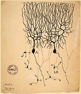
Encyclopedia
Neurons are a major class of cells in the nervous system. In
vertebrates, neurons are found in the
brain, the
spinal cord and in the
nerves and
ganglia of the peripheral nervous system. Their main role is to process and transmit information. Morphologically, a prototypical neuron is composed of a cell body, a
dendritic tree and an axon. In the classical view of the neuron, the cell body and dendritic tree receive inputs from other neurons, and axon transmits output signals. Neurons have
excitable membranes, which allow them to generate and propagate
electrical impulses. Neurons make connections with other neurons and transmit information to them via
synaptic transmission. Different types of neurons have different shapes, possess specific electrical properties adopted for their function and use different
neurotransmitters.
History
The concept of a neuron as the primary functional unit of the nervous system was derived from the work of the Spanish anatomist
Santiago Ramón y Cajal in the early 20th century. Cajal proposed that neurons were discrete cells which communicated with each other via specialized junctions, or spaces, between cells. This became known as the
neuron doctrine, one of the central tenets of modern neuroscience. To observe the structure of individual neurons, Cajal used
a silver staining method developed by his rival,
Camillo Golgi. When the Golgi stain is applied to neurons, it binds the cell's
microtubules and gives stained cells a black outline when light is shone through them.
Anatomy and histology
Neurons are highly specialized, and they differ widely in appearance and electrochemical properties. Neurons have cellular extensions known as
processes which they use to send and receive information. Neurons are typically 4 to 100 micrometres in diameter, the size varies depending on the type of neuron and the species it is from.
- The soma, or 'cell body', is the central part of the cell, where the nucleus is located and where most protein synthesis occurs. The nucleus ranges from 3 to 18 micrometers in diameter.
- The dendrite is a branching arbor of cellular extensions. Most neurons have several dendrites with profuse dendritic branches. The overall shape and structure of a neuron's dendrites is called its dendritic tree, and is traditionally thought to be the main information receiving network for the neuron. However, information outflow can also occur.
- The axon is a finer, cable-like projection which can extend tens, hundreds, or even tens of thousands of times the diameter of the soma in length. The axon carries nerve signals away from the soma . Many neurons have only one axon, but this axon may - and usually will - undergo extensive branching, enabling communication with many target cells. The part of the axon where it emerges from the soma is called the 'axon hillock'. Besides being an anatomical structure, the axon hillock is also the part of the neuron that has the greatest density of voltage-dependent sodium channels. Thus it has the most hyperpolarized action potential threshold of any part of the neuron. In other words, it is the most easily-excited part of the neuron, and thus serves as the spike initiation zone for the axon. While the axon and axon hillock are generally considered places of information outflow, this region can receive input from other neurons as well.
- The axon terminal a specialized structure at the end of the axon that is used to release neurotransmitter and communicate with target neurons.
Although the canonical view of the neuron attributes dedicated functions to its various anatomical components, dendrites and axons very often act contrary to their so-called main function.
Axons and dendrites in the central nervous system are typically only about a micrometer thick, while some in the peripheral nervous system are much thicker. The soma is usually about 10–25 micrometers in diameter and often is not much larger than the cell nucleus it contains. The longest axon of a human motoneuron can be over a meter long, reaching from the base of the spine to the toes, while
giraffes have single axons running along the whole length of their necks, several meters in length. Much of what we know about axonal function comes from studying the squid giant axon, an ideal experimental preparation because of its relatively immense size .
Classes
Structural classification
Most neurons can be anatomically characterized as:
- Unipolar or Pseudounipolar- dendrite and axon emerging from same process.
- Bipolar - single axon and single dendrite on opposite ends of the soma.
- Multipolar - more than two dendrites
- Golgi I- neurons with long-projecting axonal processes.
- Golgi II- neurons whose axonal process projects locally.
Some unique neuronal types can be identified according to their location in the nervous system and distinct shape. Some examples are basket, Betz, medium spiny,
Purkinje, pyramidal and Renshaw cells.
Functional classification
- Afferent neurons convey information from tissues and organs into the central nervous system.
- Efferent neurons transmit signals from the central nervous system to the effector cells and are sometimes called motor neurons.
- Interneuron
...
s connect neurons within specific regions of the central nervous system.
Afferent and
efferent can also refer to neurons which convey information from one region of the brain to another.
Classification by action on other neurons- Excitatory neurons evoke excitation of their target neurons. Excitatory neurons in the brain are often glutamatergic. Spinal motoneurons use acetylcholine as their neurotransmitter.
- Inhibitory neurons evoke inhibition of their target neurons. Inhibitory neurons are often interneurons. The output of some brain structures are inhibitory. The primary inhibitory neurotransmitters are GABA and glycine.
- Modulatory neurons evoke more complex effects termed neuromodulation. These neurons use such neurotransmitters as dopamine, acetylcholine, serotonin and others.
Classification by discharge patternsNeurons can be classified according to their
electrophysiological characteristics:
- Tonic or regular spiking. Some neurons are typically constantly active. Example: interneurons in neurostriatum.
- Phasic or bursting. Neurons that fire in bursts are called phasic.
- Fast spiking. Some neurons are notable for their fast firing rates, for example some types of cortical inhibitory interneurons, cells in globus pallidus.
- Thin-spike. Action potentials of some neurons are more narrow compared to the others. For example, interneurons in prefrontal cortex are thin-spike neurons.
Classification by neurotransmitter releasedSome examples are cholinergic, GABA-ergic, glutamatergic and dopaminergic neurons.
Connectivity
Neurons communicate with one another via
synapses, where the axon terminal of one cell impinges upon a dendrite or soma of another . Neurons such as
Purkinje cells in the
cerebellum can have over 1000 dendritic branches, making connections with tens of thousands of other cells; other neurons, such as the magnocellular neurons of the supraoptic nucleus, have only one or two dendrites, each of which receives thousands of synapses. Synapses can be excitatory or inhibitory and will either increase or decrease activity in the target neuron. Some neurons also communicate via
electrical synapses, which are direct, electrically-conductive
junctions between cells.
In a chemical synapse, the process of synaptic transmission is as follows: when an action potential reaches the axon terminal, it opens voltage-gated calcium channels, allowing calcium ions to enter the terminal. Calcium causes synaptic vesicles filled with neurotransmitter molecules to fuse with the membrane, releasing their contents into the synaptic cleft. The neurotransmitters diffuse across the synaptic cleft and activate receptors on the postsynaptic neuron.
The
human brain has a huge number of synapses. Each of 100 billion neurons has on average 7,000 synaptic connections to other neurons. Most authorities estimate that the brain of a three-year-old child has about 1,000 trillion synapses. This number declines with age, stabilizing by adulthood. Estimates vary for an adult, ranging from 100 to 500 trillion synapses.
Adaptations to carrying action potentials
The cell membrane in the axon and soma contain voltage-gated ion channels which allow the neuron to generate and propagate an electrical impulse .
Substantial early knowledge of neuron electrical activity came from experiments with squid giant axons. In 1937, John Zachary Young suggested that the giant squid axon can be used to study neuronal electrical properties . As they are much larger than human neurons, but similar in nature, it was easier to study them with the technology of that time. By inserting
electrodes into the giant squid axons, accurate measurements could be made of the
membrane potential.
Electrical activity can be produced in neurons by a number of stimuli. Pressure, stretch, chemical transmitters, and electrical current passing across the nerve membrane as a result of a difference in voltage can all initiate nerve activity .
The narrow cross-section of axons lessens the metabolic expense of carrying action potentials, but thicker axons convey impulses more rapidly. To minimize metabolic expense while maintaining rapid conduction, many neurons have insulating sheaths of
myelin around their axons. The sheaths are formed by
glial cells:
oligodendrocytes in the central nervous system and
Schwann cells in the peripheral nervous system. The sheath enables action potentials to travel faster than in unmyelinated axons of the same diameter, whilst using less energy. The myelin sheath in peripheral nerves normally runs along the axon in sections about 1 mm long, punctuated by unsheathed nodes of Ranvier which contain a high density of voltage-gated ion channels.
Multiple sclerosis is a neurological disorder that results from abnormal demyelination of peripheral nerves. Neurons with demyelinated axons do not conduct electrical signals properly.
Some neurons do not generate action potentials, but instead generate a graded electrical signal, which in turn causes graded neurotransmitter release. Such nonspiking neurons tend to be sensory neurons or interneurons, because they cannot carry signals long distances.
Histology and internal structure
Nerve cell bodies stained with basophilic dyes show numerous microscopic clumps of
Nissl substance , which consists of rough
endoplasmic reticulum and associated
ribosomes. The prominence of the Nissl substance can be explained by the fact that nerve cells are metabolically very active, and hence are involved in large amounts of protein synthesis.
The cell body of a neuron is supported by a complex meshwork of structural proteins called
neurofilaments, which are assembled into larger
neurofibrils. Some neurons also contain pigment granules, such as
neuromelanin and
lipofuscin .
There are different internal structural characteristics between axons and dendrites. Axons typically almost never contain
ribosomes, except some in the initial segment. Dendrites contain granular
endoplasmic reticulum or
ribosomes, with diminishing amounts with distance from the cell body.
Challenges to the neuron doctrine
The
neuron doctrine is a central tenet of modern neuroscience, but recent studies suggest that this doctrine needs to be revised.
First, electrical synapses are more common in the central nervous system than previously thought. Thus, rather than functioning as individual units, in some parts of the brain large ensembles of neurons may be active simultaneously to process neural information.
Second, dendrites, like axons, also have voltage-gated ion channels and can generate electrical potentials that carry information to and from the soma. This challenges the view that dendrites are simply passive recipients of information and axons the sole transmitters. It also suggests that the neuron is not simply active as a single element, but that complex computations can occur within a single neuron.
Third, the role of
glia in processing neural information has begun to be appreciated. Neurons and glia make up the two chief cell types of the central nervous system. There are far more glial cells than neurons: glia outnumber neurons by as many as 10:1. Recent experimental results have suggested that glia play a vital role in information processing.
Finally, recent research has challenged the historical view that neurogenesis, or the generation of new neurons, does not occur in adult mammalian brains. It is now known that the adult brain continuously creates new neurons in the
hippocampus and in an area contributing to the olfactory bulb. This research has shown that neurogenesis is environment-dependent , age-related, upregulated by a number of growth factors, and halted by survival-type stress factors.
Neurons in the brain
The number of neurons in the brain varies dramatically from species to species. The human brain has about 100 billion neurons and 100 trillion synapses. By contrast, the nematode worm has 302 neurons. Scientists have mapped all of the nematode's neurons. As a result, such worms are ideal candidates for neurobiological experiments and tests. Many properties of neurons, from the type of neurotransmitters used to ion channel composition, are maintained across species, allowing scientists to study processes occurring in more complex organisms in much simpler experimental systems.
See also
Sources
- Kandel E.R., Schwartz, J.H., Jessell, T.M. 2000. Principles of Neural Science, 4th ed., McGraw-Hill, New York.
- Bullock, T.H., Bennett, M.V.L., Johnston, D., Josephson, R., Marder, E., Fields R.D. 2005. The Neuron Doctrine, Redux, Science, V.310, p. 791-793.
- Ramón y Cajal, S. 1933 Histology, 10th ed., Wood, Baltimore.
- Roberts A., Bush B.M.H. 1981. Neurones Without Impulses. Cambridge University Press, Cambridge.
- Peters, A., Palay, S.L., Webster, H, D., 1991 The Fine Structure of the Nervous System, 3rd ed., Oxford, New York.
External links
- UC San Diego images of neurons.
- .