

Superior colliculus
Encyclopedia
The optic tectum or simply tectum is a paired structure that forms a major component of the vertebrate midbrain. In mammals this structure is more commonly called the superior colliculus (Latin
, higher hill), but, even in mammals, the adjective tectal is commonly used. The tectum is a layered structure, with a number of layers that vary by species. The superficial layers are sensory-related, and receive input from the eyes as well as other sensory systems. The deep layers are motor-related, capable of activating eye movements as well as other responses. There are also intermediate layers, with multi-sensory cells and motor properties.
The general function of the tectal system is to direct behavioral responses toward specific points in egocentric ("body-centered") space. Each layer of the tectum contains a topographic map
of the surrounding world in retinotopic
coordinates, and activation of neurons at a particular point in the map evokes a response directed toward the corresponding point in space. In primates, the tectum ("superior colliculus") has been studied mainly with respect to its role in directing eye movements. Visual input from the retina, or "command" input from the cerebral cortex, create a "bump" of activity in the tectal map, which, if strong enough, induces a saccadic eye movement
. Even in primates, however, the tectum is also involved in generating spatially directed head turns, arm-reaching movements, and shifts in attention that do not involve any overt movements. In other species, the tectum is involved in a wide range of responses, including whole-body turns in walking rats, swimming fishes, or flying birds; tongue-strikes toward prey in frogs; fang-strikes in snakes; etc.
In some non-mammal species, including fish and birds, the tectum is one of the largest components of the brain. In mammals, and especially primates, the massive expansion of the cerebral cortex reduces the tectum ("superior colliculus") to a much smaller fraction of the whole brain. Even there, however, it remains very important in terms of function as the primary integrating center for eye movements.
Note on terminology: the use by the literature of different terms for mammals and non-mammals, for what is really the same structure, creates problems for an article that attempts to encompass the full range of vertebrate species. There does not seem to be any way to handle this without causing either confusion or annoyance to some readers. The approach taken in this article is to follow the literature by using the term "superior colliculus" when discussing mammals, and "optic tectum" when discussing either specific non-mammalian species or vertebrates in general.
 The optic tectum is one of the fundamental components of the vertebrate brain, existing across the full range of species from hagfish to human. (See the brain
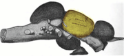
article for background.) Some aspects of the structure are very consistent, including a structure composed of a number of layers, with a dense input from the optic tracts to the superficial layers and another strong input conveying somatosensory input to deeper layers. Other aspects are highly variable, such as the total number of layers (from 3 in the African lungfish to 15 in the goldfish), and the number of different types of cells (from 2 in the lungfish to 27 in the house sparrow). In hagfish, lamprey, and shark it is a relatively small structure, but in teleost fish it is greatly expanded, in some cases becoming the largest structure in the brain. (See the adjoining drawing of a codfish brain.) In amphibians, reptiles, and especially birds it is also a very significant component, but in mammals it is dwarfed by the massive expansion of the cerebral cortex.
and tectum. In a study of the lamprey tectum published in 2007, they found that electrical stimulation could elicit eye movements, lateral bending movements, or swimming activity, and that the type, amplitude, and direction of movement varied as a function of the location within the tectum that was stimulated. These findings were interpreted as consistent with the idea that the tectum generates goal-directed locomotion in the lamprey as it does in other species.
s are not, in fact, blind, but they depend much more on echolocation than vision for navigation and prey capture. They obtain information about the surrounding world by emitting sonar chirps and then listening for the echoes. Their brains are highly specialized for this process, and some of these specializations appear in the superior colliculus. In bats, the retinal projection occupies only a thin zone just beneath the surface, but there are extensive inputs from auditory areas, and outputs to motor areas capable of orienting the ears, head, or body. Echoes coming from different directions activate neurons at different locations in the collicular layers, and activation of collicular neurons influences the chirps that the bats emit. Thus, there is a strong case that the superior colliculus performs the same sorts of functions for the auditory-guided behaviors of bats that it performs for the visual-guided behaviors of other species.
Bat
s are usually classified into two main groups: Microchiroptera (the most numerous, and commonly found throughout the world), and Megachiroptera (fruit bats, found in Asia, Africa and Australasia). With one exception, Megabats do not echolocate, and rely on a developed sense of vision to navigate. The visual receptive fields of neurons in the superior colliculus in these animals form a precise map of the retina
, similar to that found in cats and primates.
and surround the pineal gland
in the mesencephalon
of vertebrate
brains. It comprises the caudal aspect of the midbrain, posterior to the periaqueductal gray
and immediately superior to the inferior colliculus
. The inferior and superior colliculi are known collectively as the corpora quadrigemina
(Latin, quadruplet bodies).
 The microstructure of the optic tectum / superior colliculus varies across species. As a general rule, there is always a clear distinction between superficial layers, which receive input primarily from the visual system and show primarily visual responses, and deeper layers, which receive many types of input and project to numerous motor-related brain areas. The distinction between these two zones is so clear and consistent that some anatomists have suggested that they should be considered separate brain structures.
In mammals, neuroanatomists conventionally identify seven layers The top three layers are called superficial:
Next come two intermediate layers:
Finally come the two deep layers:
The superficial layers receive input mainly from the retina, vision-related areas of the cerebral cortex, and two tectal-related structures called the pretectum
and parabigeminal nucleus. The retinal input encompasses the entire superficial zone, and is bilateral, although the contralateral portion is more extensive. The cortical input comes most heavily from the primary visual cortex (area 17), the secondary visual cortex (areas 18
and 19
), and the frontal eye fields
. The parabigeminal nucleus plays a very important role in tectal function that will be described below.
In contrast to the vision-dominated inputs to the superficial layers, the intermediate and deep layers receive inputs from a very diverse set of sensory and motor structures. Most areas of the cerebral cortex project to these layers, although the input from "association" areas tends to be heavier than the input from primary sensory or motor areas. However, the cortical areas involved, and the strength of their relative projections differs across species. Another important input comes from the substantia nigra
, pars reticulata
, a component of the basal ganglia. This projection uses the inhibitory neurotransmitter GABA, and is thought to exert a "gating" effect on the superior colliculus. The intermediate and deep layers also receive input from the spinal trigeminal nucleus
, which conveys somatosensory information from the face, as well as the hypothalamus
, zona incerta
, thalamus
, and inferior colliculus
.
In addition to their distinctive inputs, the superficial and deep zones of the superior colliculus also have distinctive outputs. One of the most important outputs goes to the pulvinar
and lateral intermediate areas of the thalamus, which in turn project to areas of the cerebral cortex that are involved in controlling eye movements. There are also projections from the superficial zone to the pretectal nuclei, lateral geniculate nucleus
of the thalamus, and the parabigeminal nucleus. The projections from the deeper layers are more extensive. There are two large descending pathways, traveling to the brainstem and spinal cord, and numerous ascending projections to a variety of sensory and motor centers, including several that are involved in generating eye movements.
All species that have been examined — including mammals and non-mammals — show compartmentalization, but there are some systematic differences in the details of the arrangement. In species with a streak-type retina (mainly species with laterally placed eyes, such as rabbits and deer), the compartments cover the full extent of the SC. In species with a centrally placed fovea, however, the compartmentalization breaks down in the front (rostral) part of the SC. This portion of the SC contains many "fixation" neurons that fire continually while the eyes remain fixed in a constant position.
 The optic tectum is closely associated with an adjoining structure called nucleus isthmii, which has drawn great interest recently because of new evidence that it makes a very important contribution to tectal function. In mammals, where the term superior colliculus is generally used instead of optic tectum, this area is called the parabigeminal nucleus. Once again, this is simply a case of two different names being used for the same structure. The nucleus isthmii is divided into two parts, called pars magnocellularis (Imc; "the part with the large cells") and pars parvocellularis (Ipc; "the part with the small cells"). Imc is also sometimes called pars semilunaris, because it is shaped like a half-moon, or rather crescent-moon, in cross-section.
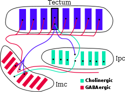
As illustrated in the adjoining diagram, connections between the three areas — tectum, Ipc, and Imc — are topographic. Neurons in the superficial layers of the tectum project to corresponding points in Ipc and Imc. The projections to Ipc are tightly focused, while the projections to Imc are somewhat more diffuse. Ipc gives rise to tightly focused cholinergic projections both to Imc and the tectum. In the tectum, the cholinergic inputs from Ipc ramify to give rise to terminals that extend across an entire column, from top to bottom. Imc, in contrast, gives rise to GABAergic projections to Ipc and tectum that spread very broadly in the lateral dimensions, encompassing most of the retinotopic map. Thus, the tectum-Ipc-Imc circuit causes tectal activity to produce recurrent feedback that involves tightly focused excitation of a small column of neighboring tectal neurons, together with global inhibition of distant tectal neurons.
In the late 1990s, however, experiments using animals whose heads were free to move showed clearly that the SC actually produces gaze shifts, usually composed of combined head and eye movements, rather than eye movements per se. This discovery reawakened interest in the full breadth of functions of the superior colliculus, and led to studies of multisensory integration in a variety of species and situations. Nevertheless, the role of the SC in controlling eye movements is understood in much greater depth than any other function.
Behavioral studies have shown that the SC is not needed for object recognition, but plays a critical role in the ability to direct behaviors toward specific objects, and can support this ability even in the absence of the cerebral cortex. Thus, cats with major damage to the visual cortex cannot recognize objects, but may still be able to follow and orient toward moving stimuli, although more slowly than usual. If one half of the SC is removed, however, the cats will circle constantly toward the side of the lesion, and orient compulsively toward objects located there, but fail to orient at all toward objects located in the opposite hemifield. These deficits diminish over time but never disappear.
Each of the two colliculi — one on each side of the brain — contains a two-dimensional map representing half of the visual field. The fovea
— the region of maximum sensitivity — is represented at the front edge of the map, and the periphery at the back edge. Eye movements are evoked by activity in the deep layers of the SC. During fixation, neurons near the front edge — the foveal zone — are tonically active. During smooth pursuit, neurons a small distance from the front edge are activated, leading to small eye movements. For saccades, neurons are activated in a region that represents the point to which the saccade will be directed. Just prior to a saccade, activity rapidly builds up at the target location and decreases in other parts of the SC. The coding is rather broad, so that for any given saccade the activity profile forms a "hill" that encompasses a substantial fraction of the collicular map: The location of the peak of this "hill" represents the saccade target.
The SC encodes the target of a gaze shift, but it does not seem to specify the precise movements needed to get there. The decomposition of a gaze shift into head and eye movements and the precise trajectory of the eye during a saccade depend on integration of collicular and non-collicular signals by downstream motor areas, in ways that are not yet well understood. Regardless of how the movement is evoked or performed, the SC encodes it in "retinotopic" coordinates: that is, a given SC activation pattern specifies a given offset from the current gaze direction, irrespective of the initial position of the eyes.
There has been some controversy about whether the SC merely commands eye movements, and leaves the execution to other structures, or whether it actively participates in the performance of a saccade. In 1991, Munoz et al., on the basis of data they collected, argued that, during a saccade, the "hill" of activity in the SC moves gradually, to reflect the changing offset of the eye from the target location while the saccade is progressing. At present, however, the predominant view is that, although the "hill" does shift slightly during a saccade, it does not shift in the steady and proportionate way that the "moving hill" hypothesis predicts.
The output from the motor sector of the SC goes to a set of midbrain and brainstem nuclei, which transform the "place" code used by the SC into the "rate" code used by oculomotor neurons. Eye movements are generated by six muscles, arranged in three orthogonally-aligned pairs. Thus, at the level of the final common path, eye movements are encoded in essentially a Cartesian coordinate system.
Although the SC receives a strong input directly from the retina, in primates it is largely under the control of the cerebral cortex, which contains several areas that are involved in determining eye movements. The frontal eye fields
, a portion of the motor cortex, are involved in triggering intentional saccades, and an adjoining area, the supplementary eye fields, are involved in organizing groups of saccades into sequences. The parietal eye fields, farther back in the brain, are involved mainly in reflexive saccades, made in response to changes in the view.
The SC only receives visual
inputs in its superficial layers, whereas the deeper layers of the colliculus receive also auditory and somatosensory inputs and are connected to many sensorimotor areas of the brain. The colliculus as a whole is thought to help orient the head and eyes toward something seen and heard.
The superior colliculus also receives auditory information from the inferior colliculus. This auditory information is integrated with the visual information already present to produce the ventriloquist effect.
superior colliculus is unique among mammals, in that it does not contain a complete map of the visual field seen by the contralateral eye. Instead, like the visual cortex
and lateral geniculate nucleus
, each colliculus represents only the contralateral half of the visual field
, up to the midline, and excludes a representation of the ipsilateral half. This functional characteristic is explained by the absence, in primates, of anatomical connections between the retinal ganglion cells in the temporal half of the retina
and the contralateral superior colliculus. In other mammals, the retinal ganglion cells throughout the contralateral retina project to the contralateral colliculus. This distinction between primates and non-primates has been one of the key lines of evidence in support of the flying primates theory
proposed by Australian neuroscientist Jack Pettigrew
in 1986, after he discovered that flying foxes (megabat
s) resemble primates in terms of the pattern of anatomical connections between the retina and superior colliculus.
s that can detect infrared radiation, such as pythons and pit vipers, the initial neural input is through the trigeminal nerve
instead of the optic tract
. The rest of the processing is similar to that of the visual sense and, thus, involves the optic tectum.
Latin
Latin is an Italic language originally spoken in Latium and Ancient Rome. It, along with most European languages, is a descendant of the ancient Proto-Indo-European language. Although it is considered a dead language, a number of scholars and members of the Christian clergy speak it fluently, and...
, higher hill), but, even in mammals, the adjective tectal is commonly used. The tectum is a layered structure, with a number of layers that vary by species. The superficial layers are sensory-related, and receive input from the eyes as well as other sensory systems. The deep layers are motor-related, capable of activating eye movements as well as other responses. There are also intermediate layers, with multi-sensory cells and motor properties.
The general function of the tectal system is to direct behavioral responses toward specific points in egocentric ("body-centered") space. Each layer of the tectum contains a topographic map
Topographic map
A topographic map is a type of map characterized by large-scale detail and quantitative representation of relief, usually using contour lines in modern mapping, but historically using a variety of methods. Traditional definitions require a topographic map to show both natural and man-made features...
of the surrounding world in retinotopic
Retinotopy
Retinotopy describes the spatial organization of the neuronal responses to visual stimuli. In many locations within the brain, adjacent neurons have receptive fields that include slightly different, but overlapping portions of the visual field. The position of the center of these receptive fields...
coordinates, and activation of neurons at a particular point in the map evokes a response directed toward the corresponding point in space. In primates, the tectum ("superior colliculus") has been studied mainly with respect to its role in directing eye movements. Visual input from the retina, or "command" input from the cerebral cortex, create a "bump" of activity in the tectal map, which, if strong enough, induces a saccadic eye movement
Saccade
A saccade is a fast movement of an eye, head or other part of an animal's body or device. It can also be a fast shift in frequency of an emitted signal or other quick change. Saccades are quick, simultaneous movements of both eyes in the same direction...
. Even in primates, however, the tectum is also involved in generating spatially directed head turns, arm-reaching movements, and shifts in attention that do not involve any overt movements. In other species, the tectum is involved in a wide range of responses, including whole-body turns in walking rats, swimming fishes, or flying birds; tongue-strikes toward prey in frogs; fang-strikes in snakes; etc.
In some non-mammal species, including fish and birds, the tectum is one of the largest components of the brain. In mammals, and especially primates, the massive expansion of the cerebral cortex reduces the tectum ("superior colliculus") to a much smaller fraction of the whole brain. Even there, however, it remains very important in terms of function as the primary integrating center for eye movements.
Note on terminology: the use by the literature of different terms for mammals and non-mammals, for what is really the same structure, creates problems for an article that attempts to encompass the full range of vertebrate species. There does not seem to be any way to handle this without causing either confusion or annoyance to some readers. The approach taken in this article is to follow the literature by using the term "superior colliculus" when discussing mammals, and "optic tectum" when discussing either specific non-mammalian species or vertebrates in general.
Evolution and comparative anatomy
Brain
The brain is the center of the nervous system in all vertebrate and most invertebrate animals—only a few primitive invertebrates such as sponges, jellyfish, sea squirts and starfishes do not have one. It is located in the head, usually close to primary sensory apparatus such as vision, hearing,...
article for background.) Some aspects of the structure are very consistent, including a structure composed of a number of layers, with a dense input from the optic tracts to the superficial layers and another strong input conveying somatosensory input to deeper layers. Other aspects are highly variable, such as the total number of layers (from 3 in the African lungfish to 15 in the goldfish), and the number of different types of cells (from 2 in the lungfish to 27 in the house sparrow). In hagfish, lamprey, and shark it is a relatively small structure, but in teleost fish it is greatly expanded, in some cases becoming the largest structure in the brain. (See the adjoining drawing of a codfish brain.) In amphibians, reptiles, and especially birds it is also a very significant component, but in mammals it is dwarfed by the massive expansion of the cerebral cortex.
Lamprey
The lamprey has been extensively studied because it has a relatively simple brain that is thought in many respects to reflect the brain structure of early vertebrate ancestors. Beginning in the 1970s, Sten Grillner and his colleagues at the Karolinska Institute in Stockholm have used the lamprey as a model system to work out the fundamental principles of motor control in vertebrates, starting in the spinal cord and working upward into the brain. In a series of studies, they found that neural circuits within the spinal cord are capable of generating the rhythmic motor patterns that underlie swimming, that these circuits are controlled by specific locomotor areas in the brainstem and midbrain, and that these areas in turn are controlled by higher brain structures including the basal gangliaBasal ganglia
The basal ganglia are a group of nuclei of varied origin in the brains of vertebrates that act as a cohesive functional unit. They are situated at the base of the forebrain and are strongly connected with the cerebral cortex, thalamus and other brain areas...
and tectum. In a study of the lamprey tectum published in 2007, they found that electrical stimulation could elicit eye movements, lateral bending movements, or swimming activity, and that the type, amplitude, and direction of movement varied as a function of the location within the tectum that was stimulated. These findings were interpreted as consistent with the idea that the tectum generates goal-directed locomotion in the lamprey as it does in other species.
Bats
BatBat
Bats are mammals of the order Chiroptera "hand" and pteron "wing") whose forelimbs form webbed wings, making them the only mammals naturally capable of true and sustained flight. By contrast, other mammals said to fly, such as flying squirrels, gliding possums, and colugos, glide rather than fly,...
s are not, in fact, blind, but they depend much more on echolocation than vision for navigation and prey capture. They obtain information about the surrounding world by emitting sonar chirps and then listening for the echoes. Their brains are highly specialized for this process, and some of these specializations appear in the superior colliculus. In bats, the retinal projection occupies only a thin zone just beneath the surface, but there are extensive inputs from auditory areas, and outputs to motor areas capable of orienting the ears, head, or body. Echoes coming from different directions activate neurons at different locations in the collicular layers, and activation of collicular neurons influences the chirps that the bats emit. Thus, there is a strong case that the superior colliculus performs the same sorts of functions for the auditory-guided behaviors of bats that it performs for the visual-guided behaviors of other species.
Bat
Bat
Bats are mammals of the order Chiroptera "hand" and pteron "wing") whose forelimbs form webbed wings, making them the only mammals naturally capable of true and sustained flight. By contrast, other mammals said to fly, such as flying squirrels, gliding possums, and colugos, glide rather than fly,...
s are usually classified into two main groups: Microchiroptera (the most numerous, and commonly found throughout the world), and Megachiroptera (fruit bats, found in Asia, Africa and Australasia). With one exception, Megabats do not echolocate, and rely on a developed sense of vision to navigate. The visual receptive fields of neurons in the superior colliculus in these animals form a precise map of the retina
Retina
The vertebrate retina is a light-sensitive tissue lining the inner surface of the eye. The optics of the eye create an image of the visual world on the retina, which serves much the same function as the film in a camera. Light striking the retina initiates a cascade of chemical and electrical...
, similar to that found in cats and primates.
Structure and relations
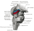
The two superior colliculi sit below the thalamusThalamus
The thalamus is a midline paired symmetrical structure within the brains of vertebrates, including humans. It is situated between the cerebral cortex and midbrain, both in terms of location and neurological connections...
and surround the pineal gland
Pineal gland
The pineal gland is a small endocrine gland in the vertebrate brain. It produces the serotonin derivative melatonin, a hormone that affects the modulation of wake/sleep patterns and seasonal functions...
in the mesencephalon
Mesencephalon
The midbrain or mesencephalon is a portion of the central nervous system associated with vision, hearing, motor control, sleep/wake, arousal , and temperature regulation....
of vertebrate
Vertebrate
Vertebrates are animals that are members of the subphylum Vertebrata . Vertebrates are the largest group of chordates, with currently about 58,000 species described. Vertebrates include the jawless fishes, bony fishes, sharks and rays, amphibians, reptiles, mammals, and birds...
brains. It comprises the caudal aspect of the midbrain, posterior to the periaqueductal gray
Periaqueductal gray
Periaqueductal gray is the gray matter located around the cerebral aqueduct within the tegmentum of the midbrain. It plays a role in the descending modulation of pain and in defensive behaviour...
and immediately superior to the inferior colliculus
Inferior colliculus
The inferior colliculus is the principal midbrain nucleus of the auditory pathway and receives input from several more peripheral brainstem nuclei in the auditory pathway, as well as inputs from the auditory cortex...
. The inferior and superior colliculi are known collectively as the corpora quadrigemina
Corpora quadrigemina
In the brain, the corpora quadrigemina are the four colliculi—two inferior, two superior—located on the tectum of the dorsal aspect of the midbrain.They are respectively named the inferior and superior colliculus....
(Latin, quadruplet bodies).
Neural circuit
In mammals, neuroanatomists conventionally identify seven layers The top three layers are called superficial:
- Lamina I or SZ, the stratum zonale, is a thin layer consisting of small myelinated axons together with marginal and horizontal cells.
- Lamina II or SGS, the stratum griseum superficiale ("superficial gray"), contains many neurons of various shapes and sizes.
- Lamina III or SO, the stratum opticum ("optic layer"), consists mainly of axons coming from the optic tractOptic tractThe optic tract is a part of the visual system in the brain.It is a continuation of the optic nerve and runs from the optic chiasm to the lateral geniculate nucleus....
.
Next come two intermediate layers:
- Lamina IV or SGI, the stratum griseum intermediale ("intermediate gray"), is the thickest layer, and is filled with many neurons of many sizes. This layer is often as thick as all the other layers together. It is often subdivided into "upper" and "lower" parts.
- Lamina V or SAI, the stratum album intermediale ("intermediate white"), consists mainly of fibers from various sources.
Finally come the two deep layers:
- Lamina VI or SGP, the stratum griseum profundum ("deep gray"), consists of loosely packed neurons and myelinated fibers.
- Lamina VII or SAP, the stratum album profundum ("deep white"), lying directly above the periaqueductal grayPeriaqueductal grayPeriaqueductal gray is the gray matter located around the cerebral aqueduct within the tegmentum of the midbrain. It plays a role in the descending modulation of pain and in defensive behaviour...
, consists entirely of fibers.
The superficial layers receive input mainly from the retina, vision-related areas of the cerebral cortex, and two tectal-related structures called the pretectum
Pretectum
The pretectum, also known as the pretectal area, is a region of neurons found between the thalamus and midbrain. It receives binocular sensory input from retinal ganglion cells of the eyes, and is the region responsible for maintaining the pupillary light reflex.-Outputs:The pretectum, after...
and parabigeminal nucleus. The retinal input encompasses the entire superficial zone, and is bilateral, although the contralateral portion is more extensive. The cortical input comes most heavily from the primary visual cortex (area 17), the secondary visual cortex (areas 18
Brodmann area 18
-Human:Brodmann area 18, or BA18, is part of the occipital cortex in the human brain. It accounts for the bulk of the volume of the occipital lobe....
and 19
Brodmann area 19
-Human:Brodmann area 19, or BA19, is part of the occipital lobe cortex in the human brain. Along with area 18, it comprises the extrastriate cortex...
), and the frontal eye fields
Frontal eye fields
The frontal eye fields is a region located in the premotor cortex, which is part of the frontal cortex of the primate brain.-Function:...
. The parabigeminal nucleus plays a very important role in tectal function that will be described below.
In contrast to the vision-dominated inputs to the superficial layers, the intermediate and deep layers receive inputs from a very diverse set of sensory and motor structures. Most areas of the cerebral cortex project to these layers, although the input from "association" areas tends to be heavier than the input from primary sensory or motor areas. However, the cortical areas involved, and the strength of their relative projections differs across species. Another important input comes from the substantia nigra
Substantia nigra
The substantia nigra is a brain structure located in the mesencephalon that plays an important role in reward, addiction, and movement. Substantia nigra is Latin for "black substance", as parts of the substantia nigra appear darker than neighboring areas due to high levels of melanin in...
, pars reticulata
Pars reticulata
-Anatomy:Neurons in the pars reticulata are much less densely packed than those in the pars compacta . They are smaller and thinner than the dopaminergic neurons and conversely identical and morphologically similar to the pallidal neurons...
, a component of the basal ganglia. This projection uses the inhibitory neurotransmitter GABA, and is thought to exert a "gating" effect on the superior colliculus. The intermediate and deep layers also receive input from the spinal trigeminal nucleus
Spinal trigeminal nucleus
The spinal trigeminal nucleus is a nucleus in the medulla that receives information about deep/crude touch, pain, and temperature from the ipsilateral face. The facial, glossopharyngeal, and vagus nerves also convey pain information from their areas to the spinal trigeminal nucleus.This nucleus...
, which conveys somatosensory information from the face, as well as the hypothalamus
Hypothalamus
The Hypothalamus is a portion of the brain that contains a number of small nuclei with a variety of functions...
, zona incerta
Zona incerta
The zona incerta is a horizontally elongated region of gray matter cells in the subthalamus below the thalamus. Its connections project extensively over the brain from the cerebral cortex down into the spinal cord....
, thalamus
Thalamus
The thalamus is a midline paired symmetrical structure within the brains of vertebrates, including humans. It is situated between the cerebral cortex and midbrain, both in terms of location and neurological connections...
, and inferior colliculus
Inferior colliculus
The inferior colliculus is the principal midbrain nucleus of the auditory pathway and receives input from several more peripheral brainstem nuclei in the auditory pathway, as well as inputs from the auditory cortex...
.
In addition to their distinctive inputs, the superficial and deep zones of the superior colliculus also have distinctive outputs. One of the most important outputs goes to the pulvinar
Pulvinar
The pulvinar nuclei are a collection of nuclei located in the pulvinar thalamus. The pulvinar part is the most posterior region of the thalamus....
and lateral intermediate areas of the thalamus, which in turn project to areas of the cerebral cortex that are involved in controlling eye movements. There are also projections from the superficial zone to the pretectal nuclei, lateral geniculate nucleus
Lateral geniculate nucleus
The lateral geniculate nucleus is the primary relay center for visual information received from the retina of the eye. The LGN is found inside the thalamus of the brain....
of the thalamus, and the parabigeminal nucleus. The projections from the deeper layers are more extensive. There are two large descending pathways, traveling to the brainstem and spinal cord, and numerous ascending projections to a variety of sensory and motor centers, including several that are involved in generating eye movements.
Mosaic structure
On detailed examination the collicular layers are actually not smooth sheets, but divided into a honeycomb arrangement of discrete columns. The clearest indication of columnar structure comes from the cholinergic inputs arising from the parabigeminal nucleus, whose terminals form evenly spaced clusters that extend from top to bottom of the tectum. Several other neurochemical markers including calretinin, parvalbumin, GAP-43, and NMDA receptors, and connections with numerous other brain structures in the brainstem and diencephalon, also show a corresponding inhomogeneity. The total number of columns has been estimated at around 100. The functional significance of this columnar architecture is not clear, but it is interesting that recent evidence has implicated the cholinergic inputs as part of a recurrent circuit producing winner-take-all dynamics within the tectum, as described in more detail below.All species that have been examined — including mammals and non-mammals — show compartmentalization, but there are some systematic differences in the details of the arrangement. In species with a streak-type retina (mainly species with laterally placed eyes, such as rabbits and deer), the compartments cover the full extent of the SC. In species with a centrally placed fovea, however, the compartmentalization breaks down in the front (rostral) part of the SC. This portion of the SC contains many "fixation" neurons that fire continually while the eyes remain fixed in a constant position.
Nucleus Isthmii/Parabigeminalis
As illustrated in the adjoining diagram, connections between the three areas — tectum, Ipc, and Imc — are topographic. Neurons in the superficial layers of the tectum project to corresponding points in Ipc and Imc. The projections to Ipc are tightly focused, while the projections to Imc are somewhat more diffuse. Ipc gives rise to tightly focused cholinergic projections both to Imc and the tectum. In the tectum, the cholinergic inputs from Ipc ramify to give rise to terminals that extend across an entire column, from top to bottom. Imc, in contrast, gives rise to GABAergic projections to Ipc and tectum that spread very broadly in the lateral dimensions, encompassing most of the retinotopic map. Thus, the tectum-Ipc-Imc circuit causes tectal activity to produce recurrent feedback that involves tightly focused excitation of a small column of neighboring tectal neurons, together with global inhibition of distant tectal neurons.
Function
The history of investigation of the optic tectum has been marked by several large shifts in opinion. Before about 1970, most studies involved non-mammals — fish, frogs, birds - that is, species in which the tectum is the dominant structure that receives input from the eyes. The general view then was that the tectum, in these species, is the main visual center in the non-mammalian brain, and, as a consequence, is involved in a wide variety of behaviors. From the 1970s to 1990s, however, neural recordings from mammals, mostly monkeys, focused primarily on the role of the superior colliculus in controlling eye movements. This line of investigation came to dominate the literature to such a degree that the majority opinion was that eye-movement control is the only important function in mammals, a view still reflected in many current textbooks.In the late 1990s, however, experiments using animals whose heads were free to move showed clearly that the SC actually produces gaze shifts, usually composed of combined head and eye movements, rather than eye movements per se. This discovery reawakened interest in the full breadth of functions of the superior colliculus, and led to studies of multisensory integration in a variety of species and situations. Nevertheless, the role of the SC in controlling eye movements is understood in much greater depth than any other function.
Behavioral studies have shown that the SC is not needed for object recognition, but plays a critical role in the ability to direct behaviors toward specific objects, and can support this ability even in the absence of the cerebral cortex. Thus, cats with major damage to the visual cortex cannot recognize objects, but may still be able to follow and orient toward moving stimuli, although more slowly than usual. If one half of the SC is removed, however, the cats will circle constantly toward the side of the lesion, and orient compulsively toward objects located there, but fail to orient at all toward objects located in the opposite hemifield. These deficits diminish over time but never disappear.
Eye movements
In primates, eye movements can be divided into several types: fixation, in which the eyes are directed toward a motionless object, with eye movements only to compensate for movements of the head; smooth pursuit, in which the eyes move steadily to track a moving object; saccades, in which the eyes move very rapidly from one location to another; and vergence, in which the eyes move simultaneously in opposite directions to obtain or maintain single binocular vision. The superior colliculus is involved in all of these, but its role in saccades has been studied most intensively.Each of the two colliculi — one on each side of the brain — contains a two-dimensional map representing half of the visual field. The fovea
Fovea
The fovea centralis, also generally known as the fovea , is a part of the eye, located in the center of the macula region of the retina....
— the region of maximum sensitivity — is represented at the front edge of the map, and the periphery at the back edge. Eye movements are evoked by activity in the deep layers of the SC. During fixation, neurons near the front edge — the foveal zone — are tonically active. During smooth pursuit, neurons a small distance from the front edge are activated, leading to small eye movements. For saccades, neurons are activated in a region that represents the point to which the saccade will be directed. Just prior to a saccade, activity rapidly builds up at the target location and decreases in other parts of the SC. The coding is rather broad, so that for any given saccade the activity profile forms a "hill" that encompasses a substantial fraction of the collicular map: The location of the peak of this "hill" represents the saccade target.
The SC encodes the target of a gaze shift, but it does not seem to specify the precise movements needed to get there. The decomposition of a gaze shift into head and eye movements and the precise trajectory of the eye during a saccade depend on integration of collicular and non-collicular signals by downstream motor areas, in ways that are not yet well understood. Regardless of how the movement is evoked or performed, the SC encodes it in "retinotopic" coordinates: that is, a given SC activation pattern specifies a given offset from the current gaze direction, irrespective of the initial position of the eyes.
There has been some controversy about whether the SC merely commands eye movements, and leaves the execution to other structures, or whether it actively participates in the performance of a saccade. In 1991, Munoz et al., on the basis of data they collected, argued that, during a saccade, the "hill" of activity in the SC moves gradually, to reflect the changing offset of the eye from the target location while the saccade is progressing. At present, however, the predominant view is that, although the "hill" does shift slightly during a saccade, it does not shift in the steady and proportionate way that the "moving hill" hypothesis predicts.
The output from the motor sector of the SC goes to a set of midbrain and brainstem nuclei, which transform the "place" code used by the SC into the "rate" code used by oculomotor neurons. Eye movements are generated by six muscles, arranged in three orthogonally-aligned pairs. Thus, at the level of the final common path, eye movements are encoded in essentially a Cartesian coordinate system.
Although the SC receives a strong input directly from the retina, in primates it is largely under the control of the cerebral cortex, which contains several areas that are involved in determining eye movements. The frontal eye fields
Frontal eye fields
The frontal eye fields is a region located in the premotor cortex, which is part of the frontal cortex of the primate brain.-Function:...
, a portion of the motor cortex, are involved in triggering intentional saccades, and an adjoining area, the supplementary eye fields, are involved in organizing groups of saccades into sequences. The parietal eye fields, farther back in the brain, are involved mainly in reflexive saccades, made in response to changes in the view.
The SC only receives visual
Visual perception
Visual perception is the ability to interpret information and surroundings from the effects of visible light reaching the eye. The resulting perception is also known as eyesight, sight, or vision...
inputs in its superficial layers, whereas the deeper layers of the colliculus receive also auditory and somatosensory inputs and are connected to many sensorimotor areas of the brain. The colliculus as a whole is thought to help orient the head and eyes toward something seen and heard.
The superior colliculus also receives auditory information from the inferior colliculus. This auditory information is integrated with the visual information already present to produce the ventriloquist effect.
Primates
It is usually accepted that the primatePrimate
A primate is a mammal of the order Primates , which contains prosimians and simians. Primates arose from ancestors that lived in the trees of tropical forests; many primate characteristics represent adaptations to life in this challenging three-dimensional environment...
superior colliculus is unique among mammals, in that it does not contain a complete map of the visual field seen by the contralateral eye. Instead, like the visual cortex
Visual cortex
The visual cortex of the brain is the part of the cerebral cortex responsible for processing visual information. It is located in the occipital lobe, in the back of the brain....
and lateral geniculate nucleus
Lateral geniculate nucleus
The lateral geniculate nucleus is the primary relay center for visual information received from the retina of the eye. The LGN is found inside the thalamus of the brain....
, each colliculus represents only the contralateral half of the visual field
Visual field
The term visual field is sometimes used as a synonym to field of view, though they do not designate the same thing. The visual field is the "spatial array of visual sensations available to observation in introspectionist psychological experiments", while 'field of view' "refers to the physical...
, up to the midline, and excludes a representation of the ipsilateral half. This functional characteristic is explained by the absence, in primates, of anatomical connections between the retinal ganglion cells in the temporal half of the retina
Retina
The vertebrate retina is a light-sensitive tissue lining the inner surface of the eye. The optics of the eye create an image of the visual world on the retina, which serves much the same function as the film in a camera. Light striking the retina initiates a cascade of chemical and electrical...
and the contralateral superior colliculus. In other mammals, the retinal ganglion cells throughout the contralateral retina project to the contralateral colliculus. This distinction between primates and non-primates has been one of the key lines of evidence in support of the flying primates theory
Flying primates theory
The flying primates theory conjectures that megabats, a sub-group of Chiroptera , form an evolutionary sister group of Primates. This theory started with Carl Linnaeus, and was again advanced by J.D Smith in 1980...
proposed by Australian neuroscientist Jack Pettigrew
Jack Pettigrew
John Douglas Pettigrew is Emeritus Professor of Physiology and Director of the Vision, Touch and Hearing Research Centre at the University of Queensland in Australia.Professor Pettigrew's research interest is in comparative neuroscience...
in 1986, after he discovered that flying foxes (megabat
Megabat
Megabats constitute the suborder Megachiroptera, family Pteropodidae of the order Chiroptera . They are also called fruit bats, old world fruit bats, or flying foxes.-Description:...
s) resemble primates in terms of the pattern of anatomical connections between the retina and superior colliculus.
Other vertebrates
In snakeSnake
Snakes are elongate, legless, carnivorous reptiles of the suborder Serpentes that can be distinguished from legless lizards by their lack of eyelids and external ears. Like all squamates, snakes are ectothermic, amniote vertebrates covered in overlapping scales...
s that can detect infrared radiation, such as pythons and pit vipers, the initial neural input is through the trigeminal nerve
Trigeminal nerve
The trigeminal nerve contains both sensory and motor fibres. It is responsible for sensation in the face and certain motor functions such as biting, chewing, and swallowing. Sensory information from the face and body is processed by parallel pathways in the central nervous system...
instead of the optic tract
Optic tract
The optic tract is a part of the visual system in the brain.It is a continuation of the optic nerve and runs from the optic chiasm to the lateral geniculate nucleus....
. The rest of the processing is similar to that of the visual sense and, thus, involves the optic tectum.
See also
- inferior colliculusInferior colliculusThe inferior colliculus is the principal midbrain nucleus of the auditory pathway and receives input from several more peripheral brainstem nuclei in the auditory pathway, as well as inputs from the auditory cortex...
- Lateral geniculate nucleusLateral geniculate nucleusThe lateral geniculate nucleus is the primary relay center for visual information received from the retina of the eye. The LGN is found inside the thalamus of the brain....
- List of regions in the human brain