
Receptive field
Encyclopedia
The receptive field of a sensory neuron
is a region of space in which the presence of a stimulus
will alter the firing of that neuron. Receptive fields have been identified for neurons of the auditory system
, the somatosensory system
, and the visual system
.
The concept of receptive fields can be extended to further up the neural system; if many sensory receptors all form synapses with a single cell
further up, they collectively form the receptive field of that cell. For example, the receptive field of a ganglion cell
in the retina
of the eye
is composed of input from all of the photoreceptors which synapse with it, and a group of ganglion cells in turn forms the receptive field for a cell in the brain. This process is called convergence
.
. Researchers rarely equate auditory receptive fields to particular regions of the sensory epithelium
such as, in the case of mammals, hair cell
s in the cochlea
.
or of internal organs. Some types of mechanoreceptor
s have large receptive fields, while others have smaller ones.
Large receptive fields allow the cell to detect changes over a wider area, but lead to a less precise perception. Thus, the fingers, which require the ability to detect fine detail, have many, densely packed (up to 500 per cubic cm) mechanoreceptors with small receptive fields (around 10 square mm), while the back and legs, for example, have fewer receptors with large receptive fields. Receptors with large receptive fields usually have a "hot spot", an area within the receptive field (usually in the center, directly over the receptor) where stimulation produces the most intense response.
Tactile-sense-related cortical neurons have receptive fields on the skin that can be modified by experience or by injury to sensory nerves resulting in changes in the field's size and position. In general these neurons have relatively large receptive fields (much larger than those of dorsal root ganglion cells. However, the neurons are able to discriminate fine detail due to patterns of excitation and inhibition relative to the field which leads to spatial resolution.
. For example, the receptive field of a single photoreceptor is a cone-shaped volume comprising all the visual directions in which light will alter the firing of that cell. Its apex
is located in the center of the lens
and its base essentially at infinity
in visual space. Traditionally, visual receptive fields were portrayed in two dimensions (e.g., as circles, squares, or rectangles), but these are simply slices, cut along the screen on which the researcher presented the stimulus, of the volume of space to which a particular cell will respond. In the case of binocular
neurons in the visual cortex
, receptive fields do not extend to optical infinity. Instead, they are restricted to a certain interval of distance from the animal, or from where the eyes are fixating (see Panum's area).
The receptive field is often identified as the region of the retina
where the action of light
alters the firing of the neuron. In retinal ganglion cells (see below), this area of the retina would encompass all the photoreceptors, all the rod
s and cone
s from one eye
that are connected to this particular ganglion cell via bipolar cell
s, horizontal cell
s, and amacrine cell
s. In binocular neurons in the visual cortex, it is necessary to specify the corresponding area in both retinas (one in each eye). Although these can be mapped separately in each retina by shutting one or the other eye, the full influence on the neuron's firing is revealed only when both eyes are open.
Hubel and Wiesel (e.g., Hubel, 1963) advanced the theory that receptive fields of cells at one level of the visual system are formed from input by cells at a lower level of the visual system. In this way, small, simple receptive fields could be combined to form large, complex receptive fields. Later theorists elaborated this simple, hierarchical arrangement by allowing cells at one level of the visual system to be influenced by feedback from higher levels.
Receptive fields have been mapped for all levels of the visual system from photoreceptors, to retinal ganglion cells, to lateral geniculate nucleus cells, to visual cortex cells, to extrastriate cortical cells. Studies based on perception do not give the full picture of the understanding of visual phenomena, so the electrophysiological tools must be used, as the retina, after all, is an outgrowth of the brain.
 Each ganglion cell or optic nerve fiber bears a receptive field, increasing with intensifying light. In the largest field, the light had to be more intense at the periphery of the field than at the center, showing that some synaptic pathways are more preferred than others.
The organization of ganglion cells' receptive fields, composed of inputs from many rods and cones, provides a way of detecting contrast, and is used for detecting objects' edges. Each receptive field is arranged into a central disk, the "center", and a concentric ring, the "surround", each region responding oppositely to light. For example, light in the centre might increase the firing of a particular ganglion cell, whereas light in the surround would decrease the firing of that cell.
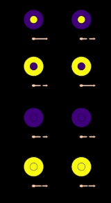
There are two types of bipolar cells: "on-center" and "off-center". An on-center cell is stimulated when the center of its receptive field is exposed to light, and is inhibited when the surround is exposed to light. Off-center cells have just the opposite reaction. On the edge between the two, in mammals, an on-off effect (i.e., discharging at switching on or off but not at a duration of either state) is present.
Stimulation of the center of an on-center cell's receptive field produces depolarization
and an increase in the firing of the ganglion cell, stimulation of the surround produces a Hyperpolarization
and a decrease in the firing of the cell, and stimulation of both the center and surround produces only a mild response (due to mutual inhibition of center and surround). An off-center cell is stimulated by activation of the surround and inhibited by stimulation of the center (see figure).
Photoreceptors that are part of the receptive fields of more than one ganglion cell are able to excite or inhibit postsynaptic neurons because they release the neurotransmitter
glutamate at their synapse
s, which can act to depolarize or to hyperpolarize a cell, depending on the ion channel
s it opens.
The center-surround receptive field organization allows ganglion cells to transmit information not merely about whether photoreceptor cells are exposed to light, but also about the differences in firing rates of cells in the center and surround. This allows them to transmit information about contrast. The size of the receptive field governs the spatial frequency
of the information: small receptive fields are stimulated by high spatial frequencies, fine detail; large receptive fields are stimulated by low spatial frequencies, coarse detail. Retinal ganglion cell receptive fields convey information about discontinuities in the distribution of light falling on the retina; these often specify the edges of objects. In dark adaptation, the peripheral opposite activity zone becomes inactive, but, since it is a diminishing of inhibition between center and periphery, the active field can actually increase, allowing more area for summation.
The receptive field tends to favor movement (such as a light or dark spot moving over the field, as in center-to-periphery (or vice versa)), as well as contours (due to their nonuniformity in the receptive fields). The center of the visual field has as much diameter as its dendrite spread, so the periphery is founded by amacrine cells connecting a wide area of bipolars to the ganglion. These amacrine cells can also inhibit signals of the periphery from being transmitted to the ganglion, thus rendering it on-center, off-periphery. In the rabbit, one direction, the "preferred," of a moving patch of light will excite a ganglion cell, whereas the opposite ("null") direction will not, also inhibiting spontaneous activity. Thus, there may be a linear nature of photoreceptors, one inhibiting its following neighbor when moving in the null direction, but arriving too late at the adjacent cell when traveling in the preferred direction.
. Receptive fields are similar to those of ganglion cells, with an antagonistic center-surround system and cells that are either on- or off center.
s, complex cell
s, and hypercomplex cell
s. Simple cell receptive fields are elongated, for example with an excitatory central oval, and an inhibitory surrounding region, or approximately rectangular, with one long side being excitatory and the other being inhibitory. Images for these receptive fields need to have a particular orientation in order to excite the cell. For complex-cell receptive fields, a correctly oriented bar of light might need to move in a particular direction in order to excite the cell. For hypercomplex receptive fields, the bar might also need to be of a particular length.
, images of faces excite the cortex more than other images. This property was one of the earliest major results obtained through fMRI (Kanwisher, McDermott and Chun, 1997); the finding was confirmed later at the neuronal level (Tsao, Freiwald, Tootell and Livingstone, 2006). In a similar vein, people have looked for other category-specific areas; some recent research for example suggests the parahippocampal place area might be somewhat specialised for buildings. However, more recent research has suggested that the fusiform face area is specialised not just for faces, but also for any discrete, within-category discrimination.
Neuron
A neuron is an electrically excitable cell that processes and transmits information by electrical and chemical signaling. Chemical signaling occurs via synapses, specialized connections with other cells. Neurons connect to each other to form networks. Neurons are the core components of the nervous...
is a region of space in which the presence of a stimulus
Stimulus (physiology)
In physiology, a stimulus is a detectable change in the internal or external environment. The ability of an organism or organ to respond to external stimuli is called sensitivity....
will alter the firing of that neuron. Receptive fields have been identified for neurons of the auditory system
Auditory system
The auditory system is the sensory system for the sense of hearing.- Outer ear :The folds of cartilage surrounding the ear canal are called the pinna...
, the somatosensory system
Somatosensory system
The somatosensory system is a diverse sensory system composed of the receptors and processing centres to produce the sensory modalities such as touch, temperature, proprioception , and nociception . The sensory receptors cover the skin and epithelia, skeletal muscles, bones and joints, internal...
, and the visual system
Visual system
The visual system is the part of the central nervous system which enables organisms to process visual detail, as well as enabling several non-image forming photoresponse functions. It interprets information from visible light to build a representation of the surrounding world...
.
The concept of receptive fields can be extended to further up the neural system; if many sensory receptors all form synapses with a single cell
Cell (biology)
The cell is the basic structural and functional unit of all known living organisms. It is the smallest unit of life that is classified as a living thing, and is often called the building block of life. The Alberts text discusses how the "cellular building blocks" move to shape developing embryos....
further up, they collectively form the receptive field of that cell. For example, the receptive field of a ganglion cell
Ganglion cell
A retinal ganglion cell is a type of neuron located near the inner surface of the retina of the eye. It receives visual information from photoreceptors via two intermediate neuron types: bipolar cells and amacrine cells...
in the retina
Retina
The vertebrate retina is a light-sensitive tissue lining the inner surface of the eye. The optics of the eye create an image of the visual world on the retina, which serves much the same function as the film in a camera. Light striking the retina initiates a cascade of chemical and electrical...
of the eye
Human eye
The human eye is an organ which reacts to light for several purposes. As a conscious sense organ, the eye allows vision. Rod and cone cells in the retina allow conscious light perception and vision including color differentiation and the perception of depth...
is composed of input from all of the photoreceptors which synapse with it, and a group of ganglion cells in turn forms the receptive field for a cell in the brain. This process is called convergence
Convergence (eye)
In ophthalmology, convergence is the simultaneous inward movement of both eyes toward each other, usually in an effort to maintain single binocular vision when viewing an object. This action is mediated by the medial rectus muscle, which is innervated by Cranial nerve III...
.
Auditory system
In the auditory system, receptive fields can correspond to volumes in auditory space, or to regions of auditory frequenciesFrequency
Frequency is the number of occurrences of a repeating event per unit time. It is also referred to as temporal frequency.The period is the duration of one cycle in a repeating event, so the period is the reciprocal of the frequency...
. Researchers rarely equate auditory receptive fields to particular regions of the sensory epithelium
Epithelium
Epithelium is one of the four basic types of animal tissue, along with connective tissue, muscle tissue and nervous tissue. Epithelial tissues line the cavities and surfaces of structures throughout the body, and also form many glands. Functions of epithelial cells include secretion, selective...
such as, in the case of mammals, hair cell
Hair cell
Hair cells are the sensory receptors of both the auditory system and the vestibular system in all vertebrates. In mammals, the auditory hair cells are located within the organ of Corti on a thin basilar membrane in the cochlea of the inner ear...
s in the cochlea
Cochlea
The cochlea is the auditory portion of the inner ear. It is a spiral-shaped cavity in the bony labyrinth, making 2.5 turns around its axis, the modiolus....
.
Somatosensory system
In the somatosensory system, receptive fields are regions of the skinSkin
-Dermis:The dermis is the layer of skin beneath the epidermis that consists of connective tissue and cushions the body from stress and strain. The dermis is tightly connected to the epidermis by a basement membrane. It also harbors many Mechanoreceptors that provide the sense of touch and heat...
or of internal organs. Some types of mechanoreceptor
Mechanoreceptor
A mechanoreceptor is a sensory receptor that responds to mechanical pressure or distortion. There are four main types in the glabrous skin of humans: Pacinian corpuscles, Meissner's corpuscles, Merkel's discs, and Ruffini corpuscles...
s have large receptive fields, while others have smaller ones.
Large receptive fields allow the cell to detect changes over a wider area, but lead to a less precise perception. Thus, the fingers, which require the ability to detect fine detail, have many, densely packed (up to 500 per cubic cm) mechanoreceptors with small receptive fields (around 10 square mm), while the back and legs, for example, have fewer receptors with large receptive fields. Receptors with large receptive fields usually have a "hot spot", an area within the receptive field (usually in the center, directly over the receptor) where stimulation produces the most intense response.
Tactile-sense-related cortical neurons have receptive fields on the skin that can be modified by experience or by injury to sensory nerves resulting in changes in the field's size and position. In general these neurons have relatively large receptive fields (much larger than those of dorsal root ganglion cells. However, the neurons are able to discriminate fine detail due to patterns of excitation and inhibition relative to the field which leads to spatial resolution.
Visual system
In the visual system, receptive fields are volumes in visual spaceVisual space
Visual space is the perceptual space housing the visual world being experienced by an aware observer; it is the subjective counterpart of the space of physical objects before an observer's eyes.-Space of Physical Objects:...
. For example, the receptive field of a single photoreceptor is a cone-shaped volume comprising all the visual directions in which light will alter the firing of that cell. Its apex
Apex (geometry)
In geometry, an apex is the vertex which is in some sense the highest of the figure to which it belongs.*In an isosceles triangle, the apex is the vertex where the two sides of equal length meet, opposite the unequal third side....
is located in the center of the lens
Lens (anatomy)
The crystalline lens is a transparent, biconvex structure in the eye that, along with the cornea, helps to refract light to be focused on the retina. The lens, by changing shape, functions to change the focal distance of the eye so that it can focus on objects at various distances, thus allowing a...
and its base essentially at infinity
Infinity
Infinity is a concept in many fields, most predominantly mathematics and physics, that refers to a quantity without bound or end. People have developed various ideas throughout history about the nature of infinity...
in visual space. Traditionally, visual receptive fields were portrayed in two dimensions (e.g., as circles, squares, or rectangles), but these are simply slices, cut along the screen on which the researcher presented the stimulus, of the volume of space to which a particular cell will respond. In the case of binocular
Binocular vision
Binocular vision is vision in which both eyes are used together. The word binocular comes from two Latin roots, bini for double, and oculus for eye. Having two eyes confers at least four advantages over having one. First, it gives a creature a spare eye in case one is damaged. Second, it gives a...
neurons in the visual cortex
Visual cortex
The visual cortex of the brain is the part of the cerebral cortex responsible for processing visual information. It is located in the occipital lobe, in the back of the brain....
, receptive fields do not extend to optical infinity. Instead, they are restricted to a certain interval of distance from the animal, or from where the eyes are fixating (see Panum's area).
The receptive field is often identified as the region of the retina
Retina
The vertebrate retina is a light-sensitive tissue lining the inner surface of the eye. The optics of the eye create an image of the visual world on the retina, which serves much the same function as the film in a camera. Light striking the retina initiates a cascade of chemical and electrical...
where the action of light
Light
Light or visible light is electromagnetic radiation that is visible to the human eye, and is responsible for the sense of sight. Visible light has wavelength in a range from about 380 nanometres to about 740 nm, with a frequency range of about 405 THz to 790 THz...
alters the firing of the neuron. In retinal ganglion cells (see below), this area of the retina would encompass all the photoreceptors, all the rod
Rod cell
Rod cells, or rods, are photoreceptor cells in the retina of the eye that can function in less intense light than can the other type of visual photoreceptor, cone cells. Named for their cylindrical shape, rods are concentrated at the outer edges of the retina and are used in peripheral vision. On...
s and cone
Cone cell
Cone cells, or cones, are photoreceptor cells in the retina of the eye that are responsible for color vision; they function best in relatively bright light, as opposed to rod cells that work better in dim light. If the retina is exposed to an intense visual stimulus, a negative afterimage will be...
s from one eye
Human eye
The human eye is an organ which reacts to light for several purposes. As a conscious sense organ, the eye allows vision. Rod and cone cells in the retina allow conscious light perception and vision including color differentiation and the perception of depth...
that are connected to this particular ganglion cell via bipolar cell
Bipolar cell
As a part of the retina, the bipolar cell exists between photoreceptors and ganglion cells. They act, directly or indirectly, to transmit signals from the photoreceptors to the ganglion cells.-Overview:...
s, horizontal cell
Horizontal cell
Horizontal cells are the laterally interconnecting neurons in the outer plexiform layer of the retina of mammalian eyes. They help integrate and regulate the input from multiple photoreceptor cells...
s, and amacrine cell
Amacrine cell
Amacrine cells are interneurons in the retina. Amacrine cells are responsible for 70% of input to retinal ganglion cells. Bipolar cells, which are responsible for the other 30% of input to retinal ganglia, are regulated by amacrine cells.-Overview:...
s. In binocular neurons in the visual cortex, it is necessary to specify the corresponding area in both retinas (one in each eye). Although these can be mapped separately in each retina by shutting one or the other eye, the full influence on the neuron's firing is revealed only when both eyes are open.
Hubel and Wiesel (e.g., Hubel, 1963) advanced the theory that receptive fields of cells at one level of the visual system are formed from input by cells at a lower level of the visual system. In this way, small, simple receptive fields could be combined to form large, complex receptive fields. Later theorists elaborated this simple, hierarchical arrangement by allowing cells at one level of the visual system to be influenced by feedback from higher levels.
Receptive fields have been mapped for all levels of the visual system from photoreceptors, to retinal ganglion cells, to lateral geniculate nucleus cells, to visual cortex cells, to extrastriate cortical cells. Studies based on perception do not give the full picture of the understanding of visual phenomena, so the electrophysiological tools must be used, as the retina, after all, is an outgrowth of the brain.
Retinal ganglion cells
The organization of ganglion cells' receptive fields, composed of inputs from many rods and cones, provides a way of detecting contrast, and is used for detecting objects' edges. Each receptive field is arranged into a central disk, the "center", and a concentric ring, the "surround", each region responding oppositely to light. For example, light in the centre might increase the firing of a particular ganglion cell, whereas light in the surround would decrease the firing of that cell.
There are two types of bipolar cells: "on-center" and "off-center". An on-center cell is stimulated when the center of its receptive field is exposed to light, and is inhibited when the surround is exposed to light. Off-center cells have just the opposite reaction. On the edge between the two, in mammals, an on-off effect (i.e., discharging at switching on or off but not at a duration of either state) is present.
Stimulation of the center of an on-center cell's receptive field produces depolarization
Depolarization
In biology, depolarization is a change in a cell's membrane potential, making it more positive, or less negative. In neurons and some other cells, a large enough depolarization may result in an action potential...
and an increase in the firing of the ganglion cell, stimulation of the surround produces a Hyperpolarization
Hyperpolarization (biology)
Hyperpolarization is a change in a cell's membrane potential that makes it more negative. It is the opposite of a depolarization.Hyperpolarization is often caused by efflux of K+ through K+ channels, or influx of Cl– through Cl– channels. On the other hand, influx of cations, e.g...
and a decrease in the firing of the cell, and stimulation of both the center and surround produces only a mild response (due to mutual inhibition of center and surround). An off-center cell is stimulated by activation of the surround and inhibited by stimulation of the center (see figure).
Photoreceptors that are part of the receptive fields of more than one ganglion cell are able to excite or inhibit postsynaptic neurons because they release the neurotransmitter
Neurotransmitter
Neurotransmitters are endogenous chemicals that transmit signals from a neuron to a target cell across a synapse. Neurotransmitters are packaged into synaptic vesicles clustered beneath the membrane on the presynaptic side of a synapse, and are released into the synaptic cleft, where they bind to...
glutamate at their synapse
Synapse
In the nervous system, a synapse is a structure that permits a neuron to pass an electrical or chemical signal to another cell...
s, which can act to depolarize or to hyperpolarize a cell, depending on the ion channel
Ion channel
Ion channels are pore-forming proteins that help establish and control the small voltage gradient across the plasma membrane of cells by allowing the flow of ions down their electrochemical gradient. They are present in the membranes that surround all biological cells...
s it opens.
The center-surround receptive field organization allows ganglion cells to transmit information not merely about whether photoreceptor cells are exposed to light, but also about the differences in firing rates of cells in the center and surround. This allows them to transmit information about contrast. The size of the receptive field governs the spatial frequency
Spatial frequency
In mathematics, physics, and engineering, spatial frequency is a characteristic of any structure that is periodic across position in space. The spatial frequency is a measure of how often sinusoidal components of the structure repeat per unit of distance. The SI unit of spatial frequency is...
of the information: small receptive fields are stimulated by high spatial frequencies, fine detail; large receptive fields are stimulated by low spatial frequencies, coarse detail. Retinal ganglion cell receptive fields convey information about discontinuities in the distribution of light falling on the retina; these often specify the edges of objects. In dark adaptation, the peripheral opposite activity zone becomes inactive, but, since it is a diminishing of inhibition between center and periphery, the active field can actually increase, allowing more area for summation.
The receptive field tends to favor movement (such as a light or dark spot moving over the field, as in center-to-periphery (or vice versa)), as well as contours (due to their nonuniformity in the receptive fields). The center of the visual field has as much diameter as its dendrite spread, so the periphery is founded by amacrine cells connecting a wide area of bipolars to the ganglion. These amacrine cells can also inhibit signals of the periphery from being transmitted to the ganglion, thus rendering it on-center, off-periphery. In the rabbit, one direction, the "preferred," of a moving patch of light will excite a ganglion cell, whereas the opposite ("null") direction will not, also inhibiting spontaneous activity. Thus, there may be a linear nature of photoreceptors, one inhibiting its following neighbor when moving in the null direction, but arriving too late at the adjacent cell when traveling in the preferred direction.
Lateral geniculate nucleus
Further along in the visual system, groups of ganglion cells form the receptive fields of cells in the lateral geniculate nucleusLateral geniculate nucleus
The lateral geniculate nucleus is the primary relay center for visual information received from the retina of the eye. The LGN is found inside the thalamus of the brain....
. Receptive fields are similar to those of ganglion cells, with an antagonistic center-surround system and cells that are either on- or off center.
Visual cortex
Receptive fields of cells in the visual cortex are larger and have more-complex stimulus requirements than retinal ganglion cells or lateral geniculate nucleus cells. Hubel and Wiesel (e.g., Hubel, 1963) classified receptive fields of cells in the visual cortex into simple cellSimple cell
A simple cell in the primary visual cortex is a cell that responds primarily to oriented edges and gratings . These cells were discovered by Torsten Wiesel and David Hubel in the late 1950s ....
s, complex cell
Complex cell
Complex cells can be found in the primary visual cortex , the secondary visual cortex , and Brodmann area 19 .Like a simple cell, a complex cell will respond primarily to oriented edges and gratings, however it has a degree of spatial invariance. This means that its receptive field cannot be...
s, and hypercomplex cell
Hypercomplex cell
Hubel and Wiesel named cells in the visual cortex that exhibit end stopping properties Hypercomplex cells, but later research has found them to be subclasses of the simple and complex cells...
s. Simple cell receptive fields are elongated, for example with an excitatory central oval, and an inhibitory surrounding region, or approximately rectangular, with one long side being excitatory and the other being inhibitory. Images for these receptive fields need to have a particular orientation in order to excite the cell. For complex-cell receptive fields, a correctly oriented bar of light might need to move in a particular direction in order to excite the cell. For hypercomplex receptive fields, the bar might also need to be of a particular length.
Extrastriate visual areas
In extrastriate visual areas, cells can have very large receptive fields requiring very complex images to excite the cell. For example in the inferotemporal cortex, receptive fields cross the midline of visual space and require images such as radial gratings or hands. It is also believed that in the fusiform face areaFusiform face area
The fusiform face area is a part of the human visual system which might be specialized for facial recognition, although there is some evidence that it also processes categorical information about other objects, particularly familiar ones.-Localization:...
, images of faces excite the cortex more than other images. This property was one of the earliest major results obtained through fMRI (Kanwisher, McDermott and Chun, 1997); the finding was confirmed later at the neuronal level (Tsao, Freiwald, Tootell and Livingstone, 2006). In a similar vein, people have looked for other category-specific areas; some recent research for example suggests the parahippocampal place area might be somewhat specialised for buildings. However, more recent research has suggested that the fusiform face area is specialised not just for faces, but also for any discrete, within-category discrimination.