
Retinal
Encyclopedia
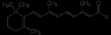
Retinal, also called retinaldehyde or vitamin A aldehyde
, is one of the many forms of vitamin A (the number of which varies from species to species). Retinal is a polyene
chromophore
, and bound to proteins called opsin
s, is the chemical basis of animal
vision. Bound to proteins called type 1 rhodopsin
s, retinal allows certain microorganisms to convert light into metabolic energy.
Vertebrate animals ingest retinal directly from meat, or produce retinal from one of four carotenoid
s (beta-carotene
, alpha-carotene
, gamma-carotene, and beta-cryptoxanthin
), which they must obtain from plants or other photosynthetic organisms (no other carotenoid
s can be converted by animals to retinal, and some carnivores cannot convert any carotenoids at all). The other main forms of vitamin A, retinol
, and a partially active form retinoic acid
, may both be produced from retinal.
Invertebrates such as insects and squid use hydroxylated forms of retinal in their visual systems, which derive from conversion from other xanthophylls.
For example
catalyzed by a beta-carotene 15,15'-monooxygenase
or a beta-carotene 15,15'-dioxygenase.
Just as carotenoids are the precursors of retinal, retinal is the precursor of the other forms of vitamin A. Retinal is interconvertible with retinol
(ROL), the transport and storage form of vitamin A
catalyzed by retinol dehydrogenase
s (RDHs) and alcohol dehydrogenase
s (ADHs).
Retinol is called vitamin A alcohol
, or more often, simply vitamin A. Retinal can also be oxidized to retinoic acid
(RA)
catalyzed by retinal dehydrogenase
s also known as retinaldehyde dehydrogenases (RALDHs)
as well as retinal oxidase
s.
Retinoic acid, sometimes called vitamin A acid
, is an important signaling molecule and hormone in vertebrate animals.
it isomerizes from the 11-cis state to the all-trans state. The absorbance spectrum of the chromophore depends on its interactions with the opsin
protein to which it is bound; different opsins produce different absorbance spectra.
s of eyes. An opsin is arranged into a bundle of seven transmembrane alpha-helices connected by six loops. In rod cells the opsin molecules are embedded in the membranes of the disks which are entirely inside of the cell. The N-terminus head of the molecule extends into the interior of the disk, and the C-terminus tail extends into the cytoplasm of the cell. In cone cells the disks are defined by the cell's plasma membrane so that the N-terminus head extends outside of the cell. Retinal binds covalently to a lysine
on the transmembrane helix nearest the C-terminus of the protein through a Schiff base
linkage. Formation of the Schiff base linkage involves removing the oxygen atom from retinal and two hydrogen atoms from the free amino group of lysine, giving H2O. Retinylidene is the divalent group formed by removing the oxygen atom from retinal, and so opsins have been called retinylidene protein
s.
Opsins are prototypical G protein-coupled receptor
s (GPCRs). Bovine rhodopsin, the opsin of the rod cells of cattle, was the first GPCR to have its X-ray structure determined.
Bovine rhodopsin contains 348 amino acid residues. The retinal chromophore binds at Lys296.
Although mammals use retinal exclusively as the opsin chromophore, other groups of animals additionally use four chromophores closely related to retinal. These are (3,4)-didehydroretinal, (3R)-3-hydroxyretinal, (3S)-3-hydroxyretinal, and (4R)-4-hydroxyretinal. Many fish and amphibians use (3,4)-didehydroretinal, also called dehydroretinal
. With the exception of the diptera
n suborder Cyclorrhapha
, the so-called higher flies, all insects examined use the R enantiomer
of 3-hydroxyretinal. The R enantiomer is to be expected if 3-hydroxyretinal is produced directly from xanthophyll
carotenoids. Cyclorrhaphans, including Drosophila
, use (3S)-3-hydroxyretinal.
Firefly squid
have been found to use (4R)-4-hydroxyretinal.
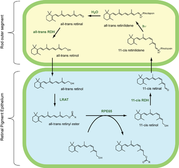 The visual cycle is a circular enzymatic pathway, which is the front-end of phototransduction. It regenerates 11-cis-retinal.
For example, the visual cycle of mammalian rod cells
Steps 3,4,5,6 occur in rod cell outer segments
; Steps 1, 2, and 7 occur in retinal pigment epithelium (RPE) cells.
 As it happens, RPE65 isomerohydrolases are homologous
with beta-carotene monooxygenases;
the homologous ninaB enzyme in Drosophila has both retinal-forming carotenoid-oxygenase activity and all-trans to 11-cis isomerase activity.
, channelrhodopsin
, and halorhodopsin
. In these molecules, light causes the all-trans-retinal to become 13-cis retinal, which then cycles back to all-trans-retinal in the dark state.
and others had outlined the visual cycle by 1958. For his work, Wald won a share of the 1967 Nobel Prize in Physiology or Medicine
with Haldan Keffer Hartline
and Ragnar Granit
.
Aldehyde
An aldehyde is an organic compound containing a formyl group. This functional group, with the structure R-CHO, consists of a carbonyl center bonded to hydrogen and an R group....
, is one of the many forms of vitamin A (the number of which varies from species to species). Retinal is a polyene
Polyene
Polyenes are poly-unsaturated organic compounds that contain one or more sequences of alternating double and single carbon-carbon bonds. These double carbon-carbon bonds interact in a process known as conjugation, which results in an overall lower energy state of the molecule.Organic compounds with...
chromophore
Chromophore
A chromophore is the part of a molecule responsible for its color. The color arises when a molecule absorbs certain wavelengths of visible light and transmits or reflects others. The chromophore is a region in the molecule where the energy difference between two different molecular orbitals falls...
, and bound to proteins called opsin
Opsin
Opsins are a group of light-sensitive 35–55 kDa membrane-bound G protein-coupled receptors of the retinylidene protein family found in photoreceptor cells of the retina. Five classical groups of opsins are involved in vision, mediating the conversion of a photon of light into an electrochemical...
s, is the chemical basis of animal
Animal
Animals are a major group of multicellular, eukaryotic organisms of the kingdom Animalia or Metazoa. Their body plan eventually becomes fixed as they develop, although some undergo a process of metamorphosis later on in their life. Most animals are motile, meaning they can move spontaneously and...
vision. Bound to proteins called type 1 rhodopsin
Rhodopsin
Rhodopsin, also known as visual purple, is a biological pigment of the retina that is responsible for both the formation of the photoreceptor cells and the first events in the perception of light. Rhodopsins belong to the G-protein coupled receptor family and are extremely sensitive to light,...
s, retinal allows certain microorganisms to convert light into metabolic energy.
Vertebrate animals ingest retinal directly from meat, or produce retinal from one of four carotenoid
Carotenoid
Carotenoids are tetraterpenoid organic pigments that are naturally occurring in the chloroplasts and chromoplasts of plants and some other photosynthetic organisms like algae, some bacteria, and some types of fungus. Carotenoids can be synthesized fats and other basic organic metabolic building...
s (beta-carotene
Beta-carotene
β-Carotene is a strongly-coloured red-orange pigment abundant in plants and fruits. It is an organic compound and chemically is classified as a hydrocarbon and specifically as a terpenoid , reflecting its derivation from isoprene units...
, alpha-carotene
Alpha-carotene
α-Carotene is a form of carotene with a β-ring at one end and an ε-ring at the other. It is the second most common form of carotene.-Human physiology:...
, gamma-carotene, and beta-cryptoxanthin
Cryptoxanthin
Cryptoxanthin is a natural carotenoid pigment. It has been isolated from a variety of sources including the petals and flowers of plants in the genus Physalis, orange rind, papaya, egg yolk, butter, apples, and bovine blood serum.-Chemistry:...
), which they must obtain from plants or other photosynthetic organisms (no other carotenoid
Carotenoid
Carotenoids are tetraterpenoid organic pigments that are naturally occurring in the chloroplasts and chromoplasts of plants and some other photosynthetic organisms like algae, some bacteria, and some types of fungus. Carotenoids can be synthesized fats and other basic organic metabolic building...
s can be converted by animals to retinal, and some carnivores cannot convert any carotenoids at all). The other main forms of vitamin A, retinol
Retinol
Retinol is one of the animal forms of vitamin A. It is a diterpenoid and an alcohol. It is convertible to other forms of vitamin A, and the retinyl ester derivative of the alcohol serves as the storage form of the vitamin in animals....
, and a partially active form retinoic acid
Retinoic acid
Retinoic acid is a metabolite of vitamin A that mediates the functions of vitamin A required for growth and development. Retinoic acid is required in chordate animals which includes all higher animals from fishes to humans...
, may both be produced from retinal.
Invertebrates such as insects and squid use hydroxylated forms of retinal in their visual systems, which derive from conversion from other xanthophylls.
Vitamin A metabolism
Living organisms produce retinal (RAL) by irreversible oxidative cleavage of carotenoids.For example
- beta-caroteneBeta-caroteneβ-Carotene is a strongly-coloured red-orange pigment abundant in plants and fruits. It is an organic compound and chemically is classified as a hydrocarbon and specifically as a terpenoid , reflecting its derivation from isoprene units...
+ O2 → 2 retinal
catalyzed by a beta-carotene 15,15'-monooxygenase
Beta-carotene 15,15'-monooxygenase
In enzymology, a beta-carotene 15,15'-monooxygenase is an enzyme that catalyzes the chemical reactionThus, the two substrates of this enzyme are beta-carotene and O2, whereas its product is retinal....
or a beta-carotene 15,15'-dioxygenase.
Just as carotenoids are the precursors of retinal, retinal is the precursor of the other forms of vitamin A. Retinal is interconvertible with retinol
Retinol
Retinol is one of the animal forms of vitamin A. It is a diterpenoid and an alcohol. It is convertible to other forms of vitamin A, and the retinyl ester derivative of the alcohol serves as the storage form of the vitamin in animals....
(ROL), the transport and storage form of vitamin A
- retinal + NADPHNicotinamide adenine dinucleotide phosphateNicotinamide adenine dinucleotide phosphate, abbreviated NADP or TPN in older notation , is a coenzyme used in anabolic reactions, such as lipid and nucleic acid synthesis, which require NADPH as a reducing agent....
+ H+ retinol + NADP+ - retinol + NADNicotinamide adenine dinucleotideNicotinamide adenine dinucleotide, abbreviated NAD, is a coenzyme found in all living cells. The compound is a dinucleotide, since it consists of two nucleotides joined through their phosphate groups. One nucleotide contains an adenine base and the other nicotinamide.In metabolism, NAD is involved...
+ retinal + NADH + H+
catalyzed by retinol dehydrogenase
Retinol dehydrogenase
In enzymology, a retinol dehydrogenase is an enzyme that catalyzes the chemical reactionThus, the two substrates of this enzyme are retinol and NAD+, whereas its 3 products are retinal, NADH, and H+....
s (RDHs) and alcohol dehydrogenase
Alcohol dehydrogenase
Alcohol dehydrogenases are a group of dehydrogenase enzymes that occur in many organisms and facilitate the interconversion between alcohols and aldehydes or ketones with the reduction of nicotinamide adenine dinucleotide...
s (ADHs).
Retinol is called vitamin A alcohol
Alcohol
In chemistry, an alcohol is an organic compound in which the hydroxy functional group is bound to a carbon atom. In particular, this carbon center should be saturated, having single bonds to three other atoms....
, or more often, simply vitamin A. Retinal can also be oxidized to retinoic acid
Retinoic acid
Retinoic acid is a metabolite of vitamin A that mediates the functions of vitamin A required for growth and development. Retinoic acid is required in chordate animals which includes all higher animals from fishes to humans...
(RA)
- retinal + NAD+ + H2O → retinoic acid + NADH + H+ (catalyzed by RALDH)
- retinal + O2 + H2O → retinoic acid + H2O2 (catalyzed by retinal oxidase)
catalyzed by retinal dehydrogenase
Retinal dehydrogenase
In enzymology, a retinal dehydrogenase is an enzyme that catalyzes the chemical reactionThe 3 substrates of this enzyme are retinal, NAD+, and H2O, whereas its 3 products are retinoic acid, NADH, and H+....
s also known as retinaldehyde dehydrogenases (RALDHs)
as well as retinal oxidase
Retinal oxidase
In enzymology, a retinal oxidase is an enzyme that catalyzes the chemical reactionThe 3 substrates of this enzyme are retinal, O2, and H2O, whereas its two products are retinoic acid and H2O2....
s.
Retinoic acid, sometimes called vitamin A acid
Carboxylic acid
Carboxylic acids are organic acids characterized by the presence of at least one carboxyl group. The general formula of a carboxylic acid is R-COOH, where R is some monovalent functional group...
, is an important signaling molecule and hormone in vertebrate animals.
Vision
Vision begins with the photoisomerization of retinal. When the 11-cis-retinal chromophore absorbs a photonPhoton
In physics, a photon is an elementary particle, the quantum of the electromagnetic interaction and the basic unit of light and all other forms of electromagnetic radiation. It is also the force carrier for the electromagnetic force...
it isomerizes from the 11-cis state to the all-trans state. The absorbance spectrum of the chromophore depends on its interactions with the opsin
Opsin
Opsins are a group of light-sensitive 35–55 kDa membrane-bound G protein-coupled receptors of the retinylidene protein family found in photoreceptor cells of the retina. Five classical groups of opsins are involved in vision, mediating the conversion of a photon of light into an electrochemical...
protein to which it is bound; different opsins produce different absorbance spectra.
Opsins
Opsins are proteins and the retinal-binding visual pigments found in the photoreceptor cells in the retinaRetina
The vertebrate retina is a light-sensitive tissue lining the inner surface of the eye. The optics of the eye create an image of the visual world on the retina, which serves much the same function as the film in a camera. Light striking the retina initiates a cascade of chemical and electrical...
s of eyes. An opsin is arranged into a bundle of seven transmembrane alpha-helices connected by six loops. In rod cells the opsin molecules are embedded in the membranes of the disks which are entirely inside of the cell. The N-terminus head of the molecule extends into the interior of the disk, and the C-terminus tail extends into the cytoplasm of the cell. In cone cells the disks are defined by the cell's plasma membrane so that the N-terminus head extends outside of the cell. Retinal binds covalently to a lysine
Lysine
Lysine is an α-amino acid with the chemical formula HO2CCH4NH2. It is an essential amino acid, which means that the human body cannot synthesize it. Its codons are AAA and AAG....
on the transmembrane helix nearest the C-terminus of the protein through a Schiff base
Schiff base
A Schiff base, named after Hugo Schiff, is a compound with a functional group that contains a carbon-nitrogen double bond with the nitrogen atom connected to an aryl or alkyl group, not hydrogen....
linkage. Formation of the Schiff base linkage involves removing the oxygen atom from retinal and two hydrogen atoms from the free amino group of lysine, giving H2O. Retinylidene is the divalent group formed by removing the oxygen atom from retinal, and so opsins have been called retinylidene protein
Retinylidene protein
Retinylidene proteins are a family of proteins that use retinal as chromophore for light reception. Proteins of this family are also called opsins...
s.
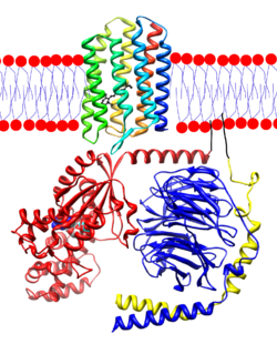
Opsins are prototypical G protein-coupled receptor
G protein-coupled receptor
G protein-coupled receptors , also known as seven-transmembrane domain receptors, 7TM receptors, heptahelical receptors, serpentine receptor, and G protein-linked receptors , comprise a large protein family of transmembrane receptors that sense molecules outside the cell and activate inside signal...
s (GPCRs). Bovine rhodopsin, the opsin of the rod cells of cattle, was the first GPCR to have its X-ray structure determined.
Bovine rhodopsin contains 348 amino acid residues. The retinal chromophore binds at Lys296.
Although mammals use retinal exclusively as the opsin chromophore, other groups of animals additionally use four chromophores closely related to retinal. These are (3,4)-didehydroretinal, (3R)-3-hydroxyretinal, (3S)-3-hydroxyretinal, and (4R)-4-hydroxyretinal. Many fish and amphibians use (3,4)-didehydroretinal, also called dehydroretinal
Dehydroretinal
Dehydroretinal is a derivative of retinal....
. With the exception of the diptera
Diptera
Diptera , or true flies, is the order of insects possessing only a single pair of wings on the mesothorax; the metathorax bears a pair of drumstick like structures called the halteres, the remnants of the hind wings. It is a large order, containing an estimated 240,000 species, although under half...
n suborder Cyclorrhapha
Cyclorrhapha
Cyclorrhapha is an unranked taxon within the infraorder Muscomorpha. They are called "Cyclorrhapha" with reference to the circular aperture through which the adult escapes the puparium...
, the so-called higher flies, all insects examined use the R enantiomer
Enantiomer
In chemistry, an enantiomer is one of two stereoisomers that are mirror images of each other that are non-superposable , much as one's left and right hands are the same except for opposite orientation. It can be clearly understood if you try to place your hands one over the other without...
of 3-hydroxyretinal. The R enantiomer is to be expected if 3-hydroxyretinal is produced directly from xanthophyll
Xanthophyll
Xanthophylls are yellow pigments that form one of two major divisions of the carotenoid group. The name is from Greek xanthos + phyllon , due to their formation of the yellow band seen in early chromatography of leaf pigments...
carotenoids. Cyclorrhaphans, including Drosophila
Drosophila
Drosophila is a genus of small flies, belonging to the family Drosophilidae, whose members are often called "fruit flies" or more appropriately pomace flies, vinegar flies, or wine flies, a reference to the characteristic of many species to linger around overripe or rotting fruit...
, use (3S)-3-hydroxyretinal.
Firefly squid
Firefly squid
The Sparkling Enope Squid , also known as the Firefly Squid, is a species of squid in the family Enoploteuthidae. It is the sole species in the genus Watasenia.-Species characteristics:...
have been found to use (4R)-4-hydroxyretinal.
Visual cycle
For example, the visual cycle of mammalian rod cells
- all-trans-retinyl ester + H2O → 11-cis-retinol + fatty acidFatty acidIn chemistry, especially biochemistry, a fatty acid is a carboxylic acid with a long unbranched aliphatic tail , which is either saturated or unsaturated. Most naturally occurring fatty acids have a chain of an even number of carbon atoms, from 4 to 28. Fatty acids are usually derived from...
; RPE65RPE65Retinal pigment epithelium-specific 65 kDa protein is a protein that in humans is encoded by the RPE65 gene.-Function:The retinal pigment epithelium-specific 65 kDa protein is located in the retinal pigment epithelium and is involved in the conversion of all-trans retinol to 11-cis retinal during...
isomerohydrolases - 11-cis-retinol + NAD+ → 11-cis-retinal + NADH + H+; 11-cis-retinol dehydrogenases
- 11-cis-retinal + aporhodopsin → rhodopsinRhodopsinRhodopsin, also known as visual purple, is a biological pigment of the retina that is responsible for both the formation of the photoreceptor cells and the first events in the perception of light. Rhodopsins belong to the G-protein coupled receptor family and are extremely sensitive to light,...
+ H2O; forms Schiff baseSchiff baseA Schiff base, named after Hugo Schiff, is a compound with a functional group that contains a carbon-nitrogen double bond with the nitrogen atom connected to an aryl or alkyl group, not hydrogen....
linkage to lysineLysineLysine is an α-amino acid with the chemical formula HO2CCH4NH2. It is an essential amino acid, which means that the human body cannot synthesize it. Its codons are AAA and AAG....
, -CH=N+H- - rhodopsin + hν → metarhodopsin II; 11-cis photoisomerizes to all-trans
- rhodopsin + hν → photorhodopsin → bathorhodopsin → lumirhodopsin → metarhodopsin I → metarhodopsin II
- metarhodopsin II + H2O → aporhodopsin + all-trans-retinal
- all-trans-retinal + NADPH + H+ → all-trans-retinol + NADP+; all-trans-retinol dehydrogenases
- all-trans-retinol + fatty acid → all-trans-retinyl ester + H2O; lecithin retinol acyltransferases (LRATs)
Steps 3,4,5,6 occur in rod cell outer segments
Rod cell
Rod cells, or rods, are photoreceptor cells in the retina of the eye that can function in less intense light than can the other type of visual photoreceptor, cone cells. Named for their cylindrical shape, rods are concentrated at the outer edges of the retina and are used in peripheral vision. On...
; Steps 1, 2, and 7 occur in retinal pigment epithelium (RPE) cells.
Homology (biology)
Homology forms the basis of organization for comparative biology. In 1843, Richard Owen defined homology as "the same organ in different animals under every variety of form and function". Organs as different as a bat's wing, a seal's flipper, a cat's paw and a human hand have a common underlying...
with beta-carotene monooxygenases;
the homologous ninaB enzyme in Drosophila has both retinal-forming carotenoid-oxygenase activity and all-trans to 11-cis isomerase activity.
Type 1 rhodopsins
All-trans-retinal is also an essential component of type I, or microbial, opsins such as bacteriorhodopsinBacteriorhodopsin
Bacteriorhodopsin is a protein used by Archaea, the most notable one being Halobacteria. It acts as a proton pump; that is, it captures light energy and uses it to move protons across the membrane out of the cell...
, channelrhodopsin
Channelrhodopsin
Channelrhodopsins are a subfamily of opsin proteins that function as light-gated ion channels. They serve as sensory photoreceptors in unicellular green algae, controlling phototaxis, i.e. movement in response to light. Expressed in cells of other organisms, they enable the use of light to control...
, and halorhodopsin
Halorhodopsin
Halorhodopsin is a light-driven ion pump, specific for chloride ions, and found in phylogenetically ancient archaea, known as halobacteria...
. In these molecules, light causes the all-trans-retinal to become 13-cis retinal, which then cycles back to all-trans-retinal in the dark state.
History
The American biochemist George WaldGeorge Wald
George Wald was an American scientist who is best known for his work with pigments in the retina. He won a share of the 1967 Nobel Prize in Physiology or Medicine with Haldan Keffer Hartline and Ragnar Granit.- Research :...
and others had outlined the visual cycle by 1958. For his work, Wald won a share of the 1967 Nobel Prize in Physiology or Medicine
Nobel Prize in Physiology or Medicine
The Nobel Prize in Physiology or Medicine administered by the Nobel Foundation, is awarded once a year for outstanding discoveries in the field of life science and medicine. It is one of five Nobel Prizes established in 1895 by Swedish chemist Alfred Nobel, the inventor of dynamite, in his will...
with Haldan Keffer Hartline
Haldan Keffer Hartline
Haldan Keffer Hartline was an American physiologist who was a co-winner of the 1967 Nobel Prize in Physiology or Medicine for his work in analyzing the neurophysiological mechanisms of vision.Hartline began his study of retinal electrophysiology as a National Research Council Fellow at Johns...
and Ragnar Granit
Ragnar Granit
Ragnar Arthur Granit was a Finnish/Swedish scientist who was awarded the Nobel Prize in Physiology or Medicine in 1967 along with Haldan Keffer Hartline and George Wald....
.
Further reading
The oceans are full of type 1 rhodopsin. Good historical review.External links
- First Steps of Vision - National Health Museum
- Vision and Light-Induced Molecular Changes
- Retinal Anatomy and Visual Capacities
- Retinal