

Axon
Encyclopedia
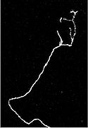
An axon is a long, slender projection of a nerve cell, or neuron
, that conducts electrical impulses
away from the neuron's cell body or soma.
An axon is one of two types of protoplasm
ic protrusions that extrude from the cell body of a neuron, the other type being dendrite
s. Axons are distinguished from dendrites by several features, including shape (dendrites often taper while axons usually maintain a constant radius), length (dendrites are restricted to a small region around the cell body while axons can be much longer), and function (dendrites usually receive signals while axons usually transmit them). All of these rules have exceptions, however.
Some types of neurons have no axon and transmit signals from their dendrites. No neuron ever has more than one axon; however in invertebrates such as insects or leeches the axon sometimes consists of several regions that function more or less independently of each other. Most axons branch, in some cases very profusely.
Axons make contact with other cells—usually other neurons but sometimes muscle or gland cells—at junctions called synapse
s. At a synapse, the membrane of the axon closely adjoins the membrane of the target cell, and special molecular structures serve to transmit electrical or electrochemical signals across the gap. Some synaptic junctions appear partway along an axon as it extends—these are called en passant ("in passing") synapses. Other synapses appear as terminals at the ends of axonal branches. A single axon, with all its branches taken together, can innervate multiple parts of the brain and generate thousands of synaptic terminals.
, and as bundles they help make up nerve
s. The length of axons is highly dependent on its location within the body. Some axons can extend up to one meter or more while others stretch to as little as one millimeter (inhibitory interneurons). The longest axons in the human body, for example, are those of the sciatic nerve
, which run from the base of the spine to the big toe of each foot. These single-cell fibers of the sciatic nerve may extend a meter or even longer. The diameter of axons is also variable. Individual axons are microscopic in diameter (typically about 1μm
across), but may be up to several feet in length. The largest mammalian axons (PNS) can reach a diameter of up to 20 μm
. The giant squid has axons that are close to 1 mm in diameter. Mammalian axonal arborization (the branching structure at the end of a nerve fiber) also differs from one nerve fiber to the next. Axons in the CNS typically model complex trees with several branch points. In comparison, the cerebellar granule cell axon is characterized by a single T-shaped branch node from which parallel fibers extend. Elaborate arborization is important for it allows for the simultaneous transmission of messages to a large number of target neurons within a single region of the brain .
In vertebrate
s, the axons of many neurons are sheathed in myelin
, which is formed by either of two types of glial cells: Schwann cell
s ensheathing peripheral
neurons and oligodendrocyte
s insulating those of the central nervous system
. Along myelinated nerve fibers, gaps in the sheath known as nodes of Ranvier
occur at evenly-spaced intervals. The myelination enables an especially rapid mode of electrical impulse propagation called saltation
. The demyelination of axons is what causes the multitude of neurological symptoms found in the disease Multiple Sclerosis
.
The axons of some neurons branch to form axon collaterals, that can be divided into a number of smaller branches called telodendria. Along these the bifurcated impulse travels simultaneously to signal more than one other cell.
initiation. It also has an important role in maintaining neuronal polarity. The exact position of the AIS along the axon differs between types of neuron and its position within a single family of neurons can vary. It has recently been discovered that the location and extent of a neuron's AIS can be altered by the neuron's level of activity and that these changes are thought to influence the excitability of the neuron.
The density of voltage-gated sodium channels is much higher here than is found in the adjacent cell body, excepting the axon hillock
.
can be described by the Hodgkin-Huxley Model
, extended to vertebrates in Frankenhaeuser-Huxley equations. Peripheral nerve fibers can be classified based on axonal conduction velocity, mylenation, fiber size etc. For example, there are slow-conducting unmyelinated C fibers and faster-conducting myelinated Aδ fiber
s. More complex mathematical modeling continues to be done today. There are several types of sensory- as well as motorfibers. Other fibers not mentioned in table are e.g. fibers of the autonomic nervous system
Lower motor neurons have two kind of fibers:
Different sensory receptors are innervated by different types of nerve fibers. Proprioceptors are innervated by type Ia, Ib and II sensory fibers, mechanoreceptor
s by type II and III sensory fibers and nociceptors and thermoreceptors by type III and IV sensory fibers.
When an axon is at rest, the internal environment is negative for the pumps ensure that sodium is kept out and potassium remains within the cell. This state is referred to as the resting potential. During the formation of an action potential, changes in electrical potential in the soma and the dendrites of the neuron travel to the axon. If at the axon the integrated signal is above the threshold, the sodium channels open . This allows for the inward flow of sodium ions, making the inside of the axon less negative. If this reversal in polarity reaches the threshold level, more gated channels open and more sodium ions are let in. This phenomenon is referred to as the action potential. Following this, the sodium gates close and the potassium gates open allowing potassium to rush out of the cell, returning a semblance of normalcy to the internal electrical environment of the cell. The potassium and sodium ions are then actively transported back to their respective locations via the sodium-potassium pumps.
This process is successive in that the opening of the sodium channels in the beginning of the axon causes neighboring sodium channels to open. During this time, the sodium channels that were first opened close and potassium channels open in this first region. In the second region, sodium is still rushing into the cell, causing the adjacent sodium channels closer to the end of the axon to open. All of the segments of the axon go through the same steps but at different times, thus allowing the action potential to be passed down the axon .
 Growing axons move through their environment via the growth cone
, which is at the tip of the axon. The growth cone has a broad sheet like extension called lamellipodia
which contain protrusions called filopodia
. The filopodia are the mechanism by which the entire process adheres to surfaces and explores the surrounding environment. Actin
plays a major role in the mobility of this system.
Environments with high levels of cell adhesion molecule
s or CAM's create an ideal environment for axonal growth. This seems to provide a "sticky" surface for axons to grow along. Examples of CAM's specific to neural systems include N-CAM
, neuroglial CAM or NgCAM, TAG-1, and MAG all of which are part of the immunoglobulin superfamily. Another set of molecules called extracellular matrix adhesion molecules also provide a sticky substrate for axons to grow along. Examples of these molecules include laminin
, fibronectin
, tenascin
, and perlecan
. Some of these are surface bound to cells and thus act as short range attractants or repellents. Others are difusible ligands and thus can have long range effects.
Cells called guidepost cells
assist in the guidance of neuronal axon growth. These cells are typically other, sometimes immature, neurons.
It has also been discovered through research that if the axons of a neuron were damaged, as long as the soma (the cell body of a neuron
) is not damaged, the axons would regenerate and remake the synaptic connections with neurons with the help of guidepost cells
. This is also referred to as neuroregeneration.
is generally credited with the discovery of the axon by distinguishing it from the dendrites. Swiss Rüdolf Albert von Kölliker and German Robert Remak were the first to identify and characterize the axon initial segment. Alan Hodgkin and Andrew Huxley
also employed the squid giant axon
(1939) and by 1952 they had obtained a full quantitative description of the ionic basis of the action potential
, leading the formulation of the Hodgkin-Huxley Model
.
Hodgkin and Huxley were awarded jointly the Nobel Prize
for this work in 1963.
The formulas detailing axonal conductance were extended to vertebrates in the Frankenhaeuser-Huxley equations. Louis-Antoine Ranvier was the first to describe the gaps or nodes found on axons and for this contribution these axonal features are now commonly referred to as the Nodes of Ranvier. Santiago Ramón y Cajal, a Spanish anatomist, proposed that axons were the output components of neurons, describing their functionality. Erlanger and Gasser earlier developed the classification system for peripheral nerve fibers, based on axonal conduction velocity, myelin
ation, fiber size etc.
Even recently our understanding of the biochemical basis for action potential propagation has advanced, and now includes many details about individual ion channel
s.
, or neurotmesis
.
Concussion is considered a mild form of diffuse axonal injury
.
Neuron
A neuron is an electrically excitable cell that processes and transmits information by electrical and chemical signaling. Chemical signaling occurs via synapses, specialized connections with other cells. Neurons connect to each other to form networks. Neurons are the core components of the nervous...
, that conducts electrical impulses
Action potential
In physiology, an action potential is a short-lasting event in which the electrical membrane potential of a cell rapidly rises and falls, following a consistent trajectory. Action potentials occur in several types of animal cells, called excitable cells, which include neurons, muscle cells, and...
away from the neuron's cell body or soma.
An axon is one of two types of protoplasm
Protoplasm
Protoplasm is the living contents of a cell that is surrounded by a plasma membrane. It is a general term of the Cytoplasm . Protoplasm is composed of a mixture of small molecules such as ions, amino acids, monosaccharides and water, and macromolecules such as nucleic acids, proteins, lipids and...
ic protrusions that extrude from the cell body of a neuron, the other type being dendrite
Dendrite
Dendrites are the branched projections of a neuron that act to conduct the electrochemical stimulation received from other neural cells to the cell body, or soma, of the neuron from which the dendrites project...
s. Axons are distinguished from dendrites by several features, including shape (dendrites often taper while axons usually maintain a constant radius), length (dendrites are restricted to a small region around the cell body while axons can be much longer), and function (dendrites usually receive signals while axons usually transmit them). All of these rules have exceptions, however.
Some types of neurons have no axon and transmit signals from their dendrites. No neuron ever has more than one axon; however in invertebrates such as insects or leeches the axon sometimes consists of several regions that function more or less independently of each other. Most axons branch, in some cases very profusely.
Axons make contact with other cells—usually other neurons but sometimes muscle or gland cells—at junctions called synapse
Synapse
In the nervous system, a synapse is a structure that permits a neuron to pass an electrical or chemical signal to another cell...
s. At a synapse, the membrane of the axon closely adjoins the membrane of the target cell, and special molecular structures serve to transmit electrical or electrochemical signals across the gap. Some synaptic junctions appear partway along an axon as it extends—these are called en passant ("in passing") synapses. Other synapses appear as terminals at the ends of axonal branches. A single axon, with all its branches taken together, can innervate multiple parts of the brain and generate thousands of synaptic terminals.
Anatomy
Axons are in effect the primary transmission lines of the nervous systemNervous system
The nervous system is an organ system containing a network of specialized cells called neurons that coordinate the actions of an animal and transmit signals between different parts of its body. In most animals the nervous system consists of two parts, central and peripheral. The central nervous...
, and as bundles they help make up nerve
Nerve
A peripheral nerve, or simply nerve, is an enclosed, cable-like bundle of peripheral axons . A nerve provides a common pathway for the electrochemical nerve impulses that are transmitted along each of the axons. Nerves are found only in the peripheral nervous system...
s. The length of axons is highly dependent on its location within the body. Some axons can extend up to one meter or more while others stretch to as little as one millimeter (inhibitory interneurons). The longest axons in the human body, for example, are those of the sciatic nerve
Sciatic nerve
The sciatic nerve is a large nerve fiber in humans and other animals. It begins in the lower back and runs through the buttock and down the lower limb...
, which run from the base of the spine to the big toe of each foot. These single-cell fibers of the sciatic nerve may extend a meter or even longer. The diameter of axons is also variable. Individual axons are microscopic in diameter (typically about 1μm
Micrometre
A micrometer , is by definition 1×10⁻⁶ of a meter .In plain English, it means one-millionth of a meter . Its unit symbol in the International System of Units is μm...
across), but may be up to several feet in length. The largest mammalian axons (PNS) can reach a diameter of up to 20 μm
Micrometre
A micrometer , is by definition 1×10⁻⁶ of a meter .In plain English, it means one-millionth of a meter . Its unit symbol in the International System of Units is μm...
. The giant squid has axons that are close to 1 mm in diameter. Mammalian axonal arborization (the branching structure at the end of a nerve fiber) also differs from one nerve fiber to the next. Axons in the CNS typically model complex trees with several branch points. In comparison, the cerebellar granule cell axon is characterized by a single T-shaped branch node from which parallel fibers extend. Elaborate arborization is important for it allows for the simultaneous transmission of messages to a large number of target neurons within a single region of the brain .
In vertebrate
Vertebrate
Vertebrates are animals that are members of the subphylum Vertebrata . Vertebrates are the largest group of chordates, with currently about 58,000 species described. Vertebrates include the jawless fishes, bony fishes, sharks and rays, amphibians, reptiles, mammals, and birds...
s, the axons of many neurons are sheathed in myelin
Myelin
Myelin is a dielectric material that forms a layer, the myelin sheath, usually around only the axon of a neuron. It is essential for the proper functioning of the nervous system. Myelin is an outgrowth of a type of glial cell. The production of the myelin sheath is called myelination...
, which is formed by either of two types of glial cells: Schwann cell
Schwann cell
Schwann cells or neurolemmocytes are the principal glia of the peripheral nervous system . Glial cells function to support neurons and in the PNS, also include satellite cells, olfactory ensheathing cells, enteric glia and glia that reside at sensory nerve endings, such as the Pacinian corpuscle...
s ensheathing peripheral
Peripheral nervous system
The peripheral nervous system consists of the nerves and ganglia outside of the brain and spinal cord. The main function of the PNS is to connect the central nervous system to the limbs and organs. Unlike the CNS, the PNS is not protected by the bone of spine and skull, or by the blood–brain...
neurons and oligodendrocyte
Oligodendrocyte
Oligodendrocytes , or oligodendroglia , are a type of brain cell. They are a variety of neuroglia. Their main function is the insulation of axons in the central nervous system of some vertebrates...
s insulating those of the central nervous system
Central nervous system
The central nervous system is the part of the nervous system that integrates the information that it receives from, and coordinates the activity of, all parts of the bodies of bilaterian animals—that is, all multicellular animals except sponges and radially symmetric animals such as jellyfish...
. Along myelinated nerve fibers, gaps in the sheath known as nodes of Ranvier
Nodes of Ranvier
Myelin sheath gaps or nodes of Ranvier are the gaps formed between the myelin sheaths generated by different cells. A myelin sheath is a many-layered coating, largely composed of a fatty substance called myelin, that wraps around the axon of a neuron and very efficiently insulates it...
occur at evenly-spaced intervals. The myelination enables an especially rapid mode of electrical impulse propagation called saltation
Saltatory conduction
Saltatory conduction is the propagation of action potentials along myelinated axons from one node of Ranvier to the next node, increasing the conduction velocity of action potentials without needing to increase the diameter of an axon.-Mechanism:Because the cytoplasm of the axon is electrically...
. The demyelination of axons is what causes the multitude of neurological symptoms found in the disease Multiple Sclerosis
Multiple sclerosis
Multiple sclerosis is an inflammatory disease in which the fatty myelin sheaths around the axons of the brain and spinal cord are damaged, leading to demyelination and scarring as well as a broad spectrum of signs and symptoms...
.
The axons of some neurons branch to form axon collaterals, that can be divided into a number of smaller branches called telodendria. Along these the bifurcated impulse travels simultaneously to signal more than one other cell.
Initial Segment
The axon initial segment (AIS) consists of a specialised complex of proteins which form part of the proximal axon of a neuron. It is unmyelinated, approximately 25μm in length and functions as the site of action potentialAction potential
In physiology, an action potential is a short-lasting event in which the electrical membrane potential of a cell rapidly rises and falls, following a consistent trajectory. Action potentials occur in several types of animal cells, called excitable cells, which include neurons, muscle cells, and...
initiation. It also has an important role in maintaining neuronal polarity. The exact position of the AIS along the axon differs between types of neuron and its position within a single family of neurons can vary. It has recently been discovered that the location and extent of a neuron's AIS can be altered by the neuron's level of activity and that these changes are thought to influence the excitability of the neuron.
The density of voltage-gated sodium channels is much higher here than is found in the adjacent cell body, excepting the axon hillock
Axon hillock
The axon hillock is a specialized part of the cell body of a neuron that connects to the axon. As a result, the axon hillock is the last site in the soma where membrane potentials propagated from synaptic inputs are summated before being transmitted to the axon. For many years it was believed...
.
Nodes of Ranvier
Nodes of Ranvier are short fragments of unmyelinated segments of the axon, which are found periodically in between the cells of the myelin sheath. These nodes are areas where the action potential is amplified using a high density of sodium (Na+) ions and is subsequently passed along the axon.Physiology
The physiologyPhysiology
Physiology is the science of the function of living systems. This includes how organisms, organ systems, organs, cells, and bio-molecules carry out the chemical or physical functions that exist in a living system. The highest honor awarded in physiology is the Nobel Prize in Physiology or...
can be described by the Hodgkin-Huxley Model
Hodgkin-Huxley model
The Hodgkin–Huxley model is a mathematical model that describes how action potentials in neurons are initiated and propagated....
, extended to vertebrates in Frankenhaeuser-Huxley equations. Peripheral nerve fibers can be classified based on axonal conduction velocity, mylenation, fiber size etc. For example, there are slow-conducting unmyelinated C fibers and faster-conducting myelinated Aδ fiber
A delta fiber
A delta fibers, or Aδ fibers, are a type of sensory fiber.They are associated with cold and pressure, and as nociceptors stimulation of them is interpreted as fast/first pain information....
s. More complex mathematical modeling continues to be done today. There are several types of sensory- as well as motorfibers. Other fibers not mentioned in table are e.g. fibers of the autonomic nervous system
Autonomic nervous system
The autonomic nervous system is the part of the peripheral nervous system that acts as a control system functioning largely below the level of consciousness, and controls visceral functions. The ANS affects heart rate, digestion, respiration rate, salivation, perspiration, diameter of the pupils,...
Motor
Lower motor neurons have two kind of fibers:
| Type | Erlanger-Gasser Classification |
Diameter | Myelin | Conduction velocity !! Associated muscle fibers | |
|---|---|---|---|---|---|
| α | Aα | 13-20 µm | Yes | 80–120 m/s | Extrafusal muscle fibers |
| γ | Aγ | 5-8 µm | Yes | 4–24 m/s | Intrafusal muscle fibers |
Sensory
Different sensory receptors are innervated by different types of nerve fibers. Proprioceptors are innervated by type Ia, Ib and II sensory fibers, mechanoreceptor
Mechanoreceptor
A mechanoreceptor is a sensory receptor that responds to mechanical pressure or distortion. There are four main types in the glabrous skin of humans: Pacinian corpuscles, Meissner's corpuscles, Merkel's discs, and Ruffini corpuscles...
s by type II and III sensory fibers and nociceptors and thermoreceptors by type III and IV sensory fibers.
| Type | Erlanger-Gasser Classification |
Diameter | Myelin | Conduction velocity !! Associated sensory receptor Sensory receptor In a sensory system, a sensory receptor is a sensory nerve ending that responds to a stimulus in the internal or external environment of an organism... s |
|
|---|---|---|---|---|---|
| Ia Type Ia sensory fiber Type Ia Sensory Fiber also called Primary Afferent Fiber is a type of sensory fiber. It is a component of a muscle fiber's muscle spindle which keeps track of how fast a muscle stretch changes .-Function of muscle spindles:... |
Aα | 13-20 µm | Yes | 80–120 m/s | Primary receptors of muscle spindle Muscle spindle Muscle spindles are sensory receptors within the belly of a muscle, which primarily detect changes in the length of this muscle. They convey length information to the central nervous system via sensory neurons. This information can be processed by the brain to determine the position of body parts... |
| Ib | Aα | 13-20 µm | Yes | 80–120 m/s | Golgi tendon organ |
| II Type II sensory fiber Type II sensory fiber is a type of sensory fiber, the second of the two main groups of stretch receptors. They are non-adapting, meaning that they keep responding even when the muscle has stopped changing its length. They are the second most highly myelinated fibers in the body.Their firing rate... |
Aβ | 6-12 µm | Yes | 33–75 m/s | Secondary receptors of muscle spindle Muscle spindle Muscle spindles are sensory receptors within the belly of a muscle, which primarily detect changes in the length of this muscle. They convey length information to the central nervous system via sensory neurons. This information can be processed by the brain to determine the position of body parts... All cutaneous mechanoreceptors |
| III | Aδ A delta fiber A delta fibers, or Aδ fibers, are a type of sensory fiber.They are associated with cold and pressure, and as nociceptors stimulation of them is interpreted as fast/first pain information.... |
1-5 µm | Thin | 3–30 m/s | Free nerve ending Free nerve ending A free nerve ending is an unspecialized, afferent nerve ending, meaning it brings information from the body's periphery toward the brain. They function as cutaneous receptors and are essentially used by vertebrates to detect pain.-Structure:... s of touch and pressure Nociceptors of neospinothalamic tract Cold thermoreceptors |
| IV | C Group C nerve fiber -Location:C fibers are found in the peripheral nerves of the somatic sensory system. They are afferent fibers, conveying input signals from the periphery to the central nervous system.-Structure:... |
0.2-1.5 µm | No | 0.5-2.0 m/s | Nociceptors of paleospinothalamic tract Warmth receptors |
Autonomic
Autonomic nervous system has two kind of peripheral fibers:| Type | Erlanger-Gasser Classification |
Diameter | Myelin | Conduction velocity |
|---|---|---|---|---|
| preganglionic fibers Preganglionic fibers In the autonomic nervous system, fibers from the CNS to the ganglion are known as preganglionic fibers.All preganglionic fibers, whether they are in the sympathetic division or in the parasympathetic division, are cholinergic .Sympathetic preganglionic fibers tend to be shorter than parasympathetic... |
B | 1-5 µm | Yes | 3–15 m/s |
| postganglionic fibers Postganglionic fibers In the autonomic nervous system, fibers from the ganglion to the effector organ are called postganglionic fibers.-Neurotransmitters:The neurotransmitters used for postganglionic fibers differ:... |
C | 0.2-1.5 µm | No | 0.5-2.0 m/s |
Action Potential
Axons allow for the conduction of information from one part of the body to another. Ion channels play a significant role in production and movement of an action potential through the cell. These channels span the axonal membrane and allow the flow of ions into and out of the cell. The two main types of channels that are critical for action potential development are voltage-gated ion channels and ion channel pumps. Axons contain both sodium and potassium voltage-gated channels and the stimulus that they respond to is that of the electrical environment within the cell. Ion channel pumps use energy to actively transport ions from one side to another (exp. sodium-potassium pump).When an axon is at rest, the internal environment is negative for the pumps ensure that sodium is kept out and potassium remains within the cell. This state is referred to as the resting potential. During the formation of an action potential, changes in electrical potential in the soma and the dendrites of the neuron travel to the axon. If at the axon the integrated signal is above the threshold, the sodium channels open . This allows for the inward flow of sodium ions, making the inside of the axon less negative. If this reversal in polarity reaches the threshold level, more gated channels open and more sodium ions are let in. This phenomenon is referred to as the action potential. Following this, the sodium gates close and the potassium gates open allowing potassium to rush out of the cell, returning a semblance of normalcy to the internal electrical environment of the cell. The potassium and sodium ions are then actively transported back to their respective locations via the sodium-potassium pumps.
This process is successive in that the opening of the sodium channels in the beginning of the axon causes neighboring sodium channels to open. During this time, the sodium channels that were first opened close and potassium channels open in this first region. In the second region, sodium is still rushing into the cell, causing the adjacent sodium channels closer to the end of the axon to open. All of the segments of the axon go through the same steps but at different times, thus allowing the action potential to be passed down the axon .
Growth and development
Growth cone
A growth cone is a dynamic, actin-supported extension of a developing axon seeking its synaptic target. Their existence was originally proposed by Spanish histologist Santiago Ramón y Cajal based upon stationary images he observed under the microscope...
, which is at the tip of the axon. The growth cone has a broad sheet like extension called lamellipodia
Lamellipodia
The lamellipodium is a cytoskeletal protein actin projection on the mobile edge of the cell. It contains a quasi-two-dimensional actin mesh; the whole structure propels the cell across a substrate...
which contain protrusions called filopodia
Filopodia
Filopodia are slender cytoplasmic projections that extend beyond the leading edge of lamellipodia in migrating cells. They contain actin filaments cross-linked into bundles by actin-binding proteins, e.g. fascin and fimbrin. Filopodia form focal adhesions with the substratum, linking it to the...
. The filopodia are the mechanism by which the entire process adheres to surfaces and explores the surrounding environment. Actin
Actin
Actin is a globular, roughly 42-kDa moonlighting protein found in all eukaryotic cells where it may be present at concentrations of over 100 μM. It is also one of the most highly-conserved proteins, differing by no more than 20% in species as diverse as algae and humans...
plays a major role in the mobility of this system.
Environments with high levels of cell adhesion molecule
Cell adhesion molecule
Cell Adhesion Molecules are proteins located on the cell surface involved with the binding with other cells or with the extracellular matrix in the process called cell adhesion....
s or CAM's create an ideal environment for axonal growth. This seems to provide a "sticky" surface for axons to grow along. Examples of CAM's specific to neural systems include N-CAM
Neural Cell Adhesion Molecule
Neural Cell Adhesion Molecule is a homophilic binding glycoprotein expressed on the surface of neurons, glia, skeletal muscle and natural killer cells...
, neuroglial CAM or NgCAM, TAG-1, and MAG all of which are part of the immunoglobulin superfamily. Another set of molecules called extracellular matrix adhesion molecules also provide a sticky substrate for axons to grow along. Examples of these molecules include laminin
Laminin
Laminins are major proteins in the basal lamina , a protein network foundation for most cells and organs...
, fibronectin
Fibronectin
Fibronectin is a high-molecular weight glycoprotein of the extracellular matrix that binds to membrane-spanning receptor proteins called integrins. In addition to integrins, fibronectin also binds extracellular matrix components such as collagen, fibrin and heparan sulfate proteoglycans...
, tenascin
Tenascin
Tenascins are extracellular matrix glycoproteins. They are abundant in the extracellular matrix of developing vertebrate embryos and they reappear around healing wounds and in the stroma of some tumors.-Types:...
, and perlecan
Perlecan
Perlecan also known as basement membrane-specific heparan sulfate proteoglycan core protein or heparan sulfate proteoglycan 2 , is a protein that in humans is encoded by the HSPG2 gene....
. Some of these are surface bound to cells and thus act as short range attractants or repellents. Others are difusible ligands and thus can have long range effects.
Cells called guidepost cells
Guidepost cells
Guidepost cells are cells which assist in the subcellular organization of both neuron axon growth and migration. These cells are typically other, sometimes immature, neurons and glial cells .-Identification:...
assist in the guidance of neuronal axon growth. These cells are typically other, sometimes immature, neurons.
It has also been discovered through research that if the axons of a neuron were damaged, as long as the soma (the cell body of a neuron
Neuron
A neuron is an electrically excitable cell that processes and transmits information by electrical and chemical signaling. Chemical signaling occurs via synapses, specialized connections with other cells. Neurons connect to each other to form networks. Neurons are the core components of the nervous...
) is not damaged, the axons would regenerate and remake the synaptic connections with neurons with the help of guidepost cells
Guidepost cells
Guidepost cells are cells which assist in the subcellular organization of both neuron axon growth and migration. These cells are typically other, sometimes immature, neurons and glial cells .-Identification:...
. This is also referred to as neuroregeneration.
History
Some of the first intracellular recordings in a nervous system were made in the late 1930s by K. Cole and H. Curtis. German anatomist Otto Friedrich Karl DeitersOtto Friedrich Karl Deiters
Otto Friedrich Karl Deiters was a German neuroanatomist. He was born in Bonn, studied at the University of Bonn, and spent most of his professional career in Bonn...
is generally credited with the discovery of the axon by distinguishing it from the dendrites. Swiss Rüdolf Albert von Kölliker and German Robert Remak were the first to identify and characterize the axon initial segment. Alan Hodgkin and Andrew Huxley
Andrew Huxley
Sir Andrew Fielding Huxley, OM, FRS is an English physiologist and biophysicist, who won the 1963 Nobel Prize in Physiology or Medicine for his experimental and mathematical work with Sir Alan Lloyd Hodgkin on the basis of nerve action potentials, the electrical impulses that enable the activity...
also employed the squid giant axon
Squid giant axon
The squid giant axon is the very large axon that controls part of the water jet propulsion system in squid. It was discovered by English zoologist and neurophysiologist John Zachary Young in 1936...
(1939) and by 1952 they had obtained a full quantitative description of the ionic basis of the action potential
Action potential
In physiology, an action potential is a short-lasting event in which the electrical membrane potential of a cell rapidly rises and falls, following a consistent trajectory. Action potentials occur in several types of animal cells, called excitable cells, which include neurons, muscle cells, and...
, leading the formulation of the Hodgkin-Huxley Model
Hodgkin-Huxley model
The Hodgkin–Huxley model is a mathematical model that describes how action potentials in neurons are initiated and propagated....
.
Hodgkin and Huxley were awarded jointly the Nobel Prize
Nobel Prize in Physiology or Medicine
The Nobel Prize in Physiology or Medicine administered by the Nobel Foundation, is awarded once a year for outstanding discoveries in the field of life science and medicine. It is one of five Nobel Prizes established in 1895 by Swedish chemist Alfred Nobel, the inventor of dynamite, in his will...
for this work in 1963.
The formulas detailing axonal conductance were extended to vertebrates in the Frankenhaeuser-Huxley equations. Louis-Antoine Ranvier was the first to describe the gaps or nodes found on axons and for this contribution these axonal features are now commonly referred to as the Nodes of Ranvier. Santiago Ramón y Cajal, a Spanish anatomist, proposed that axons were the output components of neurons, describing their functionality. Erlanger and Gasser earlier developed the classification system for peripheral nerve fibers, based on axonal conduction velocity, myelin
Myelin
Myelin is a dielectric material that forms a layer, the myelin sheath, usually around only the axon of a neuron. It is essential for the proper functioning of the nervous system. Myelin is an outgrowth of a type of glial cell. The production of the myelin sheath is called myelination...
ation, fiber size etc.
Even recently our understanding of the biochemical basis for action potential propagation has advanced, and now includes many details about individual ion channel
Ion channel
Ion channels are pore-forming proteins that help establish and control the small voltage gradient across the plasma membrane of cells by allowing the flow of ions down their electrochemical gradient. They are present in the membranes that surround all biological cells...
s.
Injury
In order of degree of severity, injury to a nerve can be described as neuropraxia, axonotmesisAxonotmesis
Axonotmesis is a disruption of nerve cell axon, with Wallerian degeneration occurring below and slightly proximal to the site of injury. If axons, and their myelin sheath are damaged, but schwann cells, the endoneurium, perineurium and epineurium remain intact is called axonotmesis. Axonotmesis is...
, or neurotmesis
Neurotmesis
Neurotmesis is part of Seddon's classification scheme used to classify nerve damage.It is the most serious nerve injury in the scheme.In this type of injury, both the nerve and the nerve sheath are disrupted....
.
Concussion is considered a mild form of diffuse axonal injury
Diffuse axonal injury
Diffuse axonal injury is one of the most common and devastating types of traumatic brain injury, meaning that damage occurs over a more widespread area than in focal brain injury. DAI, which refers to extensive lesions in white matter tracts, is one of the major causes of unconsciousness and...
.
See also
- Nerve fiberNerve fiberA nerve fiber is a threadlike extension of a nerve cell and consists of an axon and myelin sheath in the nervous system. There are nerve fibers in the central nervous system and peripheral nervous system. A nerve fiber may be myelinated and/or unmyelinated. In the central nervous system , myelin...
- NerveNerveA peripheral nerve, or simply nerve, is an enclosed, cable-like bundle of peripheral axons . A nerve provides a common pathway for the electrochemical nerve impulses that are transmitted along each of the axons. Nerves are found only in the peripheral nervous system...
- NeuronNeuronA neuron is an electrically excitable cell that processes and transmits information by electrical and chemical signaling. Chemical signaling occurs via synapses, specialized connections with other cells. Neurons connect to each other to form networks. Neurons are the core components of the nervous...
- DendriteDendriteDendrites are the branched projections of a neuron that act to conduct the electrochemical stimulation received from other neural cells to the cell body, or soma, of the neuron from which the dendrites project...
- SynapseSynapseIn the nervous system, a synapse is a structure that permits a neuron to pass an electrical or chemical signal to another cell...
- Axon guidanceAxon guidanceAxon guidance is a subfield of neural development concerning the process by which neurons send out axons to reach the correct targets...
- Pioneer axonPioneer axonPioneer axon is an axon that lays down initial growing path for the axons of other neurons to follow.-References:; covered in...
- ElectrophysiologyElectrophysiologyElectrophysiology is the study of the electrical properties of biological cells and tissues. It involves measurements of voltage change or electric current on a wide variety of scales from single ion channel proteins to whole organs like the heart...