
Cone cell
Encyclopedia
Cone cells, or cones, are photoreceptor cells in the retina
of the eye
that are responsible for color vision
; they function best in relatively bright light
, as opposed to rod cell
s that work better in dim light. If the retina is exposed to an intense visual stimulus, a negative afterimage will be formed. A negative afterimage consists of complementary colours to that of the original stimulus. Cone cells are densely packed in the fovea
, but gradually become sparser towards the periphery of the retina.
A commonly cited figure of six million in the human eye was found by Osterberg in 1935. Oyster's textbook (1999) cites work by Curcio et al. (1990) indicating an average close to 4.5 million cone cells and 90 million rod cell
s in the human retina.
Cones are less sensitive to light than the rod cell
s in the retina (which support vision at low light levels), but allow the perception of colour
. They are also able to perceive finer detail and more rapid changes in images, because their response times to stimuli are faster than those of rods. Because humans usually have three kinds of cones with different photopsin
s, which have different response curves and thus respond to variation in colour in different ways, we have trichromatic vision. Being colour blind can change this, and there have been reports of people with four or more types of cones, giving them tetrachromat
ic vision.
, 534–545 nm, and 420–440 nm, respectively.
The difference in the signals received from the three cone types allows the brain to perceive all possible colours, through the opponent process of colour vision. (Rod cells have a peak sensitivity at 498 nm, roughly halfway between the peak sensitivities of the S and M cones.)
The colour yellow, for example, is perceived when the L cones are stimulated slightly more than the M cones, and the colour red is perceived when the L cones are stimulated significantly more than the M cones. Similarly, blue and violet hues are perceived when the S receptor is stimulated more than the other two.
The S cones are most sensitive to light at wavelengths around 420 nm. However, the lens and cornea
of the human eye are increasingly absorptive to smaller wavelengths, and this sets the lower wavelength limit of human-visible light to approximately 380 nm, which is therefore called 'ultraviolet
' light. People with aphakia
, a condition where the eye lacks a lens, sometimes report the ability to see into the ultraviolet range. At moderate to bright light levels where the cones function, the eye is more sensitive to yellowish-green light than other colors because this stimulates the two most common (M and L) of the three kinds of cones almost equally. At lower light levels, where only the rod cell
s function, the sensitivity is greatest at a blueish-green wavelength.
Cones also tend to posses a significantly elevated visual acuity because each cone cell has a lone connection to the optic nerve, therefore, the cones have an easier time to tell that two stimuli are isolated.
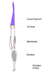 Cone cells are somewhat shorter than rods, but wider and tapered, and are much less numerous than rods in most parts of the retina, but greatly outnumber rods in the fovea
. Structurally, cone cells have a cone
-like shape at one end where a pigment filters incoming light, giving them their different response curves. They are typically 40-50 µm
long, and their diameter varies from 0.5 to 4.0 µm, being smallest and most tightly packed at the center of the eye at the fovea
. The S cones are a little larger than the others.
Photobleaching
can be used to determine cone arrangement. This is done by exposing dark-adapted retina to a certain wavelength of light that paralyzes the particular type of cone sensitive to that wavelength for up to thirty minutes from being able to dark-adapt making it appear white in contrast to the grey dark-adapted cones when a picture of the retina is taken. The results illustrate that S cones are randomly placed and appear much less frequently than the M and L cones. The ratio of M and L cones varies greatly among different people with regular vision (e.g. values of 75.8% L with 20.0% M versus 50.6% L with 44.2% M in two male subjects).
Like rods, each cone cell has a synaptic terminal, an inner segment, and an outer segment as well as an interior nucleus and various mitochondria. The synaptic terminal forms a synapse
with a neuron such as a bipolar cell
. The inner and outer segments are connected by a cilium
. The inner segment contains organelle
s and the cell's nucleus
, while the outer segment, which is pointed toward the back of the eye, contains the light-absorbing materials.
Like rods, the outer segments of cones have invaginations of their cell membrane
s that create stacks of membranous disks. Photopigments exist as transmembrane protein
s within these disks, which provide more surface area for light to affect the pigments. In cones, these disks are attached to the outer membrane, whereas they are pinched off and exist separately in rods. Neither rods nor cones divide, but their membranous disks wear out and are worn off at the end of the outer segment, to be consumed and recycled by phagocytic cells.
The response of cone cells to light is also directionally nonuniform, peaking at a direction that receives light from the center of the pupil; this effect is known as the Stiles–Crawford effect.
Retina
The vertebrate retina is a light-sensitive tissue lining the inner surface of the eye. The optics of the eye create an image of the visual world on the retina, which serves much the same function as the film in a camera. Light striking the retina initiates a cascade of chemical and electrical...
of the eye
Human eye
The human eye is an organ which reacts to light for several purposes. As a conscious sense organ, the eye allows vision. Rod and cone cells in the retina allow conscious light perception and vision including color differentiation and the perception of depth...
that are responsible for color vision
Color vision
Color vision is the capacity of an organism or machine to distinguish objects based on the wavelengths of the light they reflect, emit, or transmit...
; they function best in relatively bright light
Light
Light or visible light is electromagnetic radiation that is visible to the human eye, and is responsible for the sense of sight. Visible light has wavelength in a range from about 380 nanometres to about 740 nm, with a frequency range of about 405 THz to 790 THz...
, as opposed to rod cell
Rod cell
Rod cells, or rods, are photoreceptor cells in the retina of the eye that can function in less intense light than can the other type of visual photoreceptor, cone cells. Named for their cylindrical shape, rods are concentrated at the outer edges of the retina and are used in peripheral vision. On...
s that work better in dim light. If the retina is exposed to an intense visual stimulus, a negative afterimage will be formed. A negative afterimage consists of complementary colours to that of the original stimulus. Cone cells are densely packed in the fovea
Fovea
The fovea centralis, also generally known as the fovea , is a part of the eye, located in the center of the macula region of the retina....
, but gradually become sparser towards the periphery of the retina.
A commonly cited figure of six million in the human eye was found by Osterberg in 1935. Oyster's textbook (1999) cites work by Curcio et al. (1990) indicating an average close to 4.5 million cone cells and 90 million rod cell
Rod cell
Rod cells, or rods, are photoreceptor cells in the retina of the eye that can function in less intense light than can the other type of visual photoreceptor, cone cells. Named for their cylindrical shape, rods are concentrated at the outer edges of the retina and are used in peripheral vision. On...
s in the human retina.
Cones are less sensitive to light than the rod cell
Rod cell
Rod cells, or rods, are photoreceptor cells in the retina of the eye that can function in less intense light than can the other type of visual photoreceptor, cone cells. Named for their cylindrical shape, rods are concentrated at the outer edges of the retina and are used in peripheral vision. On...
s in the retina (which support vision at low light levels), but allow the perception of colour
Color vision
Color vision is the capacity of an organism or machine to distinguish objects based on the wavelengths of the light they reflect, emit, or transmit...
. They are also able to perceive finer detail and more rapid changes in images, because their response times to stimuli are faster than those of rods. Because humans usually have three kinds of cones with different photopsin
Photopsin
Photopsins are the photoreceptor proteins found in the cone cells of the retina that are the basis of color vision. Photopsins are very close analogs of the visual purple rhodopsin that is used in night vision...
s, which have different response curves and thus respond to variation in colour in different ways, we have trichromatic vision. Being colour blind can change this, and there have been reports of people with four or more types of cones, giving them tetrachromat
Tetrachromat
Tetrachromacy is the condition of possessing four independent channels for conveying color information, or possessing four different types of cone cells in the eye...
ic vision.
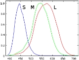
Types
Humans normally have three kinds of cones. The first responds the most to light of long wavelengths, peaking at a yellowish colour; this type is sometimes designated L for long. The second type responds the most to light of medium-wavelength, peaking at a green colour, and is abbreviated M for medium. The third type responds the most to short-wavelength light, of a bluish colour, and is designated S for short. The three types have peak wavelengths near 564–580 nmNanometre
A nanometre is a unit of length in the metric system, equal to one billionth of a metre. The name combines the SI prefix nano- with the parent unit name metre .The nanometre is often used to express dimensions on the atomic scale: the diameter...
, 534–545 nm, and 420–440 nm, respectively.
The difference in the signals received from the three cone types allows the brain to perceive all possible colours, through the opponent process of colour vision. (Rod cells have a peak sensitivity at 498 nm, roughly halfway between the peak sensitivities of the S and M cones.)
The colour yellow, for example, is perceived when the L cones are stimulated slightly more than the M cones, and the colour red is perceived when the L cones are stimulated significantly more than the M cones. Similarly, blue and violet hues are perceived when the S receptor is stimulated more than the other two.
The S cones are most sensitive to light at wavelengths around 420 nm. However, the lens and cornea
Cornea
The cornea is the transparent front part of the eye that covers the iris, pupil, and anterior chamber. Together with the lens, the cornea refracts light, with the cornea accounting for approximately two-thirds of the eye's total optical power. In humans, the refractive power of the cornea is...
of the human eye are increasingly absorptive to smaller wavelengths, and this sets the lower wavelength limit of human-visible light to approximately 380 nm, which is therefore called 'ultraviolet
Ultraviolet
Ultraviolet light is electromagnetic radiation with a wavelength shorter than that of visible light, but longer than X-rays, in the range 10 nm to 400 nm, and energies from 3 eV to 124 eV...
' light. People with aphakia
Aphakia
Aphakia is the absence of the lens of the eye, due to surgical removal, a perforating wound or ulcer, or congenital anomaly. It causes a loss of accommodation, hyperopia, and a deep anterior chamber. Complications include detachment of the vitreous or retina, and glaucoma.Aphakic people are...
, a condition where the eye lacks a lens, sometimes report the ability to see into the ultraviolet range. At moderate to bright light levels where the cones function, the eye is more sensitive to yellowish-green light than other colors because this stimulates the two most common (M and L) of the three kinds of cones almost equally. At lower light levels, where only the rod cell
Rod cell
Rod cells, or rods, are photoreceptor cells in the retina of the eye that can function in less intense light than can the other type of visual photoreceptor, cone cells. Named for their cylindrical shape, rods are concentrated at the outer edges of the retina and are used in peripheral vision. On...
s function, the sensitivity is greatest at a blueish-green wavelength.
Cones also tend to posses a significantly elevated visual acuity because each cone cell has a lone connection to the optic nerve, therefore, the cones have an easier time to tell that two stimuli are isolated.
Structure
Fovea
The fovea centralis, also generally known as the fovea , is a part of the eye, located in the center of the macula region of the retina....
. Structurally, cone cells have a cone
Cone (geometry)
A cone is an n-dimensional geometric shape that tapers smoothly from a base to a point called the apex or vertex. Formally, it is the solid figure formed by the locus of all straight line segments that join the apex to the base...
-like shape at one end where a pigment filters incoming light, giving them their different response curves. They are typically 40-50 µm
Micrometre
A micrometer , is by definition 1×10⁻⁶ of a meter .In plain English, it means one-millionth of a meter . Its unit symbol in the International System of Units is μm...
long, and their diameter varies from 0.5 to 4.0 µm, being smallest and most tightly packed at the center of the eye at the fovea
Fovea
The fovea centralis, also generally known as the fovea , is a part of the eye, located in the center of the macula region of the retina....
. The S cones are a little larger than the others.
Photobleaching
Photobleaching
Photobleaching is the photochemical destruction of a fluorophore. In microscopy, photobleaching may complicate the observation of fluorescent molecules, since they will eventually be destroyed by the light exposure necessary to stimulate them into fluorescing...
can be used to determine cone arrangement. This is done by exposing dark-adapted retina to a certain wavelength of light that paralyzes the particular type of cone sensitive to that wavelength for up to thirty minutes from being able to dark-adapt making it appear white in contrast to the grey dark-adapted cones when a picture of the retina is taken. The results illustrate that S cones are randomly placed and appear much less frequently than the M and L cones. The ratio of M and L cones varies greatly among different people with regular vision (e.g. values of 75.8% L with 20.0% M versus 50.6% L with 44.2% M in two male subjects).
Like rods, each cone cell has a synaptic terminal, an inner segment, and an outer segment as well as an interior nucleus and various mitochondria. The synaptic terminal forms a synapse
Synapse
In the nervous system, a synapse is a structure that permits a neuron to pass an electrical or chemical signal to another cell...
with a neuron such as a bipolar cell
Bipolar cell
As a part of the retina, the bipolar cell exists between photoreceptors and ganglion cells. They act, directly or indirectly, to transmit signals from the photoreceptors to the ganglion cells.-Overview:...
. The inner and outer segments are connected by a cilium
Cilium
A cilium is an organelle found in eukaryotic cells. Cilia are slender protuberances that project from the much larger cell body....
. The inner segment contains organelle
Organelle
In cell biology, an organelle is a specialized subunit within a cell that has a specific function, and is usually separately enclosed within its own lipid bilayer....
s and the cell's nucleus
Cell nucleus
In cell biology, the nucleus is a membrane-enclosed organelle found in eukaryotic cells. It contains most of the cell's genetic material, organized as multiple long linear DNA molecules in complex with a large variety of proteins, such as histones, to form chromosomes. The genes within these...
, while the outer segment, which is pointed toward the back of the eye, contains the light-absorbing materials.
Like rods, the outer segments of cones have invaginations of their cell membrane
Cell membrane
The cell membrane or plasma membrane is a biological membrane that separates the interior of all cells from the outside environment. The cell membrane is selectively permeable to ions and organic molecules and controls the movement of substances in and out of cells. It basically protects the cell...
s that create stacks of membranous disks. Photopigments exist as transmembrane protein
Transmembrane protein
A transmembrane protein is a protein that goes from one side of a membrane through to the other side of the membrane. Many TPs function as gateways or "loading docks" to deny or permit the transport of specific substances across the biological membrane, to get into the cell, or out of the cell as...
s within these disks, which provide more surface area for light to affect the pigments. In cones, these disks are attached to the outer membrane, whereas they are pinched off and exist separately in rods. Neither rods nor cones divide, but their membranous disks wear out and are worn off at the end of the outer segment, to be consumed and recycled by phagocytic cells.
The response of cone cells to light is also directionally nonuniform, peaking at a direction that receives light from the center of the pupil; this effect is known as the Stiles–Crawford effect.
See also
- Colour blindness
- Colour vision
- Cone dystrophyCone dystrophyA cone dystrophy is an inherited ocular disorder characterized by the loss of cone cells, the photoreceptors responsible for both central and color vision....
- Double coneDouble coneDouble cones , also known as twin cones in some literature, are two cone cells joined together that may also be coupled optically/electrically...
s - Tetrachromacy
- Rod cellRod cellRod cells, or rods, are photoreceptor cells in the retina of the eye that can function in less intense light than can the other type of visual photoreceptor, cone cells. Named for their cylindrical shape, rods are concentrated at the outer edges of the retina and are used in peripheral vision. On...
- Disc shedding
External links
- Cell Centered Database - Cone cell
- Webvision's Photoreceptors
- NIF Search - Cone Cell via the Neuroscience Information FrameworkNeuroscience Information FrameworkThe Neuroscience Information Framework is a repository of global neuroscience web resources, including experimental, clinical, and translational neuroscience databases, knowledge bases, atlases, and genetic/genomic resources.-Description:...
- Model and image of cone cell