
Alpha motor neuron
Encyclopedia
Alpha motor neurons are large lower motor neuron
s of the brainstem and spinal cord
. They innervate extrafusal muscle fiber
s of skeletal muscle
and are directly responsible for initiating their contraction
. Alpha motor neurons are distinct from gamma motor neurons, which innervate intrafusal muscle fiber
s of muscle spindle
s.
While their cell bodies are found in the central nervous system
(CNS), alpha motor neurons are also considered part of the somatic nervous system
—a branch of the peripheral nervous system
(PNS)—because their axon
s extend into the periphery to innervate skeletal muscle
s.
An alpha motor neuron and the muscle fibers it innervates is a motor unit
. A motor neuron pool contains the cell bodies of all the alpha motor neurons involved in contracting a single muscle.
and neck
are found in the brainstem; the remaining α-MNs innervate the rest of the body and are found in the spinal cord
. There are more α-MNs in the spinal cord than in the brainstem, as the number of α-MNs is directly proportional to the amount of fine motor control in that muscle. For example, the muscles of a single finger have more α-MNs per fiber, and more α-MNs in total, than the muscles of the quadriceps, which allows for finer control of the force a finger applies.
In general, α-MNs on one side of the brainstem or spinal cord innervate muscles on that same side of body. An exception is the trochlear nucleus
in the brainstem, which innervates the superior oblique muscle
of the eye on the opposite side of the face.
s reside within clusters of cells called nuclei
, some of which contain the cell bodies of neurons belonging to the cranial nerves. Not all cranial nerve nuclei contain α-MNs; those that do are motor nuclei, while others are sensory nuclei. Motor nuclei are found throughout the brainstem—medulla
, pons
, and midbrain—and for developmental reasons are found near the midline of the brainstem.
Generally, motor nuclei found higher in the brainstem (i.e., more rostral) innervate muscles that are higher on the face. For example, the oculomotor nucleus
contains α-MNs that innervate muscles of the eye, and is found in the midbrain, the most rostral brainstem component. By contrast, the hypoglossal nucleus
, which contains α-MNs that innervate the tongue, is found in the medulla, the most caudal (i.e., towards the bottom) of the brainstem structures.
 In the spinal cord, α-MNs are located within the gray matter
that forms the ventral horn. These α-MNs provide the motor component of the spinal nerve
s that innervate muscles of the body.
 As in the brainstem, higher segments of the spinal cord contain α-MNs that innervate muscles higher on the body. For example, the biceps brachii muscle
, a muscle of the arm, is innervated by α-MNs in spinal cord segments C5, C6, and C7, which are found rostrally in the spinal cord. On the other hand, the gastrocnemius muscle
, one of the muscles of the leg, is innervated by α-MNs within segments S1 and S2, which are found caudally in the spinal cord.
Alpha motor neurons are located in a specific region of the spinal cord's gray matter. This region is designated lamina IX in the Rexed lamina system, which classifies regions of gray matter based on their cytoarchitecture. Lamina IX is located predominantly in the medial aspect of the ventral horn, although there is some contribution to lamina IX from a collection of motor neurons located more laterally. Like other regions of the spinal cord, cells in this lamina are somatotopically organized, meaning that the position of neurons within the spinal cord is associated with what muscles they innervate. In particular, α-MNs in the medial zone of lamina IX tend to innervate proximal muscles of the body, while those in the lateral zone tend to innervate more distal muscles. There is similar somatotopy associated with α-MNs that innervate flexor and extensor muscles: α-MNs that innervate flexors
tend to be located in the dorsal portion of lamina IX; those that innervate extensors
tend to be located more ventrally.
s, sensory neuron
s, and interneuron
s. The primary output of α-MNs is to extrafusal muscle fiber
s. This afferent and efferent connectivity is required to achieve coordinated muscle activity.
Upper motor neuron
s (UMNs) send input to α-MNs via several pathways, including (but not limited to) the corticonuclear, corticospinal
, and rubrospinal tract
s. The corticonuclear and corticospinal tracts are commonly encountered in studies of upper and lower motor neuron connectivity in the control of voluntary movements.
The corticonuclear tract is so named because it connects the cerebral cortex
to cranial nerve nuclei. (The corticonuclear tract is also called the corticobulbar tract, as the brainstem is sometimes called the "bulb" of the brain.) It is via this pathway that upper motor neurons from the cortex descend from the cortex and synapse
on α-MNs of the brainstem. Similarly, UMNs of the cerebral cortex are in direct control of α-MNs of the spinal cord
via the lateral
and ventral corticospinal tracts.
The sensory input to α-MNs is extensive and has its origin in Golgi tendon organs, muscle spindle
s, mechanoreceptor
s, thermoreceptor
s, and other sensory neuron
s in the periphery. These connections provide the structure for the neural circuits that underlie reflex
es. There are several types of reflex circuits, the simplest of which consists of a single synapse between a sensory neuron and a α-MNs. The knee-jerk reflex is an example of such a monosynaptic reflex.
The most extensive input to α-MNs is from local interneuron
s, which are the most numerous type of neuron in the spinal cord
. Among their many roles, interneurons synapse on α-MNs to create more complex reflex circuitry. One type of interneuron is the Renshaw cell
, discussed later.
s. Other fibers from α-MNs synapse on Renshaw cell
s, inhibitory interneuron
s that synapse on the α-MN and limit its activity in order to prevent muscle damage.
Like other neurons, α-MNs transmit signals as action potential
s, rapid changes in electrical activity that propagate from the cell body to the end of the axon
. To increase the speed at which action potentials travel, α-MN axons have large diameters and are heavily myelin
ated by both oligodendrocyte
s and Schwann cell
s. Oligodendrocytes myelinate the part of the α-MN axon that lies in the central nervous system
(CNS), while Schwann cells myelinate the part that lies in the peripheral nervous system
(PNS). The transition between the CNS and PNS occurs at the level of the pia mater
, the innermost and most delicate layer of meningeal tissue
surrounding components of the CNS.
The axon of an α-MN connects with its extrafusal muscle fiber via a neuromuscular junction
, a specialized type of chemical synapse
that differs both in structure and function from the chemical synapses that connect neurons to each other. Both types of synapses rely on neurotransmitter
s to transduce
the electrical signal into a chemical signal and back. One way they differ is that synapses between neurons typically use glutamate or GABA
as their neurotransmitters, while the neuromuscular junction uses acetylcholine
exclusively. Acetylcholine is sensed by nicotinic acetylcholine receptor
s on extrafusal muscle fibers, causing their contraction.
Like other motor neurons, α-MNs are named after the properties of their axon
s. Alpha motor neurons have Aα axons, which are large-caliber
, heavily myelin
ated fibers that conduct action potential
s rapidly. By contrast, gamma motor neurons have Aγ axons, which are slender, lightly myelinated fibers that conduct less rapidly.
 Injury to α-MNs is the most common type of lower motor neuron lesion
. Damage may be caused by trauma
, ischemia
, and infection
, among others. In addition, certain diseases are associated with the selective loss of α-MNs. For example, poliomyelitis
is caused by a virus
that specifically targets and kills motor neurons in the ventral horn of the spinal cord. Amyotropic lateral sclerosis likewise is associated with the selective loss of motor neurons.
Paralysis
is one of the most pronounced effects of damage to α-MNs. Because α-MNs provide the only voluntary innervation to extrafusal muscle fiber
s, losing α-MNs effectively severs the connection between the brainstem and spinal cord and the muscles they innervate. Without this connection, voluntary and involuntary (reflex) muscle control is impossible. Voluntary muscle control is lost because α-MNs relay voluntary signals from upper motor neurons to muscle fibers. Loss of involuntary control results from interruption of reflex circuits such as the tonic stretch reflex
. A consequence of reflex interruption is that muscle tone
is reduced, resulting in flaccid paresis. Another consequence is the depression of deep tendon reflexes, causing hyporeflexia
.
Muscle weakness and atrophy
are inevitable consequences of α-MN lesions as well. Because muscle size and strength are related to the extent of their use, denervated muscles are prone to atrophy. A secondary cause of muscle atrophy is that denervated muscles are no longer supplied with trophic factors from the α-MNs that innervate them. Alpha motor neuron lesions also result in abnormal EMG
potentials (e.g., fibrillation potentials) and fasciculation
s, the latter being spontaneous, involuntary muscle contractions.
Diseases that impair signaling between α-MNs and extrafusal muscle fibers, namely diseases of the neuromuscular junction
have similar signs to those that occur with α-MN disease. For example, myasthenia gravis
is an autoimmune disease
that prevents signaling across the neuromuscular junction
, which results in functional denervation of muscle.
 Alpha motor neurons originate in the basal plate
, the ventral portion of the neural tube
in the developing embryo
. Sonic hedgehog
(Shh) is secreted by the nearby notochord
and other ventral structures (e.g., the floor plate
), establishing a gradient of highly concentrated Shh in the basal plate and less concentrated Shh in the alar plate
. Under the influence of Shh and other factors, some neurons of the basal plate differentiate
into α-MNs.
Like other neurons, α-MNs send axon
al projections to reach their target extrafusal muscle fiber
s via axon guidance
, a process regulated in part by neurotrophic factors released by target muscle fibers. Neurotrophic factors also ensure that each muscle fiber is innervated by the appropriate number of α-MNs. As with most types of neurons in the nervous system
, α-MNs are more numerous in early development than in adulthood. Muscle fibers secrete a limited amount of neurotrophic factors capable of sustaining only a fraction of the α-MNs that initially project to the muscle fiber. Those α-MNs that do not receive sufficient neurotrophic factors will undergo apoptosis
, a form of programmed cell death
.
Because they innervate many muscles, some clusters of α-MNs receive high concentrations of neurotrophic factors and survive this stage of neuronal pruning. This is true of the α-MNs innervating the upper and lower limbs: these α-MNs form large cell columns that contribute to the cervical
and lumbar enlargement
s of the spinal cord. In addition to receiving neurotrophic factors from muscles, α-MNs also secrete a number of trophic factors to support the muscle fibers they innervate. Reduced levels of trophic factors contributes to the muscle atrophy that follows an α-MN lesion.
Lower motor neuron
Lower motor neurons are the motor neurons connecting the brainstem and spinal cord to muscle fibers, bringing the nerve impulses from the upper motor neurons out to the muscles...
s of the brainstem and spinal cord
Spinal cord
The spinal cord is a long, thin, tubular bundle of nervous tissue and support cells that extends from the brain . The brain and spinal cord together make up the central nervous system...
. They innervate extrafusal muscle fiber
Extrafusal muscle fiber
Extrafusal muscle fiber is a term given to standard muscle fibers as to distinguish them from intrafusal muscle fibers. Extrafusal muscle fibers are innervated by alpha motor neurons and generate tension by contracting, thereby allowing for skeletal movement...
s of skeletal muscle
Skeletal muscle
Skeletal muscle is a form of striated muscle tissue existing under control of the somatic nervous system- i.e. it is voluntarily controlled. It is one of three major muscle types, the others being cardiac and smooth muscle...
and are directly responsible for initiating their contraction
Muscle contraction
Muscle fiber generates tension through the action of actin and myosin cross-bridge cycling. While under tension, the muscle may lengthen, shorten, or remain the same...
. Alpha motor neurons are distinct from gamma motor neurons, which innervate intrafusal muscle fiber
Intrafusal muscle fiber
Intrafusal muscle fibers are skeletal muscle fibers that comprise the muscle spindle and are innervated by gamma motor neurons. These fibers are a proprioceptor that detect the amount and rate of change of length in a muscle. These fibers are walled off from the rest of the muscle by a collagen...
s of muscle spindle
Muscle spindle
Muscle spindles are sensory receptors within the belly of a muscle, which primarily detect changes in the length of this muscle. They convey length information to the central nervous system via sensory neurons. This information can be processed by the brain to determine the position of body parts...
s.
While their cell bodies are found in the central nervous system
Central nervous system
The central nervous system is the part of the nervous system that integrates the information that it receives from, and coordinates the activity of, all parts of the bodies of bilaterian animals—that is, all multicellular animals except sponges and radially symmetric animals such as jellyfish...
(CNS), alpha motor neurons are also considered part of the somatic nervous system
Somatic nervous system
The somatic nervous system is the part of the peripheral nervous system associated with the voluntary control of body movements via skeletal muscles...
—a branch of the peripheral nervous system
Peripheral nervous system
The peripheral nervous system consists of the nerves and ganglia outside of the brain and spinal cord. The main function of the PNS is to connect the central nervous system to the limbs and organs. Unlike the CNS, the PNS is not protected by the bone of spine and skull, or by the blood–brain...
(PNS)—because their axon
Axon
An axon is a long, slender projection of a nerve cell, or neuron, that conducts electrical impulses away from the neuron's cell body or soma....
s extend into the periphery to innervate skeletal muscle
Skeletal muscle
Skeletal muscle is a form of striated muscle tissue existing under control of the somatic nervous system- i.e. it is voluntarily controlled. It is one of three major muscle types, the others being cardiac and smooth muscle...
s.
An alpha motor neuron and the muscle fibers it innervates is a motor unit
Motor unit
”A motor unit is a single α-motor neuron and all of the corresponding muscle fibers it innervates; all of these fibers will be of the same type . When a motor unit is activated, all of its fibers contract...
. A motor neuron pool contains the cell bodies of all the alpha motor neurons involved in contracting a single muscle.
Location
Alpha motor neurons innervating the headHead
In anatomy, the head of an animal is the rostral part that usually comprises the brain, eyes, ears, nose and mouth . Some very simple animals may not have a head, but many bilaterally symmetric forms do....
and neck
Neck
The neck is the part of the body, on many terrestrial or secondarily aquatic vertebrates, that distinguishes the head from the torso or trunk. The adjective signifying "of the neck" is cervical .-Boner anatomy: The cervical spine:The cervical portion of the human spine comprises seven boney...
are found in the brainstem; the remaining α-MNs innervate the rest of the body and are found in the spinal cord
Spinal cord
The spinal cord is a long, thin, tubular bundle of nervous tissue and support cells that extends from the brain . The brain and spinal cord together make up the central nervous system...
. There are more α-MNs in the spinal cord than in the brainstem, as the number of α-MNs is directly proportional to the amount of fine motor control in that muscle. For example, the muscles of a single finger have more α-MNs per fiber, and more α-MNs in total, than the muscles of the quadriceps, which allows for finer control of the force a finger applies.
In general, α-MNs on one side of the brainstem or spinal cord innervate muscles on that same side of body. An exception is the trochlear nucleus
Trochlear nucleus
The nucleus of the trochlear nerve is located in the midbrain, at the level of the inferior colliculus. It is a motor nucleus, so is located near the midline....
in the brainstem, which innervates the superior oblique muscle
Superior oblique muscle
For the abdominal muscle see: Abdominal external oblique muscleThe superior oblique muscle, or obliquus oculi superior, is a fusiform muscle originating in the upper, medial side of the orbit which abducts, depresses and internally rotates the eye...
of the eye on the opposite side of the face.
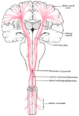
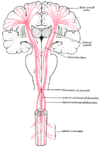
Brainstem
In the brainstem, α-MNs and other neuronNeuron
A neuron is an electrically excitable cell that processes and transmits information by electrical and chemical signaling. Chemical signaling occurs via synapses, specialized connections with other cells. Neurons connect to each other to form networks. Neurons are the core components of the nervous...
s reside within clusters of cells called nuclei
Nucleus (neuroanatomy)
In neuroanatomy, a nucleus is a brain structure consisting of a relatively compact cluster of neurons. It is one of the two most common forms of nerve cell organization, the other being layered structures such as the cerebral cortex or cerebellar cortex. In anatomical sections, a nucleus shows up...
, some of which contain the cell bodies of neurons belonging to the cranial nerves. Not all cranial nerve nuclei contain α-MNs; those that do are motor nuclei, while others are sensory nuclei. Motor nuclei are found throughout the brainstem—medulla
Medulla oblongata
The medulla oblongata is the lower half of the brainstem. In discussions of neurology and similar contexts where no ambiguity will result, it is often referred to as simply the medulla...
, pons
Pons
The pons is a structure located on the brain stem, named after the Latin word for "bridge" or the 16th-century Italian anatomist and surgeon Costanzo Varolio . It is superior to the medulla oblongata, inferior to the midbrain, and ventral to the cerebellum. In humans and other bipeds this means it...
, and midbrain—and for developmental reasons are found near the midline of the brainstem.
Generally, motor nuclei found higher in the brainstem (i.e., more rostral) innervate muscles that are higher on the face. For example, the oculomotor nucleus
Oculomotor nucleus
The fibers of the oculomotor nerve arise from a nucleus in the midbrain, which lies in the gray substance of the floor of the cerebral aqueduct and extends in front of the aqueduct for a short distance into the floor of the third ventricle...
contains α-MNs that innervate muscles of the eye, and is found in the midbrain, the most rostral brainstem component. By contrast, the hypoglossal nucleus
Hypoglossal nucleus
The hypoglossal nucleus is a cranial nerve nucleus, and it extends the length of the medulla, and being a motor nucleus, is close to the midline...
, which contains α-MNs that innervate the tongue, is found in the medulla, the most caudal (i.e., towards the bottom) of the brainstem structures.
Spinal cord
Gray Matter
"Gray Matter" is a short story by Stephen King, first published in the October 1973 issue of Cavalier magazine, and later collected in King's 1978 collection Night Shift. It is set in the same area as King's novel Dreamcatcher.-Setting:...
that forms the ventral horn. These α-MNs provide the motor component of the spinal nerve
Spinal nerve
The term spinal nerve generally refers to a mixed spinal nerve, which carries motor, sensory, and autonomic signals between the spinal cord and the body...
s that innervate muscles of the body.
Biceps brachii muscle
In human anatomy, the biceps brachii, or simply biceps in common parlance, is, as the name implies, a two-headed muscle located on the upper arm. Both heads arise on the scapula and join to form a single muscle belly which is attached to the upper forearm...
, a muscle of the arm, is innervated by α-MNs in spinal cord segments C5, C6, and C7, which are found rostrally in the spinal cord. On the other hand, the gastrocnemius muscle
Gastrocnemius muscle
In humans, the gastrocnemius muscle is a very powerful superficial pennate muscle that is in the back part of the lower leg. It runs from its two heads just above the knee to the heel, and is involved in standing, walking, running and jumping. Along with the soleus muscle it forms the calf muscle...
, one of the muscles of the leg, is innervated by α-MNs within segments S1 and S2, which are found caudally in the spinal cord.
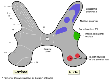
Alpha motor neurons are located in a specific region of the spinal cord's gray matter. This region is designated lamina IX in the Rexed lamina system, which classifies regions of gray matter based on their cytoarchitecture. Lamina IX is located predominantly in the medial aspect of the ventral horn, although there is some contribution to lamina IX from a collection of motor neurons located more laterally. Like other regions of the spinal cord, cells in this lamina are somatotopically organized, meaning that the position of neurons within the spinal cord is associated with what muscles they innervate. In particular, α-MNs in the medial zone of lamina IX tend to innervate proximal muscles of the body, while those in the lateral zone tend to innervate more distal muscles. There is similar somatotopy associated with α-MNs that innervate flexor and extensor muscles: α-MNs that innervate flexors
Flexion
In anatomy, flexion is a position that is made possible by the joint angle decreasing. The skeletal and muscular systems work together to move the joint into a "flexed" position. For example the elbow is flexed when the hand is brought closer to the shoulder...
tend to be located in the dorsal portion of lamina IX; those that innervate extensors
Extension (kinesiology)
In kinesiology, extension is a movement of a joint that results in increased angle between two bones or body surfaces at a joint. Extension usually results in straightening of the bones or body surfaces involved. For example, extension is produced by extending the flexed elbow. Straightening of...
tend to be located more ventrally.
Connectivity
Like other neurons, lower motor neurons have both afferent (incoming) and efferent (outgoing) connections. Alpha motor neurons receive input from a number of sources, including upper motor neuronUpper motor neuron
Upper motor neurons are motor neurons that originate in the motor region of the cerebral cortex or the brain stem and carry motor information down to the final common pathway, that is, any motor neurons that are not directly responsible for stimulating the target muscle...
s, sensory neuron
Sensory neuron
Sensory neurons are typically classified as the neurons responsible for converting external stimuli from the environment into internal stimuli. They are activated by sensory input , and send projections into the central nervous system that convey sensory information to the brain or spinal cord...
s, and interneuron
Interneuron
An interneuron is a multipolar neuron which connects afferent neurons and efferent neurons in neural pathways...
s. The primary output of α-MNs is to extrafusal muscle fiber
Extrafusal muscle fiber
Extrafusal muscle fiber is a term given to standard muscle fibers as to distinguish them from intrafusal muscle fibers. Extrafusal muscle fibers are innervated by alpha motor neurons and generate tension by contracting, thereby allowing for skeletal movement...
s. This afferent and efferent connectivity is required to achieve coordinated muscle activity.
Afferent input
| UMN origin | α-MN target | Tract name |
|---|---|---|
| Cerebral cortex Cerebral cortex The cerebral cortex is a sheet of neural tissue that is outermost to the cerebrum of the mammalian brain. It plays a key role in memory, attention, perceptual awareness, thought, language, and consciousness. It is constituted of up to six horizontal layers, each of which has a different... |
Brainstem | Corticonuclear tract |
| Cerebral cortex | Spinal cord Spinal cord The spinal cord is a long, thin, tubular bundle of nervous tissue and support cells that extends from the brain . The brain and spinal cord together make up the central nervous system... |
Corticospinal tract Corticospinal tract The corticospinal or pyramidal tract is a collection of axons that travel between the cerebral cortex of the brain and the spinal cord.... |
| Red nucleus Red nucleus The red nucleus is a structure in the rostral midbrain involved in motor coordination. It comprises a caudal magnocellular and a rostral parvocellular part.-Function:... |
Spinal cord | Rubrospinal tract Rubrospinal tract The rubrospinal tract is a part of the nervous system. It is a part of the lateral indirect extra-pyramidal tract.-Function:In humans, the rubrospinal tract is one of several major motor control pathways. It is smaller and has fewer axons than the corticospinal tract, suggesting that it is less... |
| Vestibular nuclei Vestibular nuclei The vestibular nuclei are the cranial nuclei for the vestibular nerve.In Terminologia Anatomica they are grouped in both the pons and medulla.-Subnuclei:There are 4 subnuclei; they are situated at the floor of the fourth ventricle.... |
Spinal cord | Vestibulospinal tract Vestibulospinal tract The vestibulospinal tract is a component of the extrapyramidal system and also can be classified as a component of the medial pathway. The vestibulospinal fibers relay information from nuclei to motor neurons, like other descending motor pathways. Specifically, the vestibular nuclei receive... |
| Midbrain tectum Midbrain tectum The tectum is a region of the brain, specifically the dorsal part of the mesencephalon . This is contrasted with the tegmentum, which refers to the region ventral to the ventricular system... |
Spinal cord | Tectospinal tract Tectospinal tract In humans, the tectospinal tract is a nerve pathway which coordinates head and eye movements. It is part of the indirect extrapyramidal tract... |
| Reticular formation Reticular formation The reticular formation is a part of the brain that is involved in actions such as awaking/sleeping cycle, and filtering incoming stimuli to discriminate irrelevant background stimuli... |
Spinal cord | Reticulospinal tract Reticulospinal tract The reticulospinal tract is an extrapyramidal motor tract which travels from the reticular formation.-Functions:... |
Upper motor neuron
Upper motor neuron
Upper motor neurons are motor neurons that originate in the motor region of the cerebral cortex or the brain stem and carry motor information down to the final common pathway, that is, any motor neurons that are not directly responsible for stimulating the target muscle...
s (UMNs) send input to α-MNs via several pathways, including (but not limited to) the corticonuclear, corticospinal
Corticospinal tract
The corticospinal or pyramidal tract is a collection of axons that travel between the cerebral cortex of the brain and the spinal cord....
, and rubrospinal tract
Rubrospinal tract
The rubrospinal tract is a part of the nervous system. It is a part of the lateral indirect extra-pyramidal tract.-Function:In humans, the rubrospinal tract is one of several major motor control pathways. It is smaller and has fewer axons than the corticospinal tract, suggesting that it is less...
s. The corticonuclear and corticospinal tracts are commonly encountered in studies of upper and lower motor neuron connectivity in the control of voluntary movements.
The corticonuclear tract is so named because it connects the cerebral cortex
Cerebral cortex
The cerebral cortex is a sheet of neural tissue that is outermost to the cerebrum of the mammalian brain. It plays a key role in memory, attention, perceptual awareness, thought, language, and consciousness. It is constituted of up to six horizontal layers, each of which has a different...
to cranial nerve nuclei. (The corticonuclear tract is also called the corticobulbar tract, as the brainstem is sometimes called the "bulb" of the brain.) It is via this pathway that upper motor neurons from the cortex descend from the cortex and synapse
Synapse
In the nervous system, a synapse is a structure that permits a neuron to pass an electrical or chemical signal to another cell...
on α-MNs of the brainstem. Similarly, UMNs of the cerebral cortex are in direct control of α-MNs of the spinal cord
Spinal cord
The spinal cord is a long, thin, tubular bundle of nervous tissue and support cells that extends from the brain . The brain and spinal cord together make up the central nervous system...
via the lateral
Lateral corticospinal tract
The lateral corticospinal tract is the largest part of the corticospinal tract...
and ventral corticospinal tracts.
The sensory input to α-MNs is extensive and has its origin in Golgi tendon organs, muscle spindle
Muscle spindle
Muscle spindles are sensory receptors within the belly of a muscle, which primarily detect changes in the length of this muscle. They convey length information to the central nervous system via sensory neurons. This information can be processed by the brain to determine the position of body parts...
s, mechanoreceptor
Mechanoreceptor
A mechanoreceptor is a sensory receptor that responds to mechanical pressure or distortion. There are four main types in the glabrous skin of humans: Pacinian corpuscles, Meissner's corpuscles, Merkel's discs, and Ruffini corpuscles...
s, thermoreceptor
Thermoreceptor
A thermoreceptor is a sensory receptor, or more accurately the receptive portion of a sensory neuron, that codes absolute and relative changes in temperature, primarily within the innocuous range...
s, and other sensory neuron
Sensory neuron
Sensory neurons are typically classified as the neurons responsible for converting external stimuli from the environment into internal stimuli. They are activated by sensory input , and send projections into the central nervous system that convey sensory information to the brain or spinal cord...
s in the periphery. These connections provide the structure for the neural circuits that underlie reflex
Reflex
A reflex action, also known as a reflex, is an involuntary and nearly instantaneous movement in response to a stimulus. A true reflex is a behavior which is mediated via the reflex arc; this does not apply to casual uses of the term 'reflex'.-See also:...
es. There are several types of reflex circuits, the simplest of which consists of a single synapse between a sensory neuron and a α-MNs. The knee-jerk reflex is an example of such a monosynaptic reflex.
The most extensive input to α-MNs is from local interneuron
Interneuron
An interneuron is a multipolar neuron which connects afferent neurons and efferent neurons in neural pathways...
s, which are the most numerous type of neuron in the spinal cord
Spinal cord
The spinal cord is a long, thin, tubular bundle of nervous tissue and support cells that extends from the brain . The brain and spinal cord together make up the central nervous system...
. Among their many roles, interneurons synapse on α-MNs to create more complex reflex circuitry. One type of interneuron is the Renshaw cell
Renshaw cell
Renshaw cells are inhibitory interneurons found in the gray matter of the spinal cord, and are associated in two ways with an alpha motor neuron....
, discussed later.
Efferent output
Alpha motor neurons send fibers that mainly synapse on extrafusal muscle fiberExtrafusal muscle fiber
Extrafusal muscle fiber is a term given to standard muscle fibers as to distinguish them from intrafusal muscle fibers. Extrafusal muscle fibers are innervated by alpha motor neurons and generate tension by contracting, thereby allowing for skeletal movement...
s. Other fibers from α-MNs synapse on Renshaw cell
Renshaw cell
Renshaw cells are inhibitory interneurons found in the gray matter of the spinal cord, and are associated in two ways with an alpha motor neuron....
s, inhibitory interneuron
Interneuron
An interneuron is a multipolar neuron which connects afferent neurons and efferent neurons in neural pathways...
s that synapse on the α-MN and limit its activity in order to prevent muscle damage.
Signaling
Like other neurons, α-MNs transmit signals as action potential
Action potential
In physiology, an action potential is a short-lasting event in which the electrical membrane potential of a cell rapidly rises and falls, following a consistent trajectory. Action potentials occur in several types of animal cells, called excitable cells, which include neurons, muscle cells, and...
s, rapid changes in electrical activity that propagate from the cell body to the end of the axon
Axon
An axon is a long, slender projection of a nerve cell, or neuron, that conducts electrical impulses away from the neuron's cell body or soma....
. To increase the speed at which action potentials travel, α-MN axons have large diameters and are heavily myelin
Myelin
Myelin is a dielectric material that forms a layer, the myelin sheath, usually around only the axon of a neuron. It is essential for the proper functioning of the nervous system. Myelin is an outgrowth of a type of glial cell. The production of the myelin sheath is called myelination...
ated by both oligodendrocyte
Oligodendrocyte
Oligodendrocytes , or oligodendroglia , are a type of brain cell. They are a variety of neuroglia. Their main function is the insulation of axons in the central nervous system of some vertebrates...
s and Schwann cell
Schwann cell
Schwann cells or neurolemmocytes are the principal glia of the peripheral nervous system . Glial cells function to support neurons and in the PNS, also include satellite cells, olfactory ensheathing cells, enteric glia and glia that reside at sensory nerve endings, such as the Pacinian corpuscle...
s. Oligodendrocytes myelinate the part of the α-MN axon that lies in the central nervous system
Central nervous system
The central nervous system is the part of the nervous system that integrates the information that it receives from, and coordinates the activity of, all parts of the bodies of bilaterian animals—that is, all multicellular animals except sponges and radially symmetric animals such as jellyfish...
(CNS), while Schwann cells myelinate the part that lies in the peripheral nervous system
Peripheral nervous system
The peripheral nervous system consists of the nerves and ganglia outside of the brain and spinal cord. The main function of the PNS is to connect the central nervous system to the limbs and organs. Unlike the CNS, the PNS is not protected by the bone of spine and skull, or by the blood–brain...
(PNS). The transition between the CNS and PNS occurs at the level of the pia mater
Pia mater
Pia mater often referred to as simply the pia, is the delicate innermost layer of the meninges, the membranes surrounding the brain and spinal cord. The word finds its roots in Latin, meaning literally "tender mother." The other two meningeal membranes are the dura mater and the arachnoid mater....
, the innermost and most delicate layer of meningeal tissue
Meninges
The meninges is the system of membranes which envelopes the central nervous system. The meninges consist of three layers: the dura mater, the arachnoid mater, and the pia mater. The primary function of the meninges and of the cerebrospinal fluid is to protect the central nervous system.-Dura...
surrounding components of the CNS.
The axon of an α-MN connects with its extrafusal muscle fiber via a neuromuscular junction
Neuromuscular junction
A neuromuscular junction is the synapse or junction of the axon terminal of a motor neuron with the motor end plate, the highly-excitable region of muscle fiber plasma membrane responsible for initiation of action potentials across the muscle's surface, ultimately causing the muscle to contract...
, a specialized type of chemical synapse
Chemical synapse
Chemical synapses are specialized junctions through which neurons signal to each other and to non-neuronal cells such as those in muscles or glands. Chemical synapses allow neurons to form circuits within the central nervous system. They are crucial to the biological computations that underlie...
that differs both in structure and function from the chemical synapses that connect neurons to each other. Both types of synapses rely on neurotransmitter
Neurotransmitter
Neurotransmitters are endogenous chemicals that transmit signals from a neuron to a target cell across a synapse. Neurotransmitters are packaged into synaptic vesicles clustered beneath the membrane on the presynaptic side of a synapse, and are released into the synaptic cleft, where they bind to...
s to transduce
Transduction (physiology)
In physiology, transduction is the conversion of a stimulus from one form to another.Transduction in the nervous system typically refers to stimulus alerting events wherein a mechanical/physical/etc stimulus is converted into an action potential which is transmitted along axons towards the central...
the electrical signal into a chemical signal and back. One way they differ is that synapses between neurons typically use glutamate or GABA
Gabâ
Gabâ or gabaa, for the people in many parts of the Philippines), is the concept of a non-human and non-divine, imminent retribution. A sort of negative karma, it is generally seen as an evil effect on a person because of their wrongdoings or transgressions...
as their neurotransmitters, while the neuromuscular junction uses acetylcholine
Acetylcholine
The chemical compound acetylcholine is a neurotransmitter in both the peripheral nervous system and central nervous system in many organisms including humans...
exclusively. Acetylcholine is sensed by nicotinic acetylcholine receptor
Nicotinic acetylcholine receptor
Nicotinic acetylcholine receptors, or nAChRs, are cholinergic receptors that form ligand-gated ion channels in the plasma membranes of certain neurons and on the postsynaptic side of the neuromuscular junction...
s on extrafusal muscle fibers, causing their contraction.
Like other motor neurons, α-MNs are named after the properties of their axon
Axon
An axon is a long, slender projection of a nerve cell, or neuron, that conducts electrical impulses away from the neuron's cell body or soma....
s. Alpha motor neurons have Aα axons, which are large-caliber
Caliber
In guns including firearms, caliber or calibre is the approximate internal diameter of the barrel in relation to the diameter of the projectile used in it....
, heavily myelin
Myelin
Myelin is a dielectric material that forms a layer, the myelin sheath, usually around only the axon of a neuron. It is essential for the proper functioning of the nervous system. Myelin is an outgrowth of a type of glial cell. The production of the myelin sheath is called myelination...
ated fibers that conduct action potential
Action potential
In physiology, an action potential is a short-lasting event in which the electrical membrane potential of a cell rapidly rises and falls, following a consistent trajectory. Action potentials occur in several types of animal cells, called excitable cells, which include neurons, muscle cells, and...
s rapidly. By contrast, gamma motor neurons have Aγ axons, which are slender, lightly myelinated fibers that conduct less rapidly.
Role in disease
Lesion
A lesion is any abnormality in the tissue of an organism , usually caused by disease or trauma. Lesion is derived from the Latin word laesio which means injury.- Types :...
. Damage may be caused by trauma
Physical trauma
Trauma refers to "a body wound or shock produced by sudden physical injury, as from violence or accident." It can also be described as "a physical wound or injury, such as a fracture or blow." Major trauma can result in secondary complications such as circulatory shock, respiratory failure and death...
, ischemia
Ischemia
In medicine, ischemia is a restriction in blood supply, generally due to factors in the blood vessels, with resultant damage or dysfunction of tissue. It may also be spelled ischaemia or ischæmia...
, and infection
Infection
An infection is the colonization of a host organism by parasite species. Infecting parasites seek to use the host's resources to reproduce, often resulting in disease...
, among others. In addition, certain diseases are associated with the selective loss of α-MNs. For example, poliomyelitis
Poliomyelitis
Poliomyelitis, often called polio or infantile paralysis, is an acute viral infectious disease spread from person to person, primarily via the fecal-oral route...
is caused by a virus
Virus
A virus is a small infectious agent that can replicate only inside the living cells of organisms. Viruses infect all types of organisms, from animals and plants to bacteria and archaea...
that specifically targets and kills motor neurons in the ventral horn of the spinal cord. Amyotropic lateral sclerosis likewise is associated with the selective loss of motor neurons.
Paralysis
Paralysis
Paralysis is loss of muscle function for one or more muscles. Paralysis can be accompanied by a loss of feeling in the affected area if there is sensory damage as well as motor. A study conducted by the Christopher & Dana Reeve Foundation, suggests that about 1 in 50 people have been diagnosed...
is one of the most pronounced effects of damage to α-MNs. Because α-MNs provide the only voluntary innervation to extrafusal muscle fiber
Extrafusal muscle fiber
Extrafusal muscle fiber is a term given to standard muscle fibers as to distinguish them from intrafusal muscle fibers. Extrafusal muscle fibers are innervated by alpha motor neurons and generate tension by contracting, thereby allowing for skeletal movement...
s, losing α-MNs effectively severs the connection between the brainstem and spinal cord and the muscles they innervate. Without this connection, voluntary and involuntary (reflex) muscle control is impossible. Voluntary muscle control is lost because α-MNs relay voluntary signals from upper motor neurons to muscle fibers. Loss of involuntary control results from interruption of reflex circuits such as the tonic stretch reflex
Stretch reflex
The stretch reflex is a muscle contraction in response to stretching within the muscle. It is a monosynaptic reflex which provides automatic regulation of skeletal muscle length....
. A consequence of reflex interruption is that muscle tone
Muscle tone
In physiology, medicine, and anatomy, muscle tone is the continuous and passive partial contraction of the muscles, or the muscle’s resistance to passive stretch during resting state. It helps maintain posture, and it declines during REM sleep.-Purpose:Unconscious nerve impulses maintain the...
is reduced, resulting in flaccid paresis. Another consequence is the depression of deep tendon reflexes, causing hyporeflexia
Hyporeflexia
Hyporeflexia is the condition of below normal or absent reflexes. It can be tested for by using a reflex hammer. It is the opposite of hyperreflexia....
.
Muscle weakness and atrophy
Atrophy
Atrophy is the partial or complete wasting away of a part of the body. Causes of atrophy include mutations , poor nourishment, poor circulation, loss of hormonal support, loss of nerve supply to the target organ, disuse or lack of exercise or disease intrinsic to the tissue itself...
are inevitable consequences of α-MN lesions as well. Because muscle size and strength are related to the extent of their use, denervated muscles are prone to atrophy. A secondary cause of muscle atrophy is that denervated muscles are no longer supplied with trophic factors from the α-MNs that innervate them. Alpha motor neuron lesions also result in abnormal EMG
Electromyography
Electromyography is a technique for evaluating and recording the electrical activity produced by skeletal muscles. EMG is performed using an instrument called an electromyograph, to produce a record called an electromyogram. An electromyograph detects the electrical potential generated by muscle...
potentials (e.g., fibrillation potentials) and fasciculation
Fasciculation
A fasciculation , or "muscle twitch", is a small, local, involuntary muscle contraction and relaxation visible under the skin arising from the spontaneous discharge of a bundle of skeletal muscle fibers...
s, the latter being spontaneous, involuntary muscle contractions.
Diseases that impair signaling between α-MNs and extrafusal muscle fibers, namely diseases of the neuromuscular junction
Neuromuscular junction disease
Neuromuscular junction disease is a medical condition where the normal conduction through the neuromuscular junction fails to function correctly.-Autoimmune:...
have similar signs to those that occur with α-MN disease. For example, myasthenia gravis
Myasthenia gravis
Myasthenia gravis is an autoimmune neuromuscular disease leading to fluctuating muscle weakness and fatiguability...
is an autoimmune disease
Autoimmune disease
Autoimmune diseases arise from an overactive immune response of the body against substances and tissues normally present in the body. In other words, the body actually attacks its own cells. The immune system mistakes some part of the body as a pathogen and attacks it. This may be restricted to...
that prevents signaling across the neuromuscular junction
Neuromuscular junction
A neuromuscular junction is the synapse or junction of the axon terminal of a motor neuron with the motor end plate, the highly-excitable region of muscle fiber plasma membrane responsible for initiation of action potentials across the muscle's surface, ultimately causing the muscle to contract...
, which results in functional denervation of muscle.
Development
Basal plate
Basal plate may refer to:* Basal plate , the region of the neural tube ventral to the sulcus limitans* Basal plate , between this plate and the uterine muscular fibres are the stratum spongiosum and the boundary layer...
, the ventral portion of the neural tube
Neural tube
In the developing vertebrate, the neural tube is the embryo's precursor to the central nervous system, which comprises the brain and spinal cord...
in the developing embryo
Embryo
An embryo is a multicellular diploid eukaryote in its earliest stage of development, from the time of first cell division until birth, hatching, or germination...
. Sonic hedgehog
Sonic hedgehog
Sonic hedgehog homolog is one of three proteins in the mammalian signaling pathway family called hedgehog, the others being desert hedgehog and Indian hedgehog . SHH is the best studied ligand of the hedgehog signaling pathway. It plays a key role in regulating vertebrate organogenesis, such as...
(Shh) is secreted by the nearby notochord
Notochord
The notochord is a flexible, rod-shaped body found in embryos of all chordates. It is composed of cells derived from the mesoderm and defines the primitive axis of the embryo. In some chordates, it persists throughout life as the main axial support of the body, while in most vertebrates it becomes...
and other ventral structures (e.g., the floor plate
Floor plate
The floor plate is a structure integral to the developing nervous system of vertebrate organisms. Located on the ventral midline of the embryonic neural tube, the floor plate is a specialized glial structure that spans the anteroposterior axis from the midbrain to the tail regions...
), establishing a gradient of highly concentrated Shh in the basal plate and less concentrated Shh in the alar plate
Alar plate
The alar plate is a neural structure in the embryonic nervous system, part of the dorsal side of neural tube, that involves the communication of general somatic and general visceral sensory impulses. The caudal part later becomes sensory axon part of the spinal cord.-External links:* *...
. Under the influence of Shh and other factors, some neurons of the basal plate differentiate
Cellular differentiation
In developmental biology, cellular differentiation is the process by which a less specialized cell becomes a more specialized cell type. Differentiation occurs numerous times during the development of a multicellular organism as the organism changes from a simple zygote to a complex system of...
into α-MNs.
Like other neurons, α-MNs send axon
Axon
An axon is a long, slender projection of a nerve cell, or neuron, that conducts electrical impulses away from the neuron's cell body or soma....
al projections to reach their target extrafusal muscle fiber
Extrafusal muscle fiber
Extrafusal muscle fiber is a term given to standard muscle fibers as to distinguish them from intrafusal muscle fibers. Extrafusal muscle fibers are innervated by alpha motor neurons and generate tension by contracting, thereby allowing for skeletal movement...
s via axon guidance
Axon guidance
Axon guidance is a subfield of neural development concerning the process by which neurons send out axons to reach the correct targets...
, a process regulated in part by neurotrophic factors released by target muscle fibers. Neurotrophic factors also ensure that each muscle fiber is innervated by the appropriate number of α-MNs. As with most types of neurons in the nervous system
Nervous system
The nervous system is an organ system containing a network of specialized cells called neurons that coordinate the actions of an animal and transmit signals between different parts of its body. In most animals the nervous system consists of two parts, central and peripheral. The central nervous...
, α-MNs are more numerous in early development than in adulthood. Muscle fibers secrete a limited amount of neurotrophic factors capable of sustaining only a fraction of the α-MNs that initially project to the muscle fiber. Those α-MNs that do not receive sufficient neurotrophic factors will undergo apoptosis
Apoptosis
Apoptosis is the process of programmed cell death that may occur in multicellular organisms. Biochemical events lead to characteristic cell changes and death. These changes include blebbing, cell shrinkage, nuclear fragmentation, chromatin condensation, and chromosomal DNA fragmentation...
, a form of programmed cell death
Programmed cell death
Programmed cell-death is death of a cell in any form, mediated by an intracellular program. PCD is carried out in a regulated process which generally confers advantage during an organism's life-cycle...
.
Because they innervate many muscles, some clusters of α-MNs receive high concentrations of neurotrophic factors and survive this stage of neuronal pruning. This is true of the α-MNs innervating the upper and lower limbs: these α-MNs form large cell columns that contribute to the cervical
Cervical enlargement
The cervical enlargement corresponds with the attachments of the large nerves which supply the upper limbs.It extends from about the third cervical to the second thoracic vertebra, its maximum circumference being on a level with the attachment of the sixth pair of cervical nerves.The reason behind...
and lumbar enlargement
Lumbar enlargement
The lumbar enlargement gives attachment to the nerves which supply the lower limbs.It commences about the level of T11, and reaches its maximum circumference, of about 33 mm., at L1 , below which it tapers rapidly into the conus medullaris.An analogous region for the upper limbs exists at the...
s of the spinal cord. In addition to receiving neurotrophic factors from muscles, α-MNs also secrete a number of trophic factors to support the muscle fibers they innervate. Reduced levels of trophic factors contributes to the muscle atrophy that follows an α-MN lesion.
See also
- Gamma motor neuron
- Beta motor neuronBeta motor neuronA beta motor neuron is a kind of lower motor neuron, along with alpha motor neuron and gamma motor neuron. These motor neurons innervate intrafusal fibers of muscle spindles with collaterals to extrafusal fibers . Axons of beta motor neurons are myelinated...
- Muscle spindleMuscle spindleMuscle spindles are sensory receptors within the belly of a muscle, which primarily detect changes in the length of this muscle. They convey length information to the central nervous system via sensory neurons. This information can be processed by the brain to determine the position of body parts...
- Intrafusal muscle fibre
- Extrafusal muscle fiberExtrafusal muscle fiberExtrafusal muscle fiber is a term given to standard muscle fibers as to distinguish them from intrafusal muscle fibers. Extrafusal muscle fibers are innervated by alpha motor neurons and generate tension by contracting, thereby allowing for skeletal movement...
External links
- NIF Search - Alpha Motor Neuron via the Neuroscience Information FrameworkNeuroscience Information FrameworkThe Neuroscience Information Framework is a repository of global neuroscience web resources, including experimental, clinical, and translational neuroscience databases, knowledge bases, atlases, and genetic/genomic resources.-Description:...