

Vestibulospinal tract
Encyclopedia
The vestibulospinal tract is a component of the extrapyramidal system
and also can be classified as a component of the medial pathway. The vestibulospinal fibers relay information from nuclei
to motor neurons, like other descending motor pathways. Specifically, the vestibular nuclei
receive information through the vestibulocochlear nerve
about changes in the orientation of the head. The nuclei relay motor commands through the vestibulospinal tract regarding alterations of muscle tone, extension and position to the limbs and head with a goal of supporting posture and maintaining balance of the body.
, the extrapyramidal system is a neural network
located in the brain
that is part of the motor system
involved in the coordination of movement. The system is called "extrapyramidal" to distinguish it from the tracts of the motor cortex that reach their targets by traveling through the "pyramids
" of the medulla
. The pyramidal pathways (corticospinal and some corticobulbar tracts) may directly innervate motor neurons of the spinal cord or brainstem (anterior (ventral) horn cells
or certain cranial nerve nuclei), whereas the extrapyramidal system centers around the modulation and regulation (indirect control) of anterior (ventral) horn cells. The extrapyramidal subcortical nuclei include the substantia nigra, caudate, putamen, globus pallidus, thalamus, red nucleus and subthalamic nucleus.
It has been thought that the extrapyramidal system operated independently of the pyramidal system. However, more recent research has provided a greater understanding of the integration of motor control. Motor control from both the pyramidal and extrapyramidal systems have extensive feedback loops and are heavily interconnected with each other. Motor nuclei and tracts can be classified by their functions. While the medial pathway helps control gross movements of the proximal limbs and trunk. The lateral pathway helps control precise movement of the distal portion of limbs. The vestibulospinal, as well as tectospinal
and reticulospinal
tracts are examples of components of the medial pathway.
tract consists of two sub-pathways:
is a group of descending extrapyramidal motor neurons
, or efferent fibers. This tract is found in the lateral funiculus
, a bundle of nerve roots in the the spinal cord
. The lateral vestibulospinal tract
originates in the lateral vestibular nucleus
or Deiters’ nucleus in the pons
. The Deiters' nucleus extends from pontomedullary junction to the level of Abducens nerve nucleus in the pons
.
Lateral vestibulospinal fibers descend uncrossed, or ipsilateral, in the anterior portion of the lateral funiculus
of the spinal cord. Fibers run down the total length of the spinal cord and terminate at the interneurons of laminae VII and VIII. Additionally, some neurons terminate directly on the dendrites of alpha motor neurons
in the same laminae.
is a group of descending extrapyramidal motor neurons
, or efferent fibers found in the anterior funiculus
, a bundle of nerve roots in the the spinal cord
. The medial vestibulospinal tract
originates in the medial vestibular nucleus
or Schwalbe's nucleus. The Schwalbe's nucleus extends from the rostral end of the inferior olivary nucleus
of the medulla oblongata
to the caudal portion of the pons
.
Medial vestibulospinal fibers join with the ipsilateral and contralateral medial longitudinal fasciculus
, and descend the anterior funiculus
of the spinal cord. Fibers run down to the anterior funiculus to the cervical spinal cord segments and terminate on neurons of laminae VII and VIII. Unlike the lateral vestibulospinal tract, the medial vestibulospinal tract innervates muscles that support the head. As a result, medial vestibulospinal fibers run down only to the cervical segments of the cord.
in the CNS
. The primary role of the vestibular system is to maintain head and eye coordination, upright posture and balance, and conscious realization of spatial orientation and motion. The vestibular system is able to respond correctly by recording sensory information from hairs cells in the labyrinth of the inner ear. Then the nuclei receiving these signals project out to the extraocular muscles
, spinal cord
, and cerebral cortex
to execute these functions.
One of these projections, the vestibulospinal tract, is responsible for upright posture and head stabilization. When the vestibular sensory neurons detect small movements of the body, the vestibulospinal tract commands motor signals to specific muscles to counteract these movements and re-stabilize the body. The vestibulospinal tract has two main sections: the medial vestibulospinal tract
and the lateral vestibulospinal tract
.
The medial spinal tract projects bilaterally from the medial vestibular nucleus
within the medial longitudinal fasciculus
to the ventral horns in the upper cervical cord (T6 vertebra). It promotes stabilization of head position by innervating the neck muscles, which helps with head coordination and eye movement.
The lateral vestibulospinal tract is an ipsilaterally descending tract from the vestibular system through the anterolateral white matter
. It that provides excitatory signals to interneurons, which relay the signal to the motor neurons in antigravity muscles. These antigravity muscles are extensor muscles in the legs that help maintain upright and balanced posture.
uses the vestibular organs as well as skeletal muscle in order to maintain balance, posture, and stability in an environment with gravity. These reflexes can be further broken down by timing into a dynamic reflex, static reflex or tonic reflex. It can also be categorized by the sensory input as either canal, otolith
, or both. The term vesitbulospinal reflex, is most commonly used when the sensory input evokes a response from the muscular system
below the neck. These reflexes are important in the maintenance of homeostasis
.
is a reflex that is present in newborn babies directly after birth and should be fully inhibited by 3.5 years. This reflex helps the baby master head and neck movements outside of the womb
as well as the concept of gravity
. Increased muscle tone, development of the the proprioceptive
and vestibular senses and opportunities to practice with balance are all consequences of this reflex. During early childhood, the TLR matures into more developed vestibulospinal reflexes to help with posture, head alignment and balance.
The tonic labyrinthine reflex
is found in two forms.
, which allows them orient themselves in order to land on their feet. This reflex is initiated by sensory information from the vestibular, visual
, and the somatosensory
systems and is therefore not only a vestibulospinal reflex.
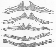 During the gastrulation
stage of vertebral development, the blastula
divides into three distinct germ layers. These three layers are the endoderm
, mesoderm
, and ectoderm
. The ectoderm
is the outermost of these layers and eventually becomes the nervous system
, the epidermis, and the lining of various external orifice
s. The next stage in the development of the nervous system is neurulation
which is the name for organogenisis
of the nervous system. This stage begins with the formation of the notochord
, a thin layer of mesodermal cells in the most dorsal
portion of the embryo. The notochord
signals the ectodermal cells above it to form the neural tube
. The formation of the neural tube is done specifically by the folding of the neural plate
into a circle which is accomplished with the help of the medial and dorsolateral hinge point cells. This structure, the neural tube, gives rise to the brain
and spinal cord
.
begins to form into spinal cord
, there are two different plates, the alar and basil plates. These two plates are separated by the sulcus limitans
. The alar plate will turn into the dorsal horn, consisting of sensory neurons
and the basil plate will turn into the ventral horn consisting of motor neurons
. The formation of the ventral horn is achieved by the secretion of sonic hedgehog
, or SHH from the notochord
during neural tube development. This induces the floor plate
to produce more SHH. The increased amount of SHH is what makes the basil plate to from motor neurons
. The vestibulospinal tract is one of the four motor tracts that send upper motor neuronal axons down the spinal cord to lower motor neurons.
, lateral vestibular nucleus
, semicircular canals or lateral vestibulospinal tract
are damaged, the person will likely sway to that side and fall when walking. Injuries to the lateral vestibulospinal tract
are most likely masked by more serious injuries to the lateral corticospinal tract
.
Resulting lesions on either the left or right vestibulocochlear nerve
, lateral vestibular nucleus
, semicircular canals or lateral vestibulospinal tract
will cause an imbalance. The healthy side "over powers" the weak side in a way that will cause the the person to veer and fall towards the injured side. Potential early onset of damage can be witnessed through a positive Romberg's test
.
Extrapyramidal system
In human anatomy, the extrapyramidal system is a neural network located in the brain that is part of the motor system involved in the coordination of movement. The system is called "extrapyramidal" to distinguish it from the tracts of the motor cortex that reach their targets by traveling through...
and also can be classified as a component of the medial pathway. The vestibulospinal fibers relay information from nuclei
Nucleus (neuroanatomy)
In neuroanatomy, a nucleus is a brain structure consisting of a relatively compact cluster of neurons. It is one of the two most common forms of nerve cell organization, the other being layered structures such as the cerebral cortex or cerebellar cortex. In anatomical sections, a nucleus shows up...
to motor neurons, like other descending motor pathways. Specifically, the vestibular nuclei
Vestibular nuclei
The vestibular nuclei are the cranial nuclei for the vestibular nerve.In Terminologia Anatomica they are grouped in both the pons and medulla.-Subnuclei:There are 4 subnuclei; they are situated at the floor of the fourth ventricle....
receive information through the vestibulocochlear nerve
Vestibulocochlear nerve
The vestibulocochlear nerve is the eighth of twelve cranial nerves, and is responsible for transmitting sound and equilibrium information from the inner ear to the brain...
about changes in the orientation of the head. The nuclei relay motor commands through the vestibulospinal tract regarding alterations of muscle tone, extension and position to the limbs and head with a goal of supporting posture and maintaining balance of the body.
Classification
The vestibulospinal tract is part of the "extrapyramidal system" of the central nervous system. In human anatomyHuman anatomy
Human anatomy is primarily the scientific study of the morphology of the human body. Anatomy is subdivided into gross anatomy and microscopic anatomy. Gross anatomy is the study of anatomical structures that can be seen by the naked eye...
, the extrapyramidal system is a neural network
Neural network
The term neural network was traditionally used to refer to a network or circuit of biological neurons. The modern usage of the term often refers to artificial neural networks, which are composed of artificial neurons or nodes...
located in the brain
Human brain
The human brain has the same general structure as the brains of other mammals, but is over three times larger than the brain of a typical mammal with an equivalent body size. Estimates for the number of neurons in the human brain range from 80 to 120 billion...
that is part of the motor system
Motor system
The motor system is the part of the central nervous system that is involved with movement. It consists of the pyramidal and extrapyramidal system....
involved in the coordination of movement. The system is called "extrapyramidal" to distinguish it from the tracts of the motor cortex that reach their targets by traveling through the "pyramids
Pyramid of medulla oblongata
The anterior or ventral portion of the medulla oblongata is named the pyramid and lies between the anterior median fissure and the antero-lateral sulcus....
" of the medulla
Medulla oblongata
The medulla oblongata is the lower half of the brainstem. In discussions of neurology and similar contexts where no ambiguity will result, it is often referred to as simply the medulla...
. The pyramidal pathways (corticospinal and some corticobulbar tracts) may directly innervate motor neurons of the spinal cord or brainstem (anterior (ventral) horn cells
Anterior horn (spinal cord)
The anterior horn of the spinal cord is the ventral grey matter section of the spinal cord. The anterior horn contains motor neurons that affect the axial muscles while the posterior horn receives information regarding touch and sensation...
or certain cranial nerve nuclei), whereas the extrapyramidal system centers around the modulation and regulation (indirect control) of anterior (ventral) horn cells. The extrapyramidal subcortical nuclei include the substantia nigra, caudate, putamen, globus pallidus, thalamus, red nucleus and subthalamic nucleus.
It has been thought that the extrapyramidal system operated independently of the pyramidal system. However, more recent research has provided a greater understanding of the integration of motor control. Motor control from both the pyramidal and extrapyramidal systems have extensive feedback loops and are heavily interconnected with each other. Motor nuclei and tracts can be classified by their functions. While the medial pathway helps control gross movements of the proximal limbs and trunk. The lateral pathway helps control precise movement of the distal portion of limbs. The vestibulospinal, as well as tectospinal
Tectospinal tract
In humans, the tectospinal tract is a nerve pathway which coordinates head and eye movements. It is part of the indirect extrapyramidal tract...
and reticulospinal
Reticulospinal tract
The reticulospinal tract is an extrapyramidal motor tract which travels from the reticular formation.-Functions:...
tracts are examples of components of the medial pathway.
Sub-pathways
The vestibulospinal tract is an upper motor neuronUpper motor neuron
Upper motor neurons are motor neurons that originate in the motor region of the cerebral cortex or the brain stem and carry motor information down to the final common pathway, that is, any motor neurons that are not directly responsible for stimulating the target muscle...
tract consists of two sub-pathways:
- Lateral Vestibulospinal TractLateral vestibulospinal tractThe lateral vestibulospinal tract is one of the descending spinal tracts of the ventromedial pathway.The lateral part of the vestibulospinal tract is the major portion and is composed of fibers originating in the lateral, superior, and inferior vestibular nuclei . It projects ipsilaterally down to...
- The lateral vestibulospinal tract innervates extension, or antigravity, muscles that are responsible for compensating movement of the body.
- Medial Vestibulospinal TractMedial vestibulospinal tractThe medial vestibulospinal tract is one of the descending spinal tracts of the ventromedial pathway.The medial part of the vestibulospinal tract is the smaller part, and is primarily made of fibers from the medial vestibular nucleus...
- The medial vestibulospinal tract innervates neck muscles that are responsible for stabilizing the head.
Lateral vestibulospinal tract
The lateral vestibulospinal tractLateral vestibulospinal tract
The lateral vestibulospinal tract is one of the descending spinal tracts of the ventromedial pathway.The lateral part of the vestibulospinal tract is the major portion and is composed of fibers originating in the lateral, superior, and inferior vestibular nuclei . It projects ipsilaterally down to...
is a group of descending extrapyramidal motor neurons
Motor neuron
In vertebrates, the term motor neuron classically applies to neurons located in the central nervous system that project their axons outside the CNS and directly or indirectly control muscles...
, or efferent fibers. This tract is found in the lateral funiculus
Lateral funiculus
The most lateral of the bundles of the anterior nerve roots is generally taken as a dividing line that separates the antero-lateral region into two parts, viz., an anterior funiculus, between the anterior median fissure and the most lateral of the anterior nerve roots; and a lateral funiculus,...
, a bundle of nerve roots in the the spinal cord
Spinal cord
The spinal cord is a long, thin, tubular bundle of nervous tissue and support cells that extends from the brain . The brain and spinal cord together make up the central nervous system...
. The lateral vestibulospinal tract
Lateral vestibulospinal tract
The lateral vestibulospinal tract is one of the descending spinal tracts of the ventromedial pathway.The lateral part of the vestibulospinal tract is the major portion and is composed of fibers originating in the lateral, superior, and inferior vestibular nuclei . It projects ipsilaterally down to...
originates in the lateral vestibular nucleus
Lateral vestibular nucleus
The lateral vestibular nucleus is the continuation upward and lateralward of the principal nucleus, and in it terminate many of the ascending branches of the vestibular nerve.-Anatomy:...
or Deiters’ nucleus in the pons
Pons
The pons is a structure located on the brain stem, named after the Latin word for "bridge" or the 16th-century Italian anatomist and surgeon Costanzo Varolio . It is superior to the medulla oblongata, inferior to the midbrain, and ventral to the cerebellum. In humans and other bipeds this means it...
. The Deiters' nucleus extends from pontomedullary junction to the level of Abducens nerve nucleus in the pons
Pons
The pons is a structure located on the brain stem, named after the Latin word for "bridge" or the 16th-century Italian anatomist and surgeon Costanzo Varolio . It is superior to the medulla oblongata, inferior to the midbrain, and ventral to the cerebellum. In humans and other bipeds this means it...
.
Lateral vestibulospinal fibers descend uncrossed, or ipsilateral, in the anterior portion of the lateral funiculus
Lateral funiculus
The most lateral of the bundles of the anterior nerve roots is generally taken as a dividing line that separates the antero-lateral region into two parts, viz., an anterior funiculus, between the anterior median fissure and the most lateral of the anterior nerve roots; and a lateral funiculus,...
of the spinal cord. Fibers run down the total length of the spinal cord and terminate at the interneurons of laminae VII and VIII. Additionally, some neurons terminate directly on the dendrites of alpha motor neurons
Alpha motor neuron
Alpha motor neurons are large lower motor neurons of the brainstem and spinal cord. They innervate extrafusal muscle fibers of skeletal muscle and are directly responsible for initiating their contraction...
in the same laminae.
Medial vestibulospinal tract
The medial vestibulospinal tractMedial vestibulospinal tract
The medial vestibulospinal tract is one of the descending spinal tracts of the ventromedial pathway.The medial part of the vestibulospinal tract is the smaller part, and is primarily made of fibers from the medial vestibular nucleus...
is a group of descending extrapyramidal motor neurons
Motor neuron
In vertebrates, the term motor neuron classically applies to neurons located in the central nervous system that project their axons outside the CNS and directly or indirectly control muscles...
, or efferent fibers found in the anterior funiculus
Anterior funiculus
The most lateral of the bundles of the anterior nerve roots is generally taken as a dividing line that separates the antero-lateral region into two parts, viz., an anterior funiculus, between the anterior median fissure and the most lateral of the anterior nerve roots; and a lateral funiculus,...
, a bundle of nerve roots in the the spinal cord
Spinal cord
The spinal cord is a long, thin, tubular bundle of nervous tissue and support cells that extends from the brain . The brain and spinal cord together make up the central nervous system...
. The medial vestibulospinal tract
Medial vestibulospinal tract
The medial vestibulospinal tract is one of the descending spinal tracts of the ventromedial pathway.The medial part of the vestibulospinal tract is the smaller part, and is primarily made of fibers from the medial vestibular nucleus...
originates in the medial vestibular nucleus
Medial vestibular nucleus
The medial vestibular nucleus is one of the vestibular nuclei. It is located in the medulla oblongata.Lateral vestibulo-spinal tract - via ventrolateral medulla and spinal cord to ventral funiculus...
or Schwalbe's nucleus. The Schwalbe's nucleus extends from the rostral end of the inferior olivary nucleus
Inferior olivary nucleus
The inferior olivary nucleus is the largest nucleus situated in the olivary body, part of the medulla oblongata.-Function:It is closely associated with the cerebellum, meaning that it is involved in control and coordination of movements, sensory processing and cognitive tasks likely by encoding the...
of the medulla oblongata
Medulla oblongata
The medulla oblongata is the lower half of the brainstem. In discussions of neurology and similar contexts where no ambiguity will result, it is often referred to as simply the medulla...
to the caudal portion of the pons
Pons
The pons is a structure located on the brain stem, named after the Latin word for "bridge" or the 16th-century Italian anatomist and surgeon Costanzo Varolio . It is superior to the medulla oblongata, inferior to the midbrain, and ventral to the cerebellum. In humans and other bipeds this means it...
.
Medial vestibulospinal fibers join with the ipsilateral and contralateral medial longitudinal fasciculus
Medial longitudinal fasciculus
The medial longitudinal fasciculus is a pair of crossed fiber tracts , one on each side of the brainstem. These bundles of axons are situated near the midline of the brainstem and are composed of both ascending and descending fibers that arise from a number of sources and terminate in different...
, and descend the anterior funiculus
Anterior funiculus
The most lateral of the bundles of the anterior nerve roots is generally taken as a dividing line that separates the antero-lateral region into two parts, viz., an anterior funiculus, between the anterior median fissure and the most lateral of the anterior nerve roots; and a lateral funiculus,...
of the spinal cord. Fibers run down to the anterior funiculus to the cervical spinal cord segments and terminate on neurons of laminae VII and VIII. Unlike the lateral vestibulospinal tract, the medial vestibulospinal tract innervates muscles that support the head. As a result, medial vestibulospinal fibers run down only to the cervical segments of the cord.
Function
The vestibulospinal tract is part of the vestibular systemVestibular system
The vestibular system, which contributes to balance in most mammals and to the sense of spatial orientation, is the sensory system that provides the leading contribution about movement and sense of balance. Together with the cochlea, a part of the auditory system, it constitutes the labyrinth of...
in the CNS
Central nervous system
The central nervous system is the part of the nervous system that integrates the information that it receives from, and coordinates the activity of, all parts of the bodies of bilaterian animals—that is, all multicellular animals except sponges and radially symmetric animals such as jellyfish...
. The primary role of the vestibular system is to maintain head and eye coordination, upright posture and balance, and conscious realization of spatial orientation and motion. The vestibular system is able to respond correctly by recording sensory information from hairs cells in the labyrinth of the inner ear. Then the nuclei receiving these signals project out to the extraocular muscles
Extraocular muscles
The extraocular muscles are the six muscles that control the movements of the eye . The actions of the extraocular muscles depend on the position of the eye at the time of muscle contraction.-List of muscles:-Importance:...
, spinal cord
Spinal cord
The spinal cord is a long, thin, tubular bundle of nervous tissue and support cells that extends from the brain . The brain and spinal cord together make up the central nervous system...
, and cerebral cortex
Cerebral cortex
The cerebral cortex is a sheet of neural tissue that is outermost to the cerebrum of the mammalian brain. It plays a key role in memory, attention, perceptual awareness, thought, language, and consciousness. It is constituted of up to six horizontal layers, each of which has a different...
to execute these functions.
One of these projections, the vestibulospinal tract, is responsible for upright posture and head stabilization. When the vestibular sensory neurons detect small movements of the body, the vestibulospinal tract commands motor signals to specific muscles to counteract these movements and re-stabilize the body. The vestibulospinal tract has two main sections: the medial vestibulospinal tract
Medial vestibulospinal tract
The medial vestibulospinal tract is one of the descending spinal tracts of the ventromedial pathway.The medial part of the vestibulospinal tract is the smaller part, and is primarily made of fibers from the medial vestibular nucleus...
and the lateral vestibulospinal tract
Lateral vestibulospinal tract
The lateral vestibulospinal tract is one of the descending spinal tracts of the ventromedial pathway.The lateral part of the vestibulospinal tract is the major portion and is composed of fibers originating in the lateral, superior, and inferior vestibular nuclei . It projects ipsilaterally down to...
.
The medial spinal tract projects bilaterally from the medial vestibular nucleus
Medial vestibular nucleus
The medial vestibular nucleus is one of the vestibular nuclei. It is located in the medulla oblongata.Lateral vestibulo-spinal tract - via ventrolateral medulla and spinal cord to ventral funiculus...
within the medial longitudinal fasciculus
Medial longitudinal fasciculus
The medial longitudinal fasciculus is a pair of crossed fiber tracts , one on each side of the brainstem. These bundles of axons are situated near the midline of the brainstem and are composed of both ascending and descending fibers that arise from a number of sources and terminate in different...
to the ventral horns in the upper cervical cord (T6 vertebra). It promotes stabilization of head position by innervating the neck muscles, which helps with head coordination and eye movement.
The lateral vestibulospinal tract is an ipsilaterally descending tract from the vestibular system through the anterolateral white matter
Lateral spinothalamic tract
The lateral spinothalamic tract , which is a part of the Anterolateral system, is a bundle of sensory axons ascending through the white matter of the spinal cord, carrying sensory information to the brain. It carries pain and temperature sensory information to the thalamus of the brain...
. It that provides excitatory signals to interneurons, which relay the signal to the motor neurons in antigravity muscles. These antigravity muscles are extensor muscles in the legs that help maintain upright and balanced posture.
Reflexes
The vestibulospinal reflexReflex
A reflex action, also known as a reflex, is an involuntary and nearly instantaneous movement in response to a stimulus. A true reflex is a behavior which is mediated via the reflex arc; this does not apply to casual uses of the term 'reflex'.-See also:...
uses the vestibular organs as well as skeletal muscle in order to maintain balance, posture, and stability in an environment with gravity. These reflexes can be further broken down by timing into a dynamic reflex, static reflex or tonic reflex. It can also be categorized by the sensory input as either canal, otolith
Otolith
An otolith, , also called statoconium or otoconium is a structure in the saccule or utricle of the inner ear, specifically in the vestibular labyrinth of vertebrates. The saccule and utricle, in turn, together make the otolith organs. They are sensitive to gravity and linear acceleration...
, or both. The term vesitbulospinal reflex, is most commonly used when the sensory input evokes a response from the muscular system
Muscular system
The muscular system is the anatomical system of a species that allows it to move. The muscular system in vertebrates is controlled through the nervous system, although some muscles can be completely autonomous.- Muscles :...
below the neck. These reflexes are important in the maintenance of homeostasis
Homeostasis
Homeostasis is the property of a system that regulates its internal environment and tends to maintain a stable, constant condition of properties like temperature or pH...
.
Example of vestibulospinal reflex
- The head is tilted to one side which stimulates both the canals and the otolithsOtolithAn otolith, , also called statoconium or otoconium is a structure in the saccule or utricle of the inner ear, specifically in the vestibular labyrinth of vertebrates. The saccule and utricle, in turn, together make the otolith organs. They are sensitive to gravity and linear acceleration...
- This movement stimulates the vestibular nerveVestibular nerveThe vestibular nerve is one of the two branches of the Vestibulocochlear nerve . It goes to the semicircular canals via the vestibular ganglion...
as well as the vestibular nucleusVestibular nucleiThe vestibular nuclei are the cranial nuclei for the vestibular nerve.In Terminologia Anatomica they are grouped in both the pons and medulla.-Subnuclei:There are 4 subnuclei; they are situated at the floor of the fourth ventricle....
. - These impulses are transmitted down both the lateral and medial vestibulospinal tracts to the spinal cordSpinal cordThe spinal cord is a long, thin, tubular bundle of nervous tissue and support cells that extends from the brain . The brain and spinal cord together make up the central nervous system...
. - The spinal cord induces extensor effects in the muscle on the side of the neck to which the head is bent, and flexor effects in the muscle in the side of the neck away from the direction of the displaced head.
Tonic labyrinthine reflex
The tonic labyrinthine reflexTonic labyrinthine reflex
The tonic labyrinthine reflex is a primitive reflex found in newborn humans. With this reflex, tilting the head back while lying on the back causes the back to stiffen and even arch backwards, the legs to straighten, stiffen, and push together, the toes to point, the arms to bend at the elbows...
is a reflex that is present in newborn babies directly after birth and should be fully inhibited by 3.5 years. This reflex helps the baby master head and neck movements outside of the womb
Uterus
The uterus or womb is a major female hormone-responsive reproductive sex organ of most mammals including humans. One end, the cervix, opens into the vagina, while the other is connected to one or both fallopian tubes, depending on the species...
as well as the concept of gravity
Gravitation
Gravitation, or gravity, is a natural phenomenon by which physical bodies attract with a force proportional to their mass. Gravitation is most familiar as the agent that gives weight to objects with mass and causes them to fall to the ground when dropped...
. Increased muscle tone, development of the the proprioceptive
Proprioception
Proprioception , from Latin proprius, meaning "one's own" and perception, is the sense of the relative position of neighbouring parts of the body and strength of effort being employed in movement...
and vestibular senses and opportunities to practice with balance are all consequences of this reflex. During early childhood, the TLR matures into more developed vestibulospinal reflexes to help with posture, head alignment and balance.
The tonic labyrinthine reflex
Tonic labyrinthine reflex
The tonic labyrinthine reflex is a primitive reflex found in newborn humans. With this reflex, tilting the head back while lying on the back causes the back to stiffen and even arch backwards, the legs to straighten, stiffen, and push together, the toes to point, the arms to bend at the elbows...
is found in two forms.
- Forward: When the head bends forward, the whole body, arms, legs and torso curl together to form the fetal positionFetal positionFetal position is a medical term used to describe the positioning of the body of a prenatal fetus as it develops...
. - Backwards: When the head is bent backward, the whole body, arms, legs and torso straighten and extend.
Righting reflex
The righting reflex is another type of reflex. This reflex positions the head or body back into its "normal" position, in response to a change in head or body position. A common example of this reflex is the cat righting reflexCat righting reflex
The cat righting reflex is a cat's innate ability to orient itself as it falls in order to land on its feet. The righting reflex begins to appear at 3-4 weeks of age, and is perfected at 7 weeks. They are able to do this as they have an unusually flexible backbone and no functional clavicle...
, which allows them orient themselves in order to land on their feet. This reflex is initiated by sensory information from the vestibular, visual
Visual system
The visual system is the part of the central nervous system which enables organisms to process visual detail, as well as enabling several non-image forming photoresponse functions. It interprets information from visible light to build a representation of the surrounding world...
, and the somatosensory
Somatosensory system
The somatosensory system is a diverse sensory system composed of the receptors and processing centres to produce the sensory modalities such as touch, temperature, proprioception , and nociception . The sensory receptors cover the skin and epithelia, skeletal muscles, bones and joints, internal...
systems and is therefore not only a vestibulospinal reflex.
CNS development
Gastrulation
Gastrulation is a phase early in the embryonic development of most animals, during which the single-layered blastula is reorganized into a trilaminar structure known as the gastrula. These three germ layers are known as the ectoderm, mesoderm, and endoderm.Gastrulation takes place after cleavage...
stage of vertebral development, the blastula
Blastula
The blastula is a hollow sphere of cells formed during an early stage of embryonic development in animals . The blastula is created when the zygote undergoes the cell division process known as cleavage. The blastula is preceded by the morula and is followed by the gastrula in the developmental...
divides into three distinct germ layers. These three layers are the endoderm
Endoderm
Endoderm is one of the three primary germ cell layers in the very early embryo. The other two layers are the ectoderm and mesoderm , with the endoderm as the intermost layer...
, mesoderm
Mesoderm
In all bilaterian animals, the mesoderm is one of the three primary germ cell layers in the very early embryo. The other two layers are the ectoderm and endoderm , with the mesoderm as the middle layer between them.The mesoderm forms mesenchyme , mesothelium, non-epithelial blood corpuscles and...
, and ectoderm
Ectoderm
The "ectoderm" is one of the three primary germ cell layers in the very early embryo. The other two layers are the mesoderm and endoderm , with the ectoderm as the most exterior layer...
. The ectoderm
Ectoderm
The "ectoderm" is one of the three primary germ cell layers in the very early embryo. The other two layers are the mesoderm and endoderm , with the ectoderm as the most exterior layer...
is the outermost of these layers and eventually becomes the nervous system
Nervous system
The nervous system is an organ system containing a network of specialized cells called neurons that coordinate the actions of an animal and transmit signals between different parts of its body. In most animals the nervous system consists of two parts, central and peripheral. The central nervous...
, the epidermis, and the lining of various external orifice
Body orifice
-External orifices:In a typical mammalian body such as the human body, the external body orifices are:* The nostrils, for breathing and the associated sense of smell.* The eyes, for the sense of sight and crying....
s. The next stage in the development of the nervous system is neurulation
Neurulation
Neurulation is the stage of organogenesis in vertebrate embryos, during which the neural tube is transformed into the primitive structures that will later develop into the central nervous system....
which is the name for organogenisis
Organogenesis
In animal development, organogenesis is the process by which the ectoderm, endoderm, and mesoderm develop into the internal organs of the organism. Internal organs initiate development in humans within the 3rd to 8th weeks in utero...
of the nervous system. This stage begins with the formation of the notochord
Notochord
The notochord is a flexible, rod-shaped body found in embryos of all chordates. It is composed of cells derived from the mesoderm and defines the primitive axis of the embryo. In some chordates, it persists throughout life as the main axial support of the body, while in most vertebrates it becomes...
, a thin layer of mesodermal cells in the most dorsal
Dorsum (anatomy)
In anatomy, the dorsum is the upper side of animals that typically run, fly, or swim in a horizontal position, and the back side of animals that walk upright. In vertebrates the dorsum contains the backbone. The term dorsal refers to anatomical structures that are either situated toward or grow...
portion of the embryo. The notochord
Notochord
The notochord is a flexible, rod-shaped body found in embryos of all chordates. It is composed of cells derived from the mesoderm and defines the primitive axis of the embryo. In some chordates, it persists throughout life as the main axial support of the body, while in most vertebrates it becomes...
signals the ectodermal cells above it to form the neural tube
Neural tube
In the developing vertebrate, the neural tube is the embryo's precursor to the central nervous system, which comprises the brain and spinal cord...
. The formation of the neural tube is done specifically by the folding of the neural plate
Neural plate
In human embryology, formation of neural plate is the first step of neurulation. It is created by a flat thickening opposite to the primitive streak of the ectoderm.-Development:...
into a circle which is accomplished with the help of the medial and dorsolateral hinge point cells. This structure, the neural tube, gives rise to the brain
Brain
The brain is the center of the nervous system in all vertebrate and most invertebrate animals—only a few primitive invertebrates such as sponges, jellyfish, sea squirts and starfishes do not have one. It is located in the head, usually close to primary sensory apparatus such as vision, hearing,...
and spinal cord
Spinal cord
The spinal cord is a long, thin, tubular bundle of nervous tissue and support cells that extends from the brain . The brain and spinal cord together make up the central nervous system...
.
Development of the vestibulospinal tract during spinal cord formation
When the neural tubeNeural tube
In the developing vertebrate, the neural tube is the embryo's precursor to the central nervous system, which comprises the brain and spinal cord...
begins to form into spinal cord
Spinal cord
The spinal cord is a long, thin, tubular bundle of nervous tissue and support cells that extends from the brain . The brain and spinal cord together make up the central nervous system...
, there are two different plates, the alar and basil plates. These two plates are separated by the sulcus limitans
Sulcus limitans
In the floor of the fourth ventricle, the sulcus limitans separates the cranial nerve motor nuclei from the sensory nuclei . In the superior part of the rhomboid fossa, it corresponds with the lateral limit of the fossa and presents a bluish-gray area, the locus ceruleus In the floor of the fourth...
. The alar plate will turn into the dorsal horn, consisting of sensory neurons
Sensory neuron
Sensory neurons are typically classified as the neurons responsible for converting external stimuli from the environment into internal stimuli. They are activated by sensory input , and send projections into the central nervous system that convey sensory information to the brain or spinal cord...
and the basil plate will turn into the ventral horn consisting of motor neurons
Motor neuron
In vertebrates, the term motor neuron classically applies to neurons located in the central nervous system that project their axons outside the CNS and directly or indirectly control muscles...
. The formation of the ventral horn is achieved by the secretion of sonic hedgehog
Sonic hedgehog
Sonic hedgehog homolog is one of three proteins in the mammalian signaling pathway family called hedgehog, the others being desert hedgehog and Indian hedgehog . SHH is the best studied ligand of the hedgehog signaling pathway. It plays a key role in regulating vertebrate organogenesis, such as...
, or SHH from the notochord
Notochord
The notochord is a flexible, rod-shaped body found in embryos of all chordates. It is composed of cells derived from the mesoderm and defines the primitive axis of the embryo. In some chordates, it persists throughout life as the main axial support of the body, while in most vertebrates it becomes...
during neural tube development. This induces the floor plate
Floor plate
The floor plate is a structure integral to the developing nervous system of vertebrate organisms. Located on the ventral midline of the embryonic neural tube, the floor plate is a specialized glial structure that spans the anteroposterior axis from the midbrain to the tail regions...
to produce more SHH. The increased amount of SHH is what makes the basil plate to from motor neurons
Motor neuron
In vertebrates, the term motor neuron classically applies to neurons located in the central nervous system that project their axons outside the CNS and directly or indirectly control muscles...
. The vestibulospinal tract is one of the four motor tracts that send upper motor neuronal axons down the spinal cord to lower motor neurons.
Injury and lesions
A typical person sways from side to side when the eyes are closed. This is the result of the vestibulospinal reflex working correctly. When an individual sways to the left side, the left lateral vestibulospinal tract is activated to bring the body back to midline. If the vestibulocochlear nerveVestibulocochlear nerve
The vestibulocochlear nerve is the eighth of twelve cranial nerves, and is responsible for transmitting sound and equilibrium information from the inner ear to the brain...
, lateral vestibular nucleus
Lateral vestibular nucleus
The lateral vestibular nucleus is the continuation upward and lateralward of the principal nucleus, and in it terminate many of the ascending branches of the vestibular nerve.-Anatomy:...
, semicircular canals or lateral vestibulospinal tract
Lateral vestibulospinal tract
The lateral vestibulospinal tract is one of the descending spinal tracts of the ventromedial pathway.The lateral part of the vestibulospinal tract is the major portion and is composed of fibers originating in the lateral, superior, and inferior vestibular nuclei . It projects ipsilaterally down to...
are damaged, the person will likely sway to that side and fall when walking. Injuries to the lateral vestibulospinal tract
Lateral vestibulospinal tract
The lateral vestibulospinal tract is one of the descending spinal tracts of the ventromedial pathway.The lateral part of the vestibulospinal tract is the major portion and is composed of fibers originating in the lateral, superior, and inferior vestibular nuclei . It projects ipsilaterally down to...
are most likely masked by more serious injuries to the lateral corticospinal tract
Lateral corticospinal tract
The lateral corticospinal tract is the largest part of the corticospinal tract...
.
Resulting lesions on either the left or right vestibulocochlear nerve
Vestibulocochlear nerve
The vestibulocochlear nerve is the eighth of twelve cranial nerves, and is responsible for transmitting sound and equilibrium information from the inner ear to the brain...
, lateral vestibular nucleus
Lateral vestibular nucleus
The lateral vestibular nucleus is the continuation upward and lateralward of the principal nucleus, and in it terminate many of the ascending branches of the vestibular nerve.-Anatomy:...
, semicircular canals or lateral vestibulospinal tract
Lateral vestibulospinal tract
The lateral vestibulospinal tract is one of the descending spinal tracts of the ventromedial pathway.The lateral part of the vestibulospinal tract is the major portion and is composed of fibers originating in the lateral, superior, and inferior vestibular nuclei . It projects ipsilaterally down to...
will cause an imbalance. The healthy side "over powers" the weak side in a way that will cause the the person to veer and fall towards the injured side. Potential early onset of damage can be witnessed through a positive Romberg's test
Romberg's test
Romberg's test or the Romberg maneuver is a test used by doctors in a neurological examination, and also as a test for drunken driving. The exam is based on the premise that a person requires at least two of the three following senses to maintain balanced while standing:Proprioception ; Vestibular...
.