

Extension (kinesiology)
Encyclopedia

Kinesiology
Kinesiology, also known as human kinetics is the scientific study of human movement. Kinesiology addresses physiological, mechanical, and psychological mechanisms. Applications of kinesiology to human health include: biomechanics and orthopedics, rehabilitation, such as physical and occupational...
, extension is a movement of a joint that results in increased angle between two bones or body surfaces at a joint. Extension usually results in straightening of the bones or body surfaces involved. For example, extension is produced by extending the flexed (bent) elbow. Straightening of the arm would require extension at the elbow joint. If the head is tilted all the way back, the neck is said to be extended.
In some anatomical muscle names, particularly with muscles of the forearm
Forearm
-See also:*Forearm flexors*Forearm muscles...
and the Cnemis, the term occurs explicitly as the second word in the Latin spelling of the name (for example, Musculus extensor carpi ulnaris).
While extension is a movement, the terms "extend" versus "flexed" refer to the final position of a body part with reference to the anatomical position of the body. For example, if an arm fully bent at the elbow is slightly extended, it will still be called "flexed" or "semiflexed", and will be called "extended" only upon complete straightening of the arm.
The movement in the opposite directions is called flexion
Flexion
In anatomy, flexion is a position that is made possible by the joint angle decreasing. The skeletal and muscular systems work together to move the joint into a "flexed" position. For example the elbow is flexed when the hand is brought closer to the shoulder...
. Flexion decreases the angle between the bones of the limb at a joint, while extension increases it.
With the foot
Foot
The foot is an anatomical structure found in many vertebrates. It is the terminal portion of a limb which bears weight and allows locomotion. In many animals with feet, the foot is a separate organ at the terminal part of the leg made up of one or more segments or bones, generally including claws...
, extension is usually called plantarflexion
Plantarflexion
Plantarflexion is the movement which increases the approximate 90 degree angle between the front part of the foot and the shin, as when depressing an automobile pedal...
.
Hyperextension
If a part of the body such as a joint is overstretched or "bent backwards" because of exaggerated extension motion, then one speaks of a hyperextension (as with the kneeKnee
The knee joint joins the thigh with the leg and consists of two articulations: one between the fibula and tibia, and one between the femur and patella. It is the largest joint in the human body and is very complicated. The knee is a mobile trocho-ginglymus , which permits flexion and extension as...
). This puts a lot of stress on the ligament
Ligament
In anatomy, the term ligament is used to denote any of three types of structures. Most commonly, it refers to fibrous tissue that connects bones to other bones and is also known as articular ligament, articular larua, fibrous ligament, or true ligament.Ligament can also refer to:* Peritoneal...
s of the joint, and need not always be a voluntary movement, but may occur as part of accidents, falls, or other causes of trauma.
Hyperextension is also sometimes defined as normal movement into the space posterior to the anatomical position.
Upper limb
- of armArmIn human anatomy, the arm is the part of the upper limb between the shoulder and the elbow joints. In other animals, the term arm can also be used for analogous structures, such as one of the paired forelimbs of a four-legged animal or the arms of cephalopods...
at shoulderShoulderThe human shoulder is made up of three bones: the clavicle , the scapula , and the humerus as well as associated muscles, ligaments and tendons. The articulations between the bones of the shoulder make up the shoulder joints. The major joint of the shoulder is the glenohumeral joint, which...
- Axilla and Shoulder
- Latissimus Dorsi
- Posterior Fibres of DeltoidDeltoid muscleIn human anatomy, the deltoid muscle is the muscle forming the rounded contour of the shoulder. Anatomically, it appears to be made up of three distinct sets of fibers though electromyography suggests that it consists of at least seven groups that can be independently coordinated by the central...
- Teres Major
- Axilla and Shoulder
- of forearmForearm-See also:*Forearm flexors*Forearm muscles...
at elbowElbowThe human elbow is the region surrounding the elbow-joint—the ginglymus or hinge joint in the middle of the arm. Three bones form the elbow joint: the humerus of the upper arm, and the paired radius and ulna of the forearm....
- Posterior compartment of the armPosterior compartment of the armThe posterior compartment of the arm is an anatomic compartment which contains muscles which are all supplied by the radial nerve. This compartment is also known as the "extensor compartment", extension being its main action.-Contents:...
- Triceps
- Anconeus
- Posterior compartment of the arm
- of handHandA hand is a prehensile, multi-fingered extremity located at the end of an arm or forelimb of primates such as humans, chimpanzees, monkeys, and lemurs...
at wristWristIn human anatomy, the wrist is variously defined as 1) the carpus or carpal bones, the complex of eight bones forming the proximal skeletal segment of the hand;...
- Posterior compartment of the forearmPosterior compartment of the forearmThe posterior compartment of the forearm contains the following muscles:-Muscles:* "E/I" refers to "extrinsic" or "intrinsic"....
- Extensor carpi radialis longus
- Extensor carpi radialis brevis
- Extensor carpi ulnaris
- Extensor digitorum
- Posterior compartment of the forearm
- of phalanges, at all joints
- Posterior compartment of the forearmPosterior compartment of the forearmThe posterior compartment of the forearm contains the following muscles:-Muscles:* "E/I" refers to "extrinsic" or "intrinsic"....
- Extensor digitorum
- Extensor digiti minimi (little finger only)
- Extensor indicis (index finger only)
- Posterior compartment of the forearm
- of phalanges, at interphalangeal joints
- Lumbricals of the handLumbricals of the handThe lumbricals are intrinsic muscles of the hand that flex the metacarpophalangeal joints and extend the interphalangeal joints.-Structure:There are four of these small, worm-like muscles on each hand. These muscles are unusual in that they do not attach to bone...
- Dorsal interossei of the handDorsal interossei of the handThe dorsal interossei of the hand are muscles that occupy the space between the metacarpals.-Structure:There are four dorsal interossei in each hand...
- Palmar interossei
- Lumbricals of the hand
- of thumbThumbThe thumb is the first digit of the hand. When a person is standing in the medical anatomical position , the thumb is the lateral-most digit...
- Extensor pollicis brevis (proximal phalange)
- Extensor pollicis longus (distal phalange)
Lower limb
- of thighThighIn humans the thigh is the area between the pelvis and the knee. Anatomically, it is part of the lower limb.The single bone in the thigh is called the femur...
/femurFemurThe femur , or thigh bone, is the most proximal bone of the leg in tetrapod vertebrates capable of walking or jumping, such as most land mammals, birds, many reptiles such as lizards, and amphibians such as frogs. In vertebrates with four legs such as dogs and horses, the femur is found only in...
at hip- Gluteus maximusGluteus maximus muscleThe gluteus maximus is the largest and most superficial of the three gluteal muscles...
- Posterior compartment of thighPosterior compartment of thighThe posterior fascial compartment of the thigh contains the knee flexors and hip extensors.It consists of the following muscles:* biceps femoris* semitendinosus* semimembranosus...
- Biceps femorisBiceps femoris muscleThe biceps femoris is a muscle of the posterior thigh. As its name implies, it has two parts, one of which forms part of the hamstrings muscle group.-Origin and insertion:It has two heads of origin;...
- SemitendinosusSemitendinosus muscleThe semitendinosus is a muscle in the back of the thigh; it is one of the hamstrings.-Structure:The semitendinosus, remarkable for the great length of its tendon of insertion, is situated at the posterior and medial aspect of the thigh ....
- SemimembranosusSemimembranosus muscleThe semimembranosus is a muscle in the back of the thigh. It is the most medial of the three hamstring muscles.-Structure:The semimembranosus, so called from its membranous tendon of origin, is situated at the back and medial side of the thigh....
- Biceps femoris
- Gluteus maximus
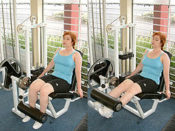
- of legHuman legThe human leg is the entire lower extremity or limb of the human body, including the foot, thigh and even the hip or gluteal region; however, the precise definition in human anatomy refers only to the section of the lower limb extending from the knee to the ankle.Legs are used for standing,...
at kneeKneeThe knee joint joins the thigh with the leg and consists of two articulations: one between the fibula and tibia, and one between the femur and patella. It is the largest joint in the human body and is very complicated. The knee is a mobile trocho-ginglymus , which permits flexion and extension as...
(L3-L4)- QuadricepsQuadriceps muscleThe quadriceps femoris , also called simply the quadriceps, quadriceps extensor, quads, is a large muscle group that includes the four prevailing muscles on the front of the thigh...
- Rectus femoris muscleRectus femoris muscleThe rectus femoris muscle is one of the four quadriceps muscles of the human body. The others are the vastus medialis, the vastus intermedius , and the vastus lateralis...
- Vastus medialisVastus medialisThe vastus medialis , often called the 'teardrop' muscle, is a medially located muscle of the quadriceps.-Function:The vasti appear to act largely in a co-ordinated manner throughout the control of knee extension...
- Vastus lateralis
- Vastus intermedius
- Rectus femoris muscle
- Quadriceps
- of toes
- Extensor hallucis longus
- Extensor digitorum longus
- Extensor digitorum brevis
- Extensor hallucis brevis