

The Lesion Project
Encyclopedia
Multiple sclerosis
is a condition in which the CNS
of a person present a special kind of distributed lesions (sclerosis) whose pathophysiology is complex and still under investigation. It is considered a pathological entity by some authors and a clinical entity by some others.
Damage occurs in two phases. First some MRI-abnormal areas with hidden damage appear in the brain and spine (NAWM, NAGM, DAWM), followed later by leaks in the blood-brain barrier where immune cells infiltrate causing the known demyelination.
MS is mainly a white matter disease, and lesions appear mainly in a peri-ventricular distribution (lesions clustered around the ventricles of the brain), but apart of the usually known white matter
demyelination, also the cortex
and deep gray matter
(GM) nuclei are affected, together with diffuse injury of the normal-appearing white matter. MS is active even during remission periods. GM atrophy is independent of the MS lesions and is associated with physical disability, fatigue, and cognitive impairment in MS
At least five characteristics are present in CNS tissues of MS patients: Inflammation
beyond classical white matter lesions, Intrathecal
Ig production with oligoclonal bands, An environment fostering immune cell persistence, Follicle
-like aggregates in the meninges
and a disruption of the blood-brain barrier
also outside of active lesions.
 Damage occurs in two phases. First some MRI-abnormal areas with hidden damage appear in the brain and spine (NAWM, NAGM, DAWM), followed later by leaks in the blood-brain barrier where immune cells infiltrate causing the known demyelination.
According to the view of most researchers, a special subset of lymphocyte
s, called T helper cell
s, specifically Th1 and Th17, play a key role in the development of the lesion. A protein called Interleukin 12 is responsible for the differentiation of naive T cells into inflammatory T cells. An over production of this protein is what causes the increased inflammation in MS patients. Under normal circumstances, these lymphocyte
s can distinguish between self and non-self. However, in a person with MS, these cells recognize healthy parts of the central nervous system as foreign and attack them as if they were an invading virus, triggering inflammatory
processes and stimulating other immune cells and soluble factors like cytokine
s and antibodies
. Many of the myelin-recognizing T cells belong to a terminally differentiated subset called co-stimulation-independent effector-memory T cells. Recently other type of immune cells, B Cell
s, have been also implicated in the pathogenesis of MS and in the degeneration of the axons.
The axon
s themselves can also be damaged by the attacks. Often, the brain is able to compensate for some of this damage, due to an ability called neuroplasticity
. MS symptoms develop as the cumulative result of multiple lesion
s in the brain and spinal cord
. This is why symptoms can vary greatly between different individuals, depending on where their lesions occur.
Repair processes, called remyelination, also play an important role in MS. Remyelination is one of the reasons why, especially in early phases of the disease, symptoms tend to decrease or disappear temporarily. Nevertheless, nerve damage and irreversible loss of neurons occur early in MS.
The oligodendrocyte
s that originally formed a myelin sheath cannot completely rebuild a destroyed myelin sheath. However, the central nervous system can recruit oligodendrocyte stem cell
s capable of proliferation and migration and differentiation into mature myelinating oligodendrocytes. The newly-formed myelin sheaths are thinner and often not as effective as the original ones. Repeated attacks lead to successively fewer effective remyelinations, until a scar-like plaque is built up around the damaged axons. Under laboratory conditions, stem cells are quite capable of proliferating and differentiating into remyelinating oligodendrocytes; it is therefore suspected that inflammatory conditions or axonal damage somehow inhibit stem cell proliferation and differentiation in affected areas
 Multiple sclerosis is considered a disease of the white matter because normally lesions appear in this area, but it is also possible to find some of them in the grey matter.
Using high field MRI system, with several variants several areas show lesions, and can be spacially classified in infratentorial, callosal, juxtacortical, periventricular, and other white matter areas. Other authors simplify this in three regions: intracortical, mixed gray-white matter, and juxtacortical. Others classify them as hippocampal, cortical, and WM lesions, and finally, others give seven areas: intracortical, mixed white matter-gray matter, juxtacortical, deep gray matter, periventricular white matter, deep white matter, and infratentorial lesions. The distribution of the lesions could be linked to the clinical evolution
Post-mortem autopsy reveal that gray matter demyelination occurs in the motor cortex
, cingulate gyrus, cerebellum
, thalamus
and spinal cord
. Cortical lesions have been observed specially in people with SPMS but they also appear in RRMS and clinically isolated syndrome. They are more frequent in men than in women and they can partly explain cognitive deficits.
It is known that two parameters of the cortical lesions, fractional anisotropy (FA) and mean diffusivity (MD), are higher in patients than in controls. They are larger in SPMS than in RRMS and most of them remain unchanged for short follow-up periods. They do not spread into the subcortical white matter and never show gadolinium enhancement. Over a one-year period, CLs can increase their number and size in a relevant proportion of MS patients, without spreading into the subcortical white matter or showing inflammatory features similar to those of white matter lesions.
Due to the distribution of the lesions, since 1916 they are also known as Dawson's fingers
. They appear around the brain blood vessels.
of cervical spinal cord is lower than normal, showing that there is damage hidden from normal MRI.
Progressive tissue loss and injury occur in the cervical cord of MS patients. These two components of cord damage are not interrelated, suggesting that a multiparametric MRI approach is needed to get estimates of such a damage. MS cord pathology is independent of brain changes, develops at different rates according to disease phenotype, and is associated to medium-term disability accrual.
Spinal cord presents grey matter lesions, that can be confirmed post-mortem and by high field MR imaging. Spinal cord grey matter lesions may be detected on MRI more readily than GM lesions in the brain, making the cord a promising site to study the grey matter demyelination.
and optic nerve
, which can be meassured by Optical coherence tomography
or by Scanning laser polarimetry
. This measure can be used to predict disease activity and to establish a differential diagnosis from Neuromyelitis optica
In vertebrate embryonic development, the retina and the optic nerve
originate as outgrowths of the developing brain
, so the retina is considered part of the central nervous system
(CNS). It is the only part of the CNS that can be imaged non-invasively in the living organism.
The retina is unique within the CNS in that it contains axons and glia but no myelin, and it is, therefore, an ideal structure within which to visualize the processes of neurodegeneration. Tissue-bound IgG was demonstrated on retinal ganglion cells in six of seven multiple sclerosis cases but not in controls.
Uveitis
and retinal phlebitis
are manifestations of MS. Trypsin
digestion with microscopic examination is a method of testing for phlebitis and the frequency found in this series is higher than in others. These lesions are similar to the perivenular cuffing that occurs in the central nervous system in MS.
Retinal vessels show narrower arterioles and wider venules even in absence of optic neuritis, possibly as a consequence of subclinical swelling of optic nerve axons, together with a higher than normal rigidity
Proton magnetic resonance
spectroscopy has shown that there is widespread neuronal loss even at the onset of MS, largely unrelated to inflammation.
A relationship between neural damage and N-Acetyl-Aspartate concentration has been established, and this could lead to new methods for early MS diagnostic through magnetic resonance spectroscopy
Axonal degeneration at CNS can be estimated by N-acetylaspartate to creatine
(NAA/Cr) ratio, both measured by with proton magnetic resonance spectroscopy.
is a condition defined by the presence of a special kind of lesions in the brain and spinal cord. Therefore it is very important to establish what those "lesions typical of MS" are. They mainly consist in demyelination
and scarring in the fatty myelin
sheaths around the axon
s of the brain and spinal cord. According with the most recent research, an active lesion is composed of different layers:
When BH's appear, around half of them revert in a month. This is considered a sign of remyelination. When they remain, this is regarded as a sign of permanent demyelination and axonal loss. This has been shown on post-mortem autopsies.
Small lesions are invisible under MRI. Therefore clinically assisted diagnostic criteria
are still required for a more accurate MS diagnosis than MRI alone.
(BBB) should not allow T-cells to enter the nervous system. BBB disruption is the moment in which T-cells cross the barrier and has always been considered one of the early problems in the MS lesions. For unknown reasons, leaks appear in the BBB during the course of MS.
Recently it has been found that BBB damage happens even in non-enhancing lesions. MS has an important vascular component.
The BBB is built up of endothelial cells lining the blood vessel
walls. After its breakdown several problems appear, such as swelling
, activation of macrophages, and more activation of cytokines and other protein
s such as matrix metalloproteinase
s which are destructive.
Whatever the demyelination process is, currently it is possible to detect lesions before demyelination, and they show clusters of activated microglia and leukocyte infiltration, together with oligodendrocytes abnormalities
As lesions appear (using MRI) in "Normal-appearing white matter" (NAWM), there is supposed to be the cause that finally triggers the BBB disruption. The damaged white matter is known as "Normal-appearing white matter" (NAWM) and is where lesions appear. These lesions form in NAWM before blood-brain barrier breakdown.
BBB can be broken centripetally or centrifugally, the first form being the most normal. Several possible biochemical disrupters have been proposed. Some hypothesis about how the BBB is compromised revolve around the presence of different compounds in the blood that could interact with the vascular vessels ony in the NAWM areas. The permeability of two cytokine
s, IL15 and LPS
, could be involved in the BBB breakdown. The BBB breakdown is responsible for monocyte
infiltration and inflammation in the brain. Monocyte migration and LFA-1
-mediated attachment to brain microvascular endothelia is regulated by SDF-1alpha through Lyn
kinase
Using iron nanoparticles, involvement of macrophages in the BBB breakdown can be detected. A special role is played by Matrix metalloproteinase
s. These are a group of proteases that increase T-cells permeability of the blood-brain barrier, specially in the case of MMP-9, and are supposed to be related to the mechanism of action of interferons.
Whether BBB dysfunction is the cause or the consequence of MS is still disputed,because activated T-Cells can cross a healthy BBB when they express adhesion proteins. Apart from that, activated T-Cells can cross a healthy BBB when they express adhesion proteins. (Adhesion molecules could also play a role in inflammation) In particular, one of these adhesion proteins involved is ALCAM
(Activated Leukocyte Cell Adhesion Molecule, also called CD166), and is under study as therapeutic target. Other protein also involved is CXCL12, which is found also in brain biopsies of inflammatory elements, and which could be related to the behavior of CXCL13
under methylprednisolone
therapy. Some molecular biochemical models for relapses have been proposed.
Normally, gadolinium enhancement is used to show BBB disruption on MRIs. Abnormal tight junctions are present in both SPMS and PPMS. They appear in active white matter lesions, and gray matter in SPMS. They persist in inactive lesions, particularly in PPMS.
A deficiency of uric acid
has been implicated in this process. Uric acid added in physiological concentrations (i.e. achieving normal concentrations) is therapeutic in MS by preventing the breakdown of the blood brain barrier through inactivation of peroxynitrite
. The low level of uric acid found in MS victims is manifestedly causative rather than a consequence of tissue damage in the white matter lesions, but not in the grey matter lesions. Besides, uric acid levels are lower during relapses.
, NAWM and normal appearing grey matter
, NAGM), but show several abnormalities under special MRI technologies:
Magnetization transfer
multi-echo T(2) relaxation
. Subjects with Long-T(2) lesions
had a significantly longer disease duration than subjects without this lesion subtype. It has been found that grey matter injury correlates with disability and that there is high oxidative stress in lesions, even in the old ones.
Diffusion tensor MRI
or Magnetic Transfer
MRI are two options to enhance MRI-hidden abnormalities discovery. This is currently an active field of research with no definitive results, but it seems that these two technologies are complementary.
Other methods of MRI allow us to get a better insight of the lesions structure. Recently MP-RAGE MRI has shown better results than PSIR and DIR for gray matter lesions. Susceptibility weighted imaging
(SWI-MRI) has shown iron (hemosiderin
) deposition in lesions, and helps to detect otherwise invisible lesions.
Brain tissues with MRI-hidden problems are usually named Normal Appearing. Exploring the normal-appearing corpus callosum
has been found a possible primary hypoperfusion, according with other findings in this same direction. Also iron (in hemosiderin
deposits and as well as in ferritin-like structures inside the macrophage) accumulation has been reported
Several findings in these areas have been shown. Post-mortem studies over NAWM and NAGM areas (Normal appearing White and Gray Matters) show several biochemical alterations, like increased protein carbonylation
and high levels of Glial fibrillary acidic protein
(GFAP), which in NAGM areas comes together with higher than normal concentration of protein carbonyl
s, suggesting reduced levels of antioxidants and the presence of small lesions. The amount of interneuronal Parvalbumin
is lower than normal in brain's motor cortex areas, and oxidative injury of oligodendrocytes and neurons could be associated with active demyelination and axonal injury.
BBB disruption takes place on NAWM areas. This can be read in different ways. Maybe some hidden changes in White Matter structure trigger the BBB disruption, or maybe the same process that creates the NAWM areas disrupts the BBB after some time.
Pre-active lesions are lesions in an early stage of development. They resolve sometimes without further damage, and not always develop into demyelinating lesions. They present clusters of activated microglia in otherwise normal-appearing white matter.
Oligodendrocyte
abnormalities appear to be crucially involved. The earliest change reported in the lesions examined is widespread oligodendrocyte apoptosis in which T cells, macrophages, activated microglia, reactive astrocytes, and neurons appear normal. This observation points to some change in the local environment (NAWM) to which oligodendrocytes are especially susceptible and which triggers a form of apoptosis.
Water diffusivity is higher in all NAWM regions, deep gray matter regions, and some cortical gray matter region of MS patients than normal controls.
Citrullination
appears in SPMS. It seems that a defect of sphingolipid
metabolism modifies the properties of normal appearing white matter. Related to these, peptidylarginine deiminase 2
is increased in patients with MS, and is related to arginine de-imination.
NAWM shows a decreased perfusion
which does not appear to be secondary to axonal loss. The reduced perfusion of the NAWM in MS might be caused by a widespread astrocyte
dysfunction, possibly related to a deficiency in astrocytic beta(2)-adrenergic receptors and a reduced formation of cAMP
, resulting in a reduced uptake of K(+) at the nodes of Ranvier
and a reduced release of K(+) in the perivascular spaces. This would be consistent again with cases of Chronic cerebrospinal venous insufficiency
.
White matter lesions appear in NAWM areas, and their behavior can be predicted by MRI parameters as MTR (magnetization transfer ratio). This MTR parameter is related to axonal density.
It also seems that myelin basic protein (MBP) from multiple sclerosis (MS) patients contains lower levels of phosphorylation at Thr97 than normal individuals.
could be involved, but is not likely. Involvement of the deep gray matter (DGM), suggested by magnetic resonance imaging, is confirmed, and most DGM lesions involve both GM and white matter. Inflammation in DGM lesions is intermediate between the destructive inflammation of white matter lesions and the minimal inflammation of cortical lesions.
Iron depositions appear in deep gray matter by magnetic field correlation MRI
pointed to venous obstructions.
Some authors like Franz Schelling proposed a mechanical damage procedure based on violent blood reflux. Later the focus moved to softer hemodynamic abnormalities, which were shown that precede changes in sub-cortical gray matter and in substantia nigra. However, such reports of a "hemodynamic cause of MS" are not universal, and possibly not even common. At this time the evidence is largely anecdotal and some MS patients have no blood flow issues. Possibly vascular problems may be an aggravating factor, like many others in MS. Indeed the research, by demonstrating patients with no hemodynamic problems actually prove that this is not the only cause of MS.
Some morphologically special medullar lesions (wedge-shaped) have also been linked to venous insufficiency.
differentiating healthy individuals from those with multiple sclerosis
. Zamboni's results were criticized as some of his studies were not blinded and they need to be verified by further studies. the theory is considered at least defensible
A more detailed evidence of a correlation between the place and type of venous malformations imaged and the reported symptoms of multiple sclerosis in the same patients was published in 2010.
Haemodynamic problems have been found in the blood flow of MS patients using Doppler, initially using transcranial color-coded duplex
sonography (TCCS), pointing to a relationship with a vascular disease called chronic cerebro-spinal venous insufficiency (CCSVI). In 2010 there were conflicting results when evaluating the relationship between MS and CCSVI. but is important to note that positives have appeared among the blinded studies.
flow impairment. This theory could be partially consistent with the previous one
Currently a small trial with 8 participants has been performed
-1 shows maybe the most striking discordance between patients and controls, being a 224% higher in patients than controls.
Creatine
and Uric acid
levels are lower than normal, at least in women. Ex vivo CD4(+) T cells isolated from the circulation show a wrong TIM
-3 (Immunoregulation) behavior, and relapses are associated with CD8(+) T Cells
. There is a set of differentially expressed genes between MS and healthy subjects in peripheral blood T cells from clinically active MS patients. There are also differences between acute relapses and complete remissions. Platelet
s are known to have abnormal high levels.
MS patients are also known to be CD46
defective, and this leads to Interleukin
-10 (IL-10
) deficiency, being this involved in the inflammatory reactions. Levels of IL-2, IL-10, and GM-CSF are lower in MS females than normal. IL6 is higher instead. These findings do not apply to men. This IL-10 interleukin could be related to the mechanism of action of methylprednisolone
, together with CCL2
. Interleukin IL-12
is also known to be associated with relapses, but this is unlikely to be related to the response to steroids
Kallikrein
s are found in serum and are associated with secondary progressive stage. Related to this, it has been found that B1-receptors, part of the kallikrein-kinin-system
, are involved in the BBB breakdown
There is evidence of Apoptosis
-related molecules in blood and they are related to disease activity. B cells in CSF appear, and they correlate with early brain inflammation.
There is also an overexpression of IgG
-free kappa light chain protein
in both CIS and RR-MS patients, compared with control subjects, together with an increased expression of an isoforms of apolipoprotein E
in RR-MS. Expression of some specific proteins in circulating CD4+ T cells is a risk factor for conversion from CIS to clinically defined multiple sclerosis.
Recently, unique autoantibody patterns that distinguish RRMS, secondary progressive (SPMS), and primary progressive (PPMS) have been found, based on up- and down-regulation
of CNS antigens, tested by microarray
s. In particular, RRMS is characterized by autoantibodies to heat shock proteins that were not observed in PPMS or SPMS. These antibodies patterns can be used to monitor disease progression.
, possibly related to axonal degeneration. It appears together with clusterin and complement C3, markers of complement-mediated inflammatory reactions. Also Fibroblast growth factor
-2 appear higher at CSF.
CSF also shows oligoclonal bands (OCB) in the majority (around 95%) of the patients. Several studies have reported differences between patients with and without OCB with regard to clinical parameters such as age, gender, disease duration, clinical severity and several MRI characteristics, together with a varying lesion load.
Varicella-zoster virus particles have been found in CSF of patients during relapses, but this particles are virtually absent during remissions. Plasma Cells in the cerebrospinal fluid of MS patients could also be used for diagnosis, because they have been found to produce myelin-specific antibodies. As of 2011, a recently discovered myelin protein TPPP/p25
, has been found in CSF of MS patients
technology groups of molecular biomarkers can be established. For example, it is known that Anti-lipid oligoclonal IgM bands (OCMB) distinguish MS patients with early aggressive course and that these patients show a favourable response to immunomodulatory treatment.
It seems that Fas and MIF are candidate biomarkers of progressive neurodegeneration. Upregulated levels of sFas (soluble form of Fas
molecule) were found in MS patients with hypotense lesions with progressive neurodegeneration, and also levels of MIF appeared to be higher in progressive than in non-progressing patients. Serum TNF-α and CCL2 seem to reflect the presence of inflammatory responses in primary progressive MS.
Four different damage patterns have been identified by her team in the scars of the brain tissue. Understanding lesion patterns can provide information about differences in disease between individuals and enable doctors to make more accurate treatment decisions. According to one of the researchers involved in the original research "Two patterns (I and II) showed close similarities to T-cell-mediated or T-cell plus antibody-mediated autoimmune encephalomyelitis, respectively. The other patterns (III and IV) were highly suggestive of a primary oligodendrocyte dystrophy, reminiscent of virus- or toxin-induced demyelination rather than autoimmunity."
Also known as Lassmann patterns, it is believed that they may correlate with differences in disease type and prognosis, and perhaps with different responses to treatment. This report suggests that there may be several types of MS with different immune-related causes, and that MS may be a family of several diseases. The four identified patterns are http://www.neurologyreviews.com/aug02/nr_aug02_mslesion.html:
Pattern I : The scar presents T-cells and macrophages around blood vessels, with preservation of oligodendrocyte
s, but no signs of complement system
activation.
Pattern II : The scar presents T-cells and macrophages around blood vessels, with preservation of oligodendrocytes, as before, but also signs of complement system
activation can be found. Though this pattern could be considered similar to damage seen in NMO, some authors report no AQP4 damage in pattern II lesions
Pattern III : The scars are diffuse with inflammation, distal oligodendrogliopathy and microglia
l activation. There is also loss of myelin-associated glycoprotein
(MAG). The scars do not surround the blood vessels, and in fact, a rim of preserved myelin appears around the vessels. There is evidence of partial remyelinization and oligodendrocyte apoptosis. For some researchers this pattern is an early stage of the evolution of the others.
Pattern IV : The scar presents sharp borders and oligodendrocyte
degeneration, with a rim of normal appearing white matter
. There is a lack of oligodendrocytes in the center of the scar. There is no complement activation or MAG loss.
The meaning of this fact is controversial. For some investigation teams it means that MS is a heterogeneous disease. Others maintain that the shape of the scars can change with time from one type to other and this could be a marker of the disease evolution. Anyway, the heterogeneity could be true only for the early stage of the disease. Some lesions present mitocondrial defects that could distinguish types of lesions. Currently antibodies to lipids and peptides in sera, detected by microarrays, can be used as markers of the pathological subtype given by brain biopsy.
Several correlations have been studied:
Multiple sclerosis
Multiple sclerosis is an inflammatory disease in which the fatty myelin sheaths around the axons of the brain and spinal cord are damaged, leading to demyelination and scarring as well as a broad spectrum of signs and symptoms...
is a condition in which the CNS
Central nervous system
The central nervous system is the part of the nervous system that integrates the information that it receives from, and coordinates the activity of, all parts of the bodies of bilaterian animals—that is, all multicellular animals except sponges and radially symmetric animals such as jellyfish...
of a person present a special kind of distributed lesions (sclerosis) whose pathophysiology is complex and still under investigation. It is considered a pathological entity by some authors and a clinical entity by some others.
Damage occurs in two phases. First some MRI-abnormal areas with hidden damage appear in the brain and spine (NAWM, NAGM, DAWM), followed later by leaks in the blood-brain barrier where immune cells infiltrate causing the known demyelination.
MS is mainly a white matter disease, and lesions appear mainly in a peri-ventricular distribution (lesions clustered around the ventricles of the brain), but apart of the usually known white matter
White matter
White matter is one of the two components of the central nervous system and consists mostly of myelinated axons. White matter tissue of the freshly cut brain appears pinkish white to the naked eye because myelin is composed largely of lipid tissue veined with capillaries. Its white color is due to...
demyelination, also the cortex
Cerebral cortex
The cerebral cortex is a sheet of neural tissue that is outermost to the cerebrum of the mammalian brain. It plays a key role in memory, attention, perceptual awareness, thought, language, and consciousness. It is constituted of up to six horizontal layers, each of which has a different...
and deep gray matter
Gray Matter
"Gray Matter" is a short story by Stephen King, first published in the October 1973 issue of Cavalier magazine, and later collected in King's 1978 collection Night Shift. It is set in the same area as King's novel Dreamcatcher.-Setting:...
(GM) nuclei are affected, together with diffuse injury of the normal-appearing white matter. MS is active even during remission periods. GM atrophy is independent of the MS lesions and is associated with physical disability, fatigue, and cognitive impairment in MS
At least five characteristics are present in CNS tissues of MS patients: Inflammation
Inflammation
Inflammation is part of the complex biological response of vascular tissues to harmful stimuli, such as pathogens, damaged cells, or irritants. Inflammation is a protective attempt by the organism to remove the injurious stimuli and to initiate the healing process...
beyond classical white matter lesions, Intrathecal
Intrathecal
Intrathecal is an adjective that refers to something introduced into or occurring in the space under the arachnoid membrane of the brain or spinal cord...
Ig production with oligoclonal bands, An environment fostering immune cell persistence, Follicle
Follicle
Follicle may refer to:*Follicle , a small spherical group of cells containing a cavity:** Dental follicle** Hair follicle** Lymph follicle** Ovarian follicle** Thyroid follicle*Follicle...
-like aggregates in the meninges
Meninges
The meninges is the system of membranes which envelopes the central nervous system. The meninges consist of three layers: the dura mater, the arachnoid mater, and the pia mater. The primary function of the meninges and of the cerebrospinal fluid is to protect the central nervous system.-Dura...
and a disruption of the blood-brain barrier
Blood-brain barrier
The blood–brain barrier is a separation of circulating blood and the brain extracellular fluid in the central nervous system . It occurs along all capillaries and consists of tight junctions around the capillaries that do not exist in normal circulation. Endothelial cells restrict the diffusion...
also outside of active lesions.
Demyelination process and specific areas of damage
According to the view of most researchers, a special subset of lymphocyte
Lymphocyte
A lymphocyte is a type of white blood cell in the vertebrate immune system.Under the microscope, lymphocytes can be divided into large lymphocytes and small lymphocytes. Large granular lymphocytes include natural killer cells...
s, called T helper cell
T helper cell
T helper cells are a sub-group of lymphocytes, a type of white blood cell, that play an important role in the immune system, particularly in the adaptive immune system. These cells have no cytotoxic or phagocytic activity; they cannot kill infected host cells or pathogens. Rather, they help other...
s, specifically Th1 and Th17, play a key role in the development of the lesion. A protein called Interleukin 12 is responsible for the differentiation of naive T cells into inflammatory T cells. An over production of this protein is what causes the increased inflammation in MS patients. Under normal circumstances, these lymphocyte
Lymphocyte
A lymphocyte is a type of white blood cell in the vertebrate immune system.Under the microscope, lymphocytes can be divided into large lymphocytes and small lymphocytes. Large granular lymphocytes include natural killer cells...
s can distinguish between self and non-self. However, in a person with MS, these cells recognize healthy parts of the central nervous system as foreign and attack them as if they were an invading virus, triggering inflammatory
Inflammation
Inflammation is part of the complex biological response of vascular tissues to harmful stimuli, such as pathogens, damaged cells, or irritants. Inflammation is a protective attempt by the organism to remove the injurious stimuli and to initiate the healing process...
processes and stimulating other immune cells and soluble factors like cytokine
Cytokine
Cytokines are small cell-signaling protein molecules that are secreted by the glial cells of the nervous system and by numerous cells of the immune system and are a category of signaling molecules used extensively in intercellular communication...
s and antibodies
Antibody
An antibody, also known as an immunoglobulin, is a large Y-shaped protein used by the immune system to identify and neutralize foreign objects such as bacteria and viruses. The antibody recognizes a unique part of the foreign target, termed an antigen...
. Many of the myelin-recognizing T cells belong to a terminally differentiated subset called co-stimulation-independent effector-memory T cells. Recently other type of immune cells, B Cell
B cell
B cells are lymphocytes that play a large role in the humoral immune response . The principal functions of B cells are to make antibodies against antigens, perform the role of antigen-presenting cells and eventually develop into memory B cells after activation by antigen interaction...
s, have been also implicated in the pathogenesis of MS and in the degeneration of the axons.
The axon
Axon
An axon is a long, slender projection of a nerve cell, or neuron, that conducts electrical impulses away from the neuron's cell body or soma....
s themselves can also be damaged by the attacks. Often, the brain is able to compensate for some of this damage, due to an ability called neuroplasticity
Neuroplasticity
Neuroplasticity is a non-specific neuroscience term referring to the ability of the brain and nervous system in all species to change structurally and functionally as a result of input from the environment. Plasticity occurs on a variety of levels, ranging from cellular changes involved in...
. MS symptoms develop as the cumulative result of multiple lesion
Lesion
A lesion is any abnormality in the tissue of an organism , usually caused by disease or trauma. Lesion is derived from the Latin word laesio which means injury.- Types :...
s in the brain and spinal cord
Spinal cord
The spinal cord is a long, thin, tubular bundle of nervous tissue and support cells that extends from the brain . The brain and spinal cord together make up the central nervous system...
. This is why symptoms can vary greatly between different individuals, depending on where their lesions occur.
Repair processes, called remyelination, also play an important role in MS. Remyelination is one of the reasons why, especially in early phases of the disease, symptoms tend to decrease or disappear temporarily. Nevertheless, nerve damage and irreversible loss of neurons occur early in MS.
The oligodendrocyte
Oligodendrocyte
Oligodendrocytes , or oligodendroglia , are a type of brain cell. They are a variety of neuroglia. Their main function is the insulation of axons in the central nervous system of some vertebrates...
s that originally formed a myelin sheath cannot completely rebuild a destroyed myelin sheath. However, the central nervous system can recruit oligodendrocyte stem cell
Stem cell
This article is about the cell type. For the medical therapy, see Stem Cell TreatmentsStem cells are biological cells found in all multicellular organisms, that can divide and differentiate into diverse specialized cell types and can self-renew to produce more stem cells...
s capable of proliferation and migration and differentiation into mature myelinating oligodendrocytes. The newly-formed myelin sheaths are thinner and often not as effective as the original ones. Repeated attacks lead to successively fewer effective remyelinations, until a scar-like plaque is built up around the damaged axons. Under laboratory conditions, stem cells are quite capable of proliferating and differentiating into remyelinating oligodendrocytes; it is therefore suspected that inflammatory conditions or axonal damage somehow inhibit stem cell proliferation and differentiation in affected areas
Brain lesions distribution
Using high field MRI system, with several variants several areas show lesions, and can be spacially classified in infratentorial, callosal, juxtacortical, periventricular, and other white matter areas. Other authors simplify this in three regions: intracortical, mixed gray-white matter, and juxtacortical. Others classify them as hippocampal, cortical, and WM lesions, and finally, others give seven areas: intracortical, mixed white matter-gray matter, juxtacortical, deep gray matter, periventricular white matter, deep white matter, and infratentorial lesions. The distribution of the lesions could be linked to the clinical evolution
Post-mortem autopsy reveal that gray matter demyelination occurs in the motor cortex
Motor cortex
Motor cortex is a term that describes regions of the cerebral cortex involved in the planning, control, and execution of voluntary motor functions.-Anatomy of the motor cortex :The motor cortex can be divided into four main parts:...
, cingulate gyrus, cerebellum
Cerebellum
The cerebellum is a region of the brain that plays an important role in motor control. It may also be involved in some cognitive functions such as attention and language, and in regulating fear and pleasure responses, but its movement-related functions are the most solidly established...
, thalamus
Thalamus
The thalamus is a midline paired symmetrical structure within the brains of vertebrates, including humans. It is situated between the cerebral cortex and midbrain, both in terms of location and neurological connections...
and spinal cord
Spinal cord
The spinal cord is a long, thin, tubular bundle of nervous tissue and support cells that extends from the brain . The brain and spinal cord together make up the central nervous system...
. Cortical lesions have been observed specially in people with SPMS but they also appear in RRMS and clinically isolated syndrome. They are more frequent in men than in women and they can partly explain cognitive deficits.
It is known that two parameters of the cortical lesions, fractional anisotropy (FA) and mean diffusivity (MD), are higher in patients than in controls. They are larger in SPMS than in RRMS and most of them remain unchanged for short follow-up periods. They do not spread into the subcortical white matter and never show gadolinium enhancement. Over a one-year period, CLs can increase their number and size in a relevant proportion of MS patients, without spreading into the subcortical white matter or showing inflammatory features similar to those of white matter lesions.
Due to the distribution of the lesions, since 1916 they are also known as Dawson's fingers
Dawson's fingers
"Dawson's Fingers" is the name for the multiple sclerosis lesions around the ventricle-based brain veins of Multiple Sclerosis patients. The condition is supposed to be the result of inflammation or mechanical damage by blood pressurearound long axis of medular veins.Dawson's fingers spread along,...
. They appear around the brain blood vessels.
Spinal cord damage
Cervical spinal cord has been found to be affected by MS even without attacks, and damage correlates with disability. In RRMS, cervical spinal cord activity is enhanced, to compensate for the damage of other tissues. It has been shown that Fractional anisotropyFractional anisotropy
Fractional anisotropy is a scalar value between zero and one that describes the degree of anisotropy of a diffusion process. A value of zero means that diffusion is isotropic, i.e. it is unrestricted in all directions. A value of one means that diffusion occurs only along one axis and is fully...
of cervical spinal cord is lower than normal, showing that there is damage hidden from normal MRI.
Progressive tissue loss and injury occur in the cervical cord of MS patients. These two components of cord damage are not interrelated, suggesting that a multiparametric MRI approach is needed to get estimates of such a damage. MS cord pathology is independent of brain changes, develops at different rates according to disease phenotype, and is associated to medium-term disability accrual.
Spinal cord presents grey matter lesions, that can be confirmed post-mortem and by high field MR imaging. Spinal cord grey matter lesions may be detected on MRI more readily than GM lesions in the brain, making the cord a promising site to study the grey matter demyelination.
Retina and optic nerve damage
There is axonal loss in the retinaRetina
The vertebrate retina is a light-sensitive tissue lining the inner surface of the eye. The optics of the eye create an image of the visual world on the retina, which serves much the same function as the film in a camera. Light striking the retina initiates a cascade of chemical and electrical...
and optic nerve
Optic nerve
The optic nerve, also called cranial nerve 2, transmits visual information from the retina to the brain. Derived from the embryonic retinal ganglion cell, a diverticulum located in the diencephalon, the optic nerve doesn't regenerate after transection.-Anatomy:The optic nerve is the second of...
, which can be meassured by Optical coherence tomography
Optical coherence tomography
Optical coherence tomography is an optical signal acquisition and processing method. It captures micrometer-resolution, three-dimensional images from within optical scattering media . Optical coherence tomography is an interferometric technique, typically employing near-infrared light...
or by Scanning laser polarimetry
Scanning laser polarimetry
Scanning laser polarimetry is the use of polarised light to measure the thickness of the retinal nerve fiber layer as part of a glaucoma workup. The GDx-VCC is one example....
. This measure can be used to predict disease activity and to establish a differential diagnosis from Neuromyelitis optica
In vertebrate embryonic development, the retina and the optic nerve
Optic nerve
The optic nerve, also called cranial nerve 2, transmits visual information from the retina to the brain. Derived from the embryonic retinal ganglion cell, a diverticulum located in the diencephalon, the optic nerve doesn't regenerate after transection.-Anatomy:The optic nerve is the second of...
originate as outgrowths of the developing brain
Brain
The brain is the center of the nervous system in all vertebrate and most invertebrate animals—only a few primitive invertebrates such as sponges, jellyfish, sea squirts and starfishes do not have one. It is located in the head, usually close to primary sensory apparatus such as vision, hearing,...
, so the retina is considered part of the central nervous system
Central nervous system
The central nervous system is the part of the nervous system that integrates the information that it receives from, and coordinates the activity of, all parts of the bodies of bilaterian animals—that is, all multicellular animals except sponges and radially symmetric animals such as jellyfish...
(CNS). It is the only part of the CNS that can be imaged non-invasively in the living organism.
The retina is unique within the CNS in that it contains axons and glia but no myelin, and it is, therefore, an ideal structure within which to visualize the processes of neurodegeneration. Tissue-bound IgG was demonstrated on retinal ganglion cells in six of seven multiple sclerosis cases but not in controls.
Uveitis
Uveitis
Uveitis specifically refers to inflammation of the middle layer of the eye, termed the "uvea" but in common usage may refer to any inflammatory process involving the interior of the eye....
and retinal phlebitis
Phlebitis
Phlebitis is an inflammation of a vein, usually in the legs.When phlebitis is associated with the formation of blood clots , usually in the deep veins of the legs, the condition is called thrombophlebitis...
are manifestations of MS. Trypsin
Trypsin
Trypsin is a serine protease found in the digestive system of many vertebrates, where it hydrolyses proteins. Trypsin is produced in the pancreas as the inactive proenzyme trypsinogen. Trypsin cleaves peptide chains mainly at the carboxyl side of the amino acids lysine or arginine, except when...
digestion with microscopic examination is a method of testing for phlebitis and the frequency found in this series is higher than in others. These lesions are similar to the perivenular cuffing that occurs in the central nervous system in MS.
Retinal vessels show narrower arterioles and wider venules even in absence of optic neuritis, possibly as a consequence of subclinical swelling of optic nerve axons, together with a higher than normal rigidity
Neural and axonal damage
The axons of the neurons are damaged probably by B-Cells, though currently no relationship has been established with the relapses or the attacks. It seems that this damage is a primary target of the immune system, i.e. not secondary damage after attacks against myelin, though this has been disputedProton magnetic resonance
Proton NMR
Proton NMR is the application of nuclear magnetic resonance in NMR spectroscopy with respect to hydrogen-1 nuclei within the molecules of a substance, in order to determine the structure of its molecules. In samples where natural hydrogen is used, practically all of the hydrogen consists of the...
spectroscopy has shown that there is widespread neuronal loss even at the onset of MS, largely unrelated to inflammation.
A relationship between neural damage and N-Acetyl-Aspartate concentration has been established, and this could lead to new methods for early MS diagnostic through magnetic resonance spectroscopy
Axonal degeneration at CNS can be estimated by N-acetylaspartate to creatine
Creatine
Creatine is a nitrogenous organic acid that occurs naturally in vertebrates and helps to supply energy to all cells in the body, primarily muscle. This is achieved by increasing the formation of Adenosine triphosphate...
(NAA/Cr) ratio, both measured by with proton magnetic resonance spectroscopy.
Lesion structure
Multiple sclerosisMultiple sclerosis
Multiple sclerosis is an inflammatory disease in which the fatty myelin sheaths around the axons of the brain and spinal cord are damaged, leading to demyelination and scarring as well as a broad spectrum of signs and symptoms...
is a condition defined by the presence of a special kind of lesions in the brain and spinal cord. Therefore it is very important to establish what those "lesions typical of MS" are. They mainly consist in demyelination
Demyelinating disease
A demyelinating disease is any disease of the nervous system in which the myelin sheath of neurons is damaged. This impairs the conduction of signals in the affected nerves, causing impairment in sensation, movement, cognition, or other functions depending on which nerves are involved.The term...
and scarring in the fatty myelin
Myelin
Myelin is a dielectric material that forms a layer, the myelin sheath, usually around only the axon of a neuron. It is essential for the proper functioning of the nervous system. Myelin is an outgrowth of a type of glial cell. The production of the myelin sheath is called myelination...
sheaths around the axon
Axon
An axon is a long, slender projection of a nerve cell, or neuron, that conducts electrical impulses away from the neuron's cell body or soma....
s of the brain and spinal cord. According with the most recent research, an active lesion is composed of different layers:
- NAWM border with the lesion: These areas contained activated microglia, antibodies binding to astrocytes, axons,oligodendrocytes and dendritic cells along blood vessels. No T or B cells are present.
- Lesion external layer: Number of oligodendrocyte cell bodies decreases. Remaining oligodendrocytes are sometimes swollen or dying. Myelin sheaths are still intact but swollen. Small increase in microglia and T cells.
- Active layer: Phagocytic demyelinating areas: There is myelin debris taken up by local microglia and phagocytes entering from the bloodstream. More T cells in these areas, and in the space adjacent to blood vessels.
- Recently demyelinated tissue: Tissues were full of myelin-containing phagocytes. Signs of early remyelination together with small numbers of oligodendrocytes. Large numbers of T cells, B cells, and other immune cells concentrated around blood vessels.
- Inactive layer: Again activated microglia and dendritic cells were also found around blood vessels.
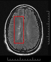
Lesions under MRI
Most MS lesions are isointense to white matter (they appear bright) on T1-weighted MRI, but some are "hypointense" (lower intensity). These are called "black holes" (BH). They appear specially in the supratentorial region of the brain.When BH's appear, around half of them revert in a month. This is considered a sign of remyelination. When they remain, this is regarded as a sign of permanent demyelination and axonal loss. This has been shown on post-mortem autopsies.
Small lesions are invisible under MRI. Therefore clinically assisted diagnostic criteria
McDonald criteria
The McDonald criteria are diagnostic criteria for multiple sclerosis . These criteria are named after neurologist W. Ian McDonald. In April 2001 an international panel in association with the National Multiple Sclerosis Society of America recommended revised diagnostic criteria for MS...
are still required for a more accurate MS diagnosis than MRI alone.
Blood-brain barrier disruption
A healthy blood-brain barrierBlood-brain barrier
The blood–brain barrier is a separation of circulating blood and the brain extracellular fluid in the central nervous system . It occurs along all capillaries and consists of tight junctions around the capillaries that do not exist in normal circulation. Endothelial cells restrict the diffusion...
(BBB) should not allow T-cells to enter the nervous system. BBB disruption is the moment in which T-cells cross the barrier and has always been considered one of the early problems in the MS lesions. For unknown reasons, leaks appear in the BBB during the course of MS.
Recently it has been found that BBB damage happens even in non-enhancing lesions. MS has an important vascular component.
The BBB is built up of endothelial cells lining the blood vessel
Blood vessel
The blood vessels are the part of the circulatory system that transports blood throughout the body. There are three major types of blood vessels: the arteries, which carry the blood away from the heart; the capillaries, which enable the actual exchange of water and chemicals between the blood and...
walls. After its breakdown several problems appear, such as swelling
Edema
Edema or oedema ; both words from the Greek , oídēma "swelling"), formerly known as dropsy or hydropsy, is an abnormal accumulation of fluid beneath the skin or in one or more cavities of the body that produces swelling...
, activation of macrophages, and more activation of cytokines and other protein
Protein
Proteins are biochemical compounds consisting of one or more polypeptides typically folded into a globular or fibrous form, facilitating a biological function. A polypeptide is a single linear polymer chain of amino acids bonded together by peptide bonds between the carboxyl and amino groups of...
s such as matrix metalloproteinase
Matrix metalloproteinase
Matrix metalloproteinases are zinc-dependent endopeptidases; other family members are adamalysins, serralysins, and astacins. The MMPs belong to a larger family of proteases known as the metzincin superfamily....
s which are destructive.
Whatever the demyelination process is, currently it is possible to detect lesions before demyelination, and they show clusters of activated microglia and leukocyte infiltration, together with oligodendrocytes abnormalities
As lesions appear (using MRI) in "Normal-appearing white matter" (NAWM), there is supposed to be the cause that finally triggers the BBB disruption. The damaged white matter is known as "Normal-appearing white matter" (NAWM) and is where lesions appear. These lesions form in NAWM before blood-brain barrier breakdown.
BBB can be broken centripetally or centrifugally, the first form being the most normal. Several possible biochemical disrupters have been proposed. Some hypothesis about how the BBB is compromised revolve around the presence of different compounds in the blood that could interact with the vascular vessels ony in the NAWM areas. The permeability of two cytokine
Cytokine
Cytokines are small cell-signaling protein molecules that are secreted by the glial cells of the nervous system and by numerous cells of the immune system and are a category of signaling molecules used extensively in intercellular communication...
s, IL15 and LPS
Lipopolysaccharide
Lipopolysaccharides , also known as lipoglycans, are large molecules consisting of a lipid and a polysaccharide joined by a covalent bond; they are found in the outer membrane of Gram-negative bacteria, act as endotoxins and elicit strong immune responses in animals.-Functions:LPS is the major...
, could be involved in the BBB breakdown. The BBB breakdown is responsible for monocyte
Monocyte
Monocytes are a type of white blood cell and are part of the innate immune system of vertebrates including all mammals , birds, reptiles, and fish. Monocytes play multiple roles in immune function...
infiltration and inflammation in the brain. Monocyte migration and LFA-1
LFA-1
Lymphocyte function-associated antigen 1, also known as LFA-1 is found on all T-cells and also on B-cells, macrophages and neutrophils and is involved in recruitment to the site of infection. It binds to ICAM-1 on antigen-presenting cells and functions as an adhesion molecule. LFA-1 is the first...
-mediated attachment to brain microvascular endothelia is regulated by SDF-1alpha through Lyn
LYN
Tyrosine-protein kinase Lyn is a protein that in humans is encoded in humans by the LYN gene.Lyn is a member of the Src family of protein tyrosine kinases, which is mainly expressed in hematopoietic cells, in neural tissues liver, and adipose tissue. In various hematopoietic cells, Lyn has emerged...
kinase
Kinase
In chemistry and biochemistry, a kinase is a type of enzyme that transfers phosphate groups from high-energy donor molecules, such as ATP, to specific substrates, a process referred to as phosphorylation. Kinases are part of the larger family of phosphotransferases...
Using iron nanoparticles, involvement of macrophages in the BBB breakdown can be detected. A special role is played by Matrix metalloproteinase
Matrix metalloproteinase
Matrix metalloproteinases are zinc-dependent endopeptidases; other family members are adamalysins, serralysins, and astacins. The MMPs belong to a larger family of proteases known as the metzincin superfamily....
s. These are a group of proteases that increase T-cells permeability of the blood-brain barrier, specially in the case of MMP-9, and are supposed to be related to the mechanism of action of interferons.
Whether BBB dysfunction is the cause or the consequence of MS is still disputed,because activated T-Cells can cross a healthy BBB when they express adhesion proteins. Apart from that, activated T-Cells can cross a healthy BBB when they express adhesion proteins. (Adhesion molecules could also play a role in inflammation) In particular, one of these adhesion proteins involved is ALCAM
ALCAM
CD166 antigen is a protein that in humans is encoded by the ALCAM gene.. It is also called CD166 .-External links:...
(Activated Leukocyte Cell Adhesion Molecule, also called CD166), and is under study as therapeutic target. Other protein also involved is CXCL12, which is found also in brain biopsies of inflammatory elements, and which could be related to the behavior of CXCL13
CXCL13
C-X-C motif chemokine 13 also known as B lymphocyte chemoattractant is a protein that in humans is encoded by the CXCL13 gene.- Function :CXCL13 is a small cytokine belonging to the CXC chemokine family...
under methylprednisolone
Methylprednisolone
Methylprednisolone is a synthetic glucocorticoid or corticosteroid drug. It is marketed in the USA and Canada under the brand names Medrol and Solu-Medrol. It is also available as a generic drug....
therapy. Some molecular biochemical models for relapses have been proposed.
Normally, gadolinium enhancement is used to show BBB disruption on MRIs. Abnormal tight junctions are present in both SPMS and PPMS. They appear in active white matter lesions, and gray matter in SPMS. They persist in inactive lesions, particularly in PPMS.
A deficiency of uric acid
Uric acid
Uric acid is a heterocyclic compound of carbon, nitrogen, oxygen, and hydrogen with the formula C5H4N4O3. It forms ions and salts known as urates and acid urates such as ammonium acid urate. Uric acid is created when the body breaks down purine nucleotides. High blood concentrations of uric acid...
has been implicated in this process. Uric acid added in physiological concentrations (i.e. achieving normal concentrations) is therapeutic in MS by preventing the breakdown of the blood brain barrier through inactivation of peroxynitrite
Peroxynitrite
Peroxynitrite is the anion with the formula ONOO−. It is an unstable structural isomer of nitrate, NO3−, which has the same formula but a different structure. Although peroxynitrous acid is highly reactive, its conjugate base peroxynitrite is stable in basic solution...
. The low level of uric acid found in MS victims is manifestedly causative rather than a consequence of tissue damage in the white matter lesions, but not in the grey matter lesions. Besides, uric acid levels are lower during relapses.
Special MRI methods
Before BBB disruption, brain tissues present normal aspect under normal MRI (Normal appearing white matterWhite matter
White matter is one of the two components of the central nervous system and consists mostly of myelinated axons. White matter tissue of the freshly cut brain appears pinkish white to the naked eye because myelin is composed largely of lipid tissue veined with capillaries. Its white color is due to...
, NAWM and normal appearing grey matter
Grey matter
Grey matter is a major component of the central nervous system, consisting of neuronal cell bodies, neuropil , glial cells and capillaries. Grey matter contains neural cell bodies, in contrast to white matter, which does not and mostly contains myelinated axon tracts...
, NAGM), but show several abnormalities under special MRI technologies:
Magnetization transfer
Magnetization transfer
Magnetization transfer , as commonly used in biomedical MRI, refers to the transfer of longitudinal magnetization from the hydrogen nuclei of water that have restricted motion to the hydrogen nuclei of water that moves with many degrees of freedom...
multi-echo T(2) relaxation
Relaxation (NMR)
In nuclear magnetic resonance spectroscopy and magnetic resonance imaging the term relaxation describes several processes by which nuclear magnetization prepared in a non-equilibrium state return to the equilibrium distribution. In other words, relaxation describes how fast spins "forget" the...
. Subjects with Long-T(2) lesions
Relaxation (NMR)
In nuclear magnetic resonance spectroscopy and magnetic resonance imaging the term relaxation describes several processes by which nuclear magnetization prepared in a non-equilibrium state return to the equilibrium distribution. In other words, relaxation describes how fast spins "forget" the...
had a significantly longer disease duration than subjects without this lesion subtype. It has been found that grey matter injury correlates with disability and that there is high oxidative stress in lesions, even in the old ones.
Diffusion tensor MRI
Diffusion MRI
Diffusion MRI is a magnetic resonance imaging method that produces in vivo images of biological tissues weighted with the local microstructural characteristics of water diffusion, which is capable of showing connections between brain regions...
or Magnetic Transfer
Magnetization transfer
Magnetization transfer , as commonly used in biomedical MRI, refers to the transfer of longitudinal magnetization from the hydrogen nuclei of water that have restricted motion to the hydrogen nuclei of water that moves with many degrees of freedom...
MRI are two options to enhance MRI-hidden abnormalities discovery. This is currently an active field of research with no definitive results, but it seems that these two technologies are complementary.
Other methods of MRI allow us to get a better insight of the lesions structure. Recently MP-RAGE MRI has shown better results than PSIR and DIR for gray matter lesions. Susceptibility weighted imaging
Susceptibility weighted imaging
Susceptibility weighted imaging , originally called BOLD venographic imaging, uses a type of contrast in magnetic resonance imaging different from traditional spin density, T1, or T2 imaging. SWI uses a fully flow compensated, long echo, gradient echo scan to acquire images...
(SWI-MRI) has shown iron (hemosiderin
Hemosiderin
thumb|Hemosiderin image of a kidney viewed under a microscope. The brown areas represent hemosiderinHemosiderin or haemosiderin is an iron-storage complex. It is always found within cells and appears to be a complex of ferritin, denatured ferritin and other material...
) deposition in lesions, and helps to detect otherwise invisible lesions.
Normal appearing brain tissues
Using several texture analysis technologies, it is possible to classify white matter areas into three categories: normal, normal-appearing and lesions. Currently is possible to detect lesions before they present demyelination, and they are called pre-active lesions. A fourth area called DAWM (diffusely abnormal white matter) has recently been proposed and can help to differentiate PPMS and SPMS. Abundant extracellular myelin in the meninges of patients with multiple sclerosis has been foundBrain tissues with MRI-hidden problems are usually named Normal Appearing. Exploring the normal-appearing corpus callosum
Corpus callosum
The corpus callosum , also known as the colossal commissure, is a wide, flat bundle of neural fibers beneath the cortex in the eutherian brain at the longitudinal fissure. It connects the left and right cerebral hemispheres and facilitates interhemispheric communication...
has been found a possible primary hypoperfusion, according with other findings in this same direction. Also iron (in hemosiderin
Hemosiderin
thumb|Hemosiderin image of a kidney viewed under a microscope. The brown areas represent hemosiderinHemosiderin or haemosiderin is an iron-storage complex. It is always found within cells and appears to be a complex of ferritin, denatured ferritin and other material...
deposits and as well as in ferritin-like structures inside the macrophage) accumulation has been reported
Several findings in these areas have been shown. Post-mortem studies over NAWM and NAGM areas (Normal appearing White and Gray Matters) show several biochemical alterations, like increased protein carbonylation
Carbonylation
Carbonylation refers to reactions that introduce carbon monoxide into organic and inorganic substrates. Carbon monoxide is abundantly available and conveniently reactive, so it is widely used as a reactant in industrial chemistry.-Organic chemistry:...
and high levels of Glial fibrillary acidic protein
Glial fibrillary acidic protein
Glial fibrillary acidic protein is an intermediate filament protein that was thought to be specific for astrocytes in the central nervous system . Later, it was shown that GFAP is also expressed by other cell types in CNS, including ependymal cells...
(GFAP), which in NAGM areas comes together with higher than normal concentration of protein carbonyl
Carbonyl
In organic chemistry, a carbonyl group is a functional group composed of a carbon atom double-bonded to an oxygen atom: C=O. It is common to several classes of organic compounds, as part of many larger functional groups....
s, suggesting reduced levels of antioxidants and the presence of small lesions. The amount of interneuronal Parvalbumin
Parvalbumin
Parvalbumin is a calcium-binding albumin protein with low molecular weight .It has three EF hand motifs and is structurally related to calmodulin and troponin C...
is lower than normal in brain's motor cortex areas, and oxidative injury of oligodendrocytes and neurons could be associated with active demyelination and axonal injury.
Normal appearing White Matter
The white matter with hidden but MRI-visible damage is known as "Normal-appearing white matter" (NAWM) and is where lesions appear.BBB disruption takes place on NAWM areas. This can be read in different ways. Maybe some hidden changes in White Matter structure trigger the BBB disruption, or maybe the same process that creates the NAWM areas disrupts the BBB after some time.
Pre-active lesions are lesions in an early stage of development. They resolve sometimes without further damage, and not always develop into demyelinating lesions. They present clusters of activated microglia in otherwise normal-appearing white matter.
Oligodendrocyte
Oligodendrocyte
Oligodendrocytes , or oligodendroglia , are a type of brain cell. They are a variety of neuroglia. Their main function is the insulation of axons in the central nervous system of some vertebrates...
abnormalities appear to be crucially involved. The earliest change reported in the lesions examined is widespread oligodendrocyte apoptosis in which T cells, macrophages, activated microglia, reactive astrocytes, and neurons appear normal. This observation points to some change in the local environment (NAWM) to which oligodendrocytes are especially susceptible and which triggers a form of apoptosis.
Water diffusivity is higher in all NAWM regions, deep gray matter regions, and some cortical gray matter region of MS patients than normal controls.
Citrullination
Citrullination
Citrullination or deimination is the term used for the post-translational modification of the amino acid arginine in a protein into the amino acid citrulline. This reaction, shown below, is performed by enzymes called peptidylarginine deiminases...
appears in SPMS. It seems that a defect of sphingolipid
Sphingolipid
Sphingolipids are a class of lipids containing a backbone of sphingoid bases, a set of aliphatic amino alcohols that includes sphingosine. They were discovered in brain extracts in the 1870s and were named for the mythological Sphinx because of their enigmatic nature. These compounds play...
metabolism modifies the properties of normal appearing white matter. Related to these, peptidylarginine deiminase 2
Protein-arginine deiminase
In enzymology, a protein-arginine deiminase is an enzyme that catalyzes a form of post translational modification called arginine de-imination or citrullination:...
is increased in patients with MS, and is related to arginine de-imination.
NAWM shows a decreased perfusion
Perfusion
In physiology, perfusion is the process of nutritive delivery of arterial blood to a capillary bed in the biological tissue. The word is derived from the French verb "perfuser" meaning to "pour over or through."...
which does not appear to be secondary to axonal loss. The reduced perfusion of the NAWM in MS might be caused by a widespread astrocyte
Astrocyte
Astrocytes , also known collectively as astroglia, are characteristic star-shaped glial cells in the brain and spinal cord...
dysfunction, possibly related to a deficiency in astrocytic beta(2)-adrenergic receptors and a reduced formation of cAMP
Cyclic adenosine monophosphate
Cyclic adenosine monophosphate is a second messenger important in many biological processes...
, resulting in a reduced uptake of K(+) at the nodes of Ranvier
Nodes of Ranvier
Myelin sheath gaps or nodes of Ranvier are the gaps formed between the myelin sheaths generated by different cells. A myelin sheath is a many-layered coating, largely composed of a fatty substance called myelin, that wraps around the axon of a neuron and very efficiently insulates it...
and a reduced release of K(+) in the perivascular spaces. This would be consistent again with cases of Chronic cerebrospinal venous insufficiency
Chronic cerebrospinal venous insufficiency
Chronic cerebrospinal venous insufficiency is a term developed by Italian researcher Paolo Zamboni in 2008 to describe compromised flow of blood in the veins draining the central nervous system...
.
White matter lesions appear in NAWM areas, and their behavior can be predicted by MRI parameters as MTR (magnetization transfer ratio). This MTR parameter is related to axonal density.
It also seems that myelin basic protein (MBP) from multiple sclerosis (MS) patients contains lower levels of phosphorylation at Thr97 than normal individuals.
Gray matter damage. Normal Appearing Gray Matter
Gray matter tissue damage dominates the pathological process as MS progresses, and underlies neurological disability. Imaging correlates of gray matter atrophy indicate that mechanisms differ in RRMS and SPMS. Epstein-Barr virusEpstein-Barr virus
The Epstein–Barr virus , also called human herpesvirus 4 , is a virus of the herpes family and is one of the most common viruses in humans. It is best known as the cause of infectious mononucleosis...
could be involved, but is not likely. Involvement of the deep gray matter (DGM), suggested by magnetic resonance imaging, is confirmed, and most DGM lesions involve both GM and white matter. Inflammation in DGM lesions is intermediate between the destructive inflammation of white matter lesions and the minimal inflammation of cortical lesions.
Iron depositions appear in deep gray matter by magnetic field correlation MRI
Diffusely abnormal white matter
Other active area of study is the Diffusely abnormal white matter (DAWM). It seems to be a reduction of myelin phospholipids that correlates with a reduction of the myelin water fraction. The DAWM consisted of extensive axonal loss, decreased myelin density, and chronic fibrillary gliosis, all of which were substantially abnormal compared with normal-appearing WM and significantly different from focal WM lesion pathology. Changes in the vasculature take place not only in focal lesions but also in DAWM as detected by postmortem MRIOrigin of the normal-appearing tissues
The cause why the normal appearing areas appear in the brain is unknown. Historically, several theories about how this happens has been presented.Old blood flow theories
Venous pathology has been associated with MS for more than a century. Pathologist Georg Eduard Rindfleisch noted in 1863 that the inflammation-associated lesions were distributed around veins. Some other authors like Tracy PutnamTracy Putnam
Tracy Jackson Putnam was the co-discoverer, together with H. Houston Merritt, of Dilantin for controlling epilepsy.He graduated from Harvard College in 1915. Later from Harvard medical school in 1920. He worked by the Boston City Hospital and in the New York Neurological Institute at Columbia...
pointed to venous obstructions.
Some authors like Franz Schelling proposed a mechanical damage procedure based on violent blood reflux. Later the focus moved to softer hemodynamic abnormalities, which were shown that precede changes in sub-cortical gray matter and in substantia nigra. However, such reports of a "hemodynamic cause of MS" are not universal, and possibly not even common. At this time the evidence is largely anecdotal and some MS patients have no blood flow issues. Possibly vascular problems may be an aggravating factor, like many others in MS. Indeed the research, by demonstrating patients with no hemodynamic problems actually prove that this is not the only cause of MS.
Endothelium theories
Other theories point to a possible primary endothelial disfunction. The importance of vascular misbehaviour in MS pathogenesis has also been independently confirmed by seven-tesla MRI. It is reported that a number of studies have provided evidence of vascular occlusion in MS, which suggest the possibility of a primary vascular injury in MS lesions or at least that they are occasionally correlated.Some morphologically special medullar lesions (wedge-shaped) have also been linked to venous insufficiency.
CCSVI
The term "chronic cerebrospinal venous insufficiency" was coined in 2008 by Paolo Zamboni, who described it in patients with multiple sclerosis. Instead of intracranial venous problems he described extracranial blockages, and he stated that the location of those obstructions seemed to influence the clinical course of the disease. According to Zamboni, CCSVI had a high sensitivity and specificitySensitivity and specificity
Sensitivity and specificity are statistical measures of the performance of a binary classification test, also known in statistics as classification function. Sensitivity measures the proportion of actual positives which are correctly identified as such Sensitivity and specificity are statistical...
differentiating healthy individuals from those with multiple sclerosis
Multiple sclerosis
Multiple sclerosis is an inflammatory disease in which the fatty myelin sheaths around the axons of the brain and spinal cord are damaged, leading to demyelination and scarring as well as a broad spectrum of signs and symptoms...
. Zamboni's results were criticized as some of his studies were not blinded and they need to be verified by further studies. the theory is considered at least defensible
A more detailed evidence of a correlation between the place and type of venous malformations imaged and the reported symptoms of multiple sclerosis in the same patients was published in 2010.
Haemodynamic problems have been found in the blood flow of MS patients using Doppler, initially using transcranial color-coded duplex
Transcranial doppler
Transcranial Doppler is a test that measures the velocity of blood flow through the brain's blood vessels. Used to help in the diagnosis of emboli, stenosis, vasospasm from a subarachnoid hemorrhage , and other problems, this relatively quick and inexpensive test is growing in popularity in the...
sonography (TCCS), pointing to a relationship with a vascular disease called chronic cerebro-spinal venous insufficiency (CCSVI). In 2010 there were conflicting results when evaluating the relationship between MS and CCSVI. but is important to note that positives have appeared among the blinded studies.
CSF flow theories
Other theories focus in the possible role of cerebrospinal fluidCerebrospinal fluid
Cerebrospinal fluid , Liquor cerebrospinalis, is a clear, colorless, bodily fluid, that occupies the subarachnoid space and the ventricular system around and inside the brain and spinal cord...
flow impairment. This theory could be partially consistent with the previous one
Currently a small trial with 8 participants has been performed
Molecular biomarkers
Diagnosis of MS has always been made by clinical examination, supported by MRI or CSF tests. According with the autoimmune hypothesis, researchers expect to find biomarkers able to yield a better diagnosis. As of 2009 no biomarker with perfect correlation has been found, but some of them have shown a special behavior.In blood
Blood serum of MS patients shows abnormalities. EndothelinEndothelin
Endothelins are proteins that constrict blood vessels and raise blood pressure. They are normally kept in balance by other mechanisms, but when they are over-expressed, they contribute to high blood pressure and heart disease....
-1 shows maybe the most striking discordance between patients and controls, being a 224% higher in patients than controls.
Creatine
Creatine
Creatine is a nitrogenous organic acid that occurs naturally in vertebrates and helps to supply energy to all cells in the body, primarily muscle. This is achieved by increasing the formation of Adenosine triphosphate...
and Uric acid
Uric acid
Uric acid is a heterocyclic compound of carbon, nitrogen, oxygen, and hydrogen with the formula C5H4N4O3. It forms ions and salts known as urates and acid urates such as ammonium acid urate. Uric acid is created when the body breaks down purine nucleotides. High blood concentrations of uric acid...
levels are lower than normal, at least in women. Ex vivo CD4(+) T cells isolated from the circulation show a wrong TIM
TIM
Tim may refer to:Names*Timothy or Tim, for short*Tim , the Australian moviePeople*Tim, the Oldenburg Baby, abortion survivor*Tim , popular K-pop solo singer*Tiny Tim , American singer, ukulele player, and musical archivist...
-3 (Immunoregulation) behavior, and relapses are associated with CD8(+) T Cells
Cytotoxic T cell
A cytotoxic T cell belongs to a sub-group of T lymphocytes that are capable of inducing the death of infected somatic or tumor cells; they kill cells that are infected with viruses , or are otherwise damaged or...
. There is a set of differentially expressed genes between MS and healthy subjects in peripheral blood T cells from clinically active MS patients. There are also differences between acute relapses and complete remissions. Platelet
Platelet
Platelets, or thrombocytes , are small,irregularly shaped clear cell fragments , 2–3 µm in diameter, which are derived from fragmentation of precursor megakaryocytes. The average lifespan of a platelet is normally just 5 to 9 days...
s are known to have abnormal high levels.
MS patients are also known to be CD46
CD46
CD46 complement regulatory protein also known as CD46 and Membrane Cofactor Protein is a protein which in humans is encoded by the CD46 gene. CD46 is an inhibitory complement receptor.- Gene :...
defective, and this leads to Interleukin
Interleukin
Interleukins are a group of cytokines that were first seen to be expressed by white blood cells . The term interleukin derives from "as a means of communication", and "deriving from the fact that many of these proteins are produced by leukocytes and act on leukocytes"...
-10 (IL-10
Interleukin 10
Interleukin-10 , also known as human cytokine synthesis inhibitory factor , is an anti-inflammatory cytokine. In humans IL-10 is encoded by the IL10 gene....
) deficiency, being this involved in the inflammatory reactions. Levels of IL-2, IL-10, and GM-CSF are lower in MS females than normal. IL6 is higher instead. These findings do not apply to men. This IL-10 interleukin could be related to the mechanism of action of methylprednisolone
Methylprednisolone
Methylprednisolone is a synthetic glucocorticoid or corticosteroid drug. It is marketed in the USA and Canada under the brand names Medrol and Solu-Medrol. It is also available as a generic drug....
, together with CCL2
CCL2
For the ICAO airport code see Candle Lake Airpark, for the diradical compound see Dichlorocarbene.Chemokine ligand 2 also known as monocyte chemotactic protein-1 or small inducible cytokine A2 is a protein that in humans is encoded by the CCL2 gene. CCL2 is a small cytokine belonging to the CC...
. Interleukin IL-12
Interleukin 12
Interleukin 12 is an interleukin that is naturally produced by dendritic cells, macrophages and human B-lymphoblastoid cells in response to antigenic stimulation.-Gene and structure:...
is also known to be associated with relapses, but this is unlikely to be related to the response to steroids
Kallikrein
Kallikrein
Kallikreins are a subgroup of serine proteases, enzymes capable of cleaving peptide bonds in proteins. In humans, plasma kallikrein has no known homologue, while tissue kallikrein-related peptidases encode a family of fifteen closely related serine proteases...
s are found in serum and are associated with secondary progressive stage. Related to this, it has been found that B1-receptors, part of the kallikrein-kinin-system
Kinin-kallikrein system
The kinin-kallikrein system or simply kinin system is a poorly understood system of blood proteins that plays a role in inflammation, blood pressure control, coagulation and pain...
, are involved in the BBB breakdown
There is evidence of Apoptosis
Apoptosis
Apoptosis is the process of programmed cell death that may occur in multicellular organisms. Biochemical events lead to characteristic cell changes and death. These changes include blebbing, cell shrinkage, nuclear fragmentation, chromatin condensation, and chromosomal DNA fragmentation...
-related molecules in blood and they are related to disease activity. B cells in CSF appear, and they correlate with early brain inflammation.
There is also an overexpression of IgG
Immunoglobulin G
Immunoglobulin G are antibody molecules. Each IgG is composed of four peptide chains — two heavy chains γ and two light chains. Each IgG has two antigen binding sites. Other immunoglobulins may be described in terms of polymers with the IgG structure considered the monomer.IgG constitutes 75%...
-free kappa light chain protein
Immunoglobulin light chain
]The immunoglobulin light chain is the small polypeptide subunit of an antibody .A typical antibody is composed of two immunoglobulin heavy chains and two Ig light chains.-In humans:...
in both CIS and RR-MS patients, compared with control subjects, together with an increased expression of an isoforms of apolipoprotein E
Apolipoprotein E
Apolipoprotein E is a class of apolipoprotein found in the chylomicron and IDLs that binds to a specific receptor on liver cells and peripheral cells. It is essential for the normal catabolism of triglyceride-rich lipoprotein constituents.-Function:...
in RR-MS. Expression of some specific proteins in circulating CD4+ T cells is a risk factor for conversion from CIS to clinically defined multiple sclerosis.
Recently, unique autoantibody patterns that distinguish RRMS, secondary progressive (SPMS), and primary progressive (PPMS) have been found, based on up- and down-regulation
Regulation of gene expression
Gene modulation redirects here. For information on therapeutic regulation of gene expression, see therapeutic gene modulation.Regulation of gene expression includes the processes that cells and viruses use to regulate the way that the information in genes is turned into gene products...
of CNS antigens, tested by microarray
Antibody microarray
An antibody microarray is a specific form of protein microarrays, a collection of capture antibodies are spotted and fixed on a solid surface, such as glass, plastic and silicon chip for the purpose of detecting antigens...
s. In particular, RRMS is characterized by autoantibodies to heat shock proteins that were not observed in PPMS or SPMS. These antibodies patterns can be used to monitor disease progression.
In blood vessels tissue
Endothelial disfunction has been reported in MS and could be used as biomarker via biopsia. Blood circulation is slower in MS patients and can be meassured using contrast or by MRIIn Cerebrospinal Fluid
It has been known for quite some time that glutamate is present at higher levels in CSF during relapses compared to healthy subjects and to MS patients before relapses. Also a specific MS protein has been found in CSF, chromogranin AChromogranin A
Chromogranin A or parathyroid secretory protein 1 is a member of the granin family of neuroendocrine secretory proteins, i.e. it is located in secretory vesicles of neurons and endocrine cells...
, possibly related to axonal degeneration. It appears together with clusterin and complement C3, markers of complement-mediated inflammatory reactions. Also Fibroblast growth factor
Fibroblast growth factor
Fibroblast growth factors, or FGFs, are a family of growth factors involved in angiogenesis, wound healing, and embryonic development. The FGFs are heparin-binding proteins and interactions with cell-surface associated heparan sulfate proteoglycans have been shown to be essential for FGF signal...
-2 appear higher at CSF.
CSF also shows oligoclonal bands (OCB) in the majority (around 95%) of the patients. Several studies have reported differences between patients with and without OCB with regard to clinical parameters such as age, gender, disease duration, clinical severity and several MRI characteristics, together with a varying lesion load.
Varicella-zoster virus particles have been found in CSF of patients during relapses, but this particles are virtually absent during remissions. Plasma Cells in the cerebrospinal fluid of MS patients could also be used for diagnosis, because they have been found to produce myelin-specific antibodies. As of 2011, a recently discovered myelin protein TPPP/p25
TPPP
Tubulin polymerization-promoting protein is a protein that in humans is encoded by the TPPP gene.This protein has been linked to multiple sclerosis myelin lesions and CSF abnormalities in multiple sclerosis patients....
, has been found in CSF of MS patients
Subgroups by molecular biomarkers
Differences have been found between the proteines expressed by patients and healthy subjects, and between attacks and remissions. Using DNA microarrayDNA microarray
A DNA microarray is a collection of microscopic DNA spots attached to a solid surface. Scientists use DNA microarrays to measure the expression levels of large numbers of genes simultaneously or to genotype multiple regions of a genome...
technology groups of molecular biomarkers can be established. For example, it is known that Anti-lipid oligoclonal IgM bands (OCMB) distinguish MS patients with early aggressive course and that these patients show a favourable response to immunomodulatory treatment.
It seems that Fas and MIF are candidate biomarkers of progressive neurodegeneration. Upregulated levels of sFas (soluble form of Fas
Fas receptor
The FAS receptor also known as apoptosis antigen 1 , cluster of differentiation 95 or tumor necrosis factor receptor superfamily member 6 is a protein that in humans is encoded by the TNFRSF6 gene....
molecule) were found in MS patients with hypotense lesions with progressive neurodegeneration, and also levels of MIF appeared to be higher in progressive than in non-progressing patients. Serum TNF-α and CCL2 seem to reflect the presence of inflammatory responses in primary progressive MS.
Heterogeneity of the disease
Multiple sclerosis has been reported to be heterogeneus in its behavior, in its underlying mechanisms and recently also in its response to medication.Four different damage patterns have been identified by her team in the scars of the brain tissue. Understanding lesion patterns can provide information about differences in disease between individuals and enable doctors to make more accurate treatment decisions. According to one of the researchers involved in the original research "Two patterns (I and II) showed close similarities to T-cell-mediated or T-cell plus antibody-mediated autoimmune encephalomyelitis, respectively. The other patterns (III and IV) were highly suggestive of a primary oligodendrocyte dystrophy, reminiscent of virus- or toxin-induced demyelination rather than autoimmunity."
Also known as Lassmann patterns, it is believed that they may correlate with differences in disease type and prognosis, and perhaps with different responses to treatment. This report suggests that there may be several types of MS with different immune-related causes, and that MS may be a family of several diseases. The four identified patterns are http://www.neurologyreviews.com/aug02/nr_aug02_mslesion.html:
Pattern I : The scar presents T-cells and macrophages around blood vessels, with preservation of oligodendrocyte
Oligodendrocyte
Oligodendrocytes , or oligodendroglia , are a type of brain cell. They are a variety of neuroglia. Their main function is the insulation of axons in the central nervous system of some vertebrates...
s, but no signs of complement system
Complement system
The complement system helps or “complements” the ability of antibodies and phagocytic cells to clear pathogens from an organism. It is part of the immune system called the innate immune system that is not adaptable and does not change over the course of an individual's lifetime...
activation.
Pattern II : The scar presents T-cells and macrophages around blood vessels, with preservation of oligodendrocytes, as before, but also signs of complement system
Complement system
The complement system helps or “complements” the ability of antibodies and phagocytic cells to clear pathogens from an organism. It is part of the immune system called the innate immune system that is not adaptable and does not change over the course of an individual's lifetime...
activation can be found. Though this pattern could be considered similar to damage seen in NMO, some authors report no AQP4 damage in pattern II lesions
Pattern III : The scars are diffuse with inflammation, distal oligodendrogliopathy and microglia
Microglia
Microglia are a type of glial cell that are the resident macrophages of the brain and spinal cord, and thus act as the first and main form of active immune defense in the central nervous system . Microglia constitute 20% of the total glial cell population within the brain...
l activation. There is also loss of myelin-associated glycoprotein
Myelin-associated glycoprotein
Myelin-associated glycoprotein is a cell membrane glycoprotein that is a member of the SIGLEC family of proteins and is a functional ligand of the NOGO-66 receptor, NgR. MAG is believed to be involved in myelination during nerve regeneration....
(MAG). The scars do not surround the blood vessels, and in fact, a rim of preserved myelin appears around the vessels. There is evidence of partial remyelinization and oligodendrocyte apoptosis. For some researchers this pattern is an early stage of the evolution of the others.
Pattern IV : The scar presents sharp borders and oligodendrocyte
Oligodendrocyte
Oligodendrocytes , or oligodendroglia , are a type of brain cell. They are a variety of neuroglia. Their main function is the insulation of axons in the central nervous system of some vertebrates...
degeneration, with a rim of normal appearing white matter
White matter
White matter is one of the two components of the central nervous system and consists mostly of myelinated axons. White matter tissue of the freshly cut brain appears pinkish white to the naked eye because myelin is composed largely of lipid tissue veined with capillaries. Its white color is due to...
. There is a lack of oligodendrocytes in the center of the scar. There is no complement activation or MAG loss.
The meaning of this fact is controversial. For some investigation teams it means that MS is a heterogeneous disease. Others maintain that the shape of the scars can change with time from one type to other and this could be a marker of the disease evolution. Anyway, the heterogeneity could be true only for the early stage of the disease. Some lesions present mitocondrial defects that could distinguish types of lesions. Currently antibodies to lipids and peptides in sera, detected by microarrays, can be used as markers of the pathological subtype given by brain biopsy.
Several correlations have been studied:
- With clinical courses: No definitive relationship between these patterns and the clinical subtypes has been established by now, but some relations have been established. All the cases with PPMS (primary progressive) had pattern IV (oligodendrocyte degeneration) in the original study and nobody with RRMS was found with this pattern. Balo concentric sclerosisBalo concentric sclerosisBalo concentric sclerosis is one of the borderline forms of multiple sclerosis.Balo concentric sclerosis is a demyelinating disease similar to standard multiple sclerosis, but with the particularity that the demyelinated tissues form concentric layers...
lesions have been classified as pattern III (distal oligodendrogliopathy). Neuromyelitis optica was associated with pattern II (complement mediated demyelination), though they show a perivascular distribution, at difference from MS pattern II lesions.
- With MRI and MRS findings: The researchers are attempting this with magnetic resonance images to confirm their initial findings of different patterns of immune pathology and any evidence of possible disease “sub-types” of underlying pathologies. It is possible that such “sub-types” of MS may evolve differently over time and may respond differently to the same therapies. Ultimately investigators could identify which individuals would do best with which treatments. It seems that Pulsed magnetization transfer imaging, diffusion Tensor MRI, and VCAM-1 enhanced MRI could be able to show the pathological differences of these patterns. Together with MRI, magnetic resonance spectroscopy will allow in the future to see the biochemical composition of the lesions.
- With CSF findings: Teams in Oxford and Germany, found correlation with CSF and progression in November 2001, and hypotheses have been made suggesting correlation between CSF findings and pathophysiological patterns. In particular, B-cell to monocyte ratio looks promising. The anti-MOG antibody has been investigated but no utility as biomarker has been found, though this is disputed. High levels of anti-nuclear antibodiesAnti-nuclear antibodyAnti-nuclear antibodies are autoantibodies directed against contents of the cell nucleus....
are found normally in patients with MS. Antibodies against Neurofascin–186 could be involved in a subtype of MS
- With responses to therapy: It is known that 30% of MS patients are non-responsive to Beta interferon. The heterogeneous response to therapy can support the idea of hetherogeneous aetiology. It has also been shown that IFN receptors and interleukinInterleukinInterleukins are a group of cytokines that were first seen to be expressed by white blood cells . The term interleukin derives from "as a means of communication", and "deriving from the fact that many of these proteins are produced by leukocytes and act on leukocytes"...
s in blood serum predicts response to IFN therapy, and interleukins IL12/IL10 ratio has been proposed as marker of clinical course. Besides:
-
- Pattern II lesions patients are responsive to plasmapheresisPlasmapheresisPlasmapheresis is the removal, treatment, and return of blood plasma from blood circulation. It is thus an extracorporeal therapy...
, while others are not. - The subtype associated with macrophage activation, T cell infiltration and expression of inflammatory mediator molecules may be most likely responsive to immunomodulation with interferon-beta or glatiramer acetate.
- People non-responsive to interferons are the most responsive to Copaxone http://www.msworld.org/Resource_Center/news/n03switch.htm
- In general, people non-responsive to a treatment is more responsive to other, and changing therapy can be effective.
- There are genetic differences between responders and not responders. Though the article points to heterogeneous metabolic reactions to interferons instead of disease heterogeneity, it has been shown that most genetic differences are not related to interferon behavior
- Pattern II lesions patients are responsive to plasmapheresis
External links
- The lesion project page
- MRI and CT of Multiple Sclerosis MedPix Image Database