

Astrocyte
Encyclopedia
Astrocytes also known collectively as astroglia, are characteristic star-shaped glial cells
in the brain
and spinal cord
. They perform many functions, including biochemical support of endothelial cells that form the blood–brain barrier, provision of nutrients to the nervous tissue, maintenance of extracellular ion balance, and a role in the repair and scarring process of the brain and spinal cord following traumatic injuries.
Research since the mid-1990s has shown that astrocytes propagate intercellular Ca2+
waves over long distances in response to stimulation, and, similar to neurons, release transmitters (called gliotransmitter
s) in a Ca2+-dependent manner. Data suggest that astrocytes also signal to neurons through Ca2+-dependent release of glutamate. Such discoveries have made astrocytes an important area of research within the field of neuroscience
.
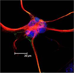
. They are also known as astrocytic glial cells. Star-shaped, their many processes envelope synapses made by neuron
s. Astrocytes are classically identified using histological analysis; many of these cells express the intermediate filament glial fibrillary acidic protein
(GFAP). Three forms of astrocytes exist in the CNS, fibrous, protoplasmic, and radial. The fibrous glia are usually located within white matter, have relatively few organelles, and exhibit long unbranched cellular processes. This type often has "vascular feet" that physically connect the cells to the outside of capillary wall when they are in close proximity to them. The protoplasmic glia are found in grey matter tissue, possess a larger quantity of organelles, and exhibit short and highly branched cellular processes. The radial glia are disposed in a plane perpendicular to axis of ventricles. One of their processes about the pia mater, while the other is deeply buried in gray matter. Radial glia are mostly present during development, playing a role in neuron migration. Mueller cells of retina and Bergmann glia cells of cerebellar cortex represent an exception, being present still during adulthood. When in proximity to the pia mater
, all three forms of astrocytes send out process to form the pia-glial membrane.
.
One study done in Shanghai had two types of hippocampal neuronal cultures: In one culture, the neuron was grown from a layer of astrocytes and the other culture was not in contact with any astrocytes, but they were instead fed a Glial Conditioned Medium (GCM), which inhibits the rapid growth of cultured astrocytes in the brains of rats in most cases.
In their results they were able to see that astrocytes had a direct role in LTP with the mixed culture (which is the culture that was grown from a layer of astrocytes) but not in GCM cultures 36
.
Recent studies have shown that astrocytes play an important function in the regulation of neural stem cell
s. Research from the Schepens Eye Research Institute at Harvard shows the human brain to abound in neural stem cells, which are kept in a dormant state by chemical signals (ephrin-A2 and ephrin-A3) from the astrocytes. The astrocytes are able to activate the stem cells to transform into working neurons by dampening the release of ephrin-A2 and ephrin-A3.
Furthermore, studies are underway to determine whether astroglia play an instrumental role in depression, based on the link between diabetes and depression. Altered CNS glucose
metabolism is seen in both these conditions, and the astroglial cells are the only cells with insulin
receptors in the brain.
In a study published in a 2011 issue of Nature Biotechnology and reported in lay science article, a group of researchers from the University of Wisconsin reports that it has been able to direct embryonic and induced human stem cells to become astrocytes.
s, creating an electrically coupled (functional) syncytium
.
An increase in intracellular calcium concentration can propagate outwards through this functional syncytium. Mechanisms of calcium wave propagation include diffusion of calcium ions and IP3 through gap junctions and extracellular ATP signalling. Calcium elevations are the primary known axis of activation in astrocytes, and are necessary and sufficient for some types of astrocytic glutamate release.
 Bergmann glia, a type of glia also known as radial epithelial cells (as named by Camillo Golgi
) or Golgi epithelial cells (GCEs; not to be mixed up with Golgi cells), are astrocytes in the cerebellum
that have their cell bodies in the Purkinje cell
layer and processes that extend into the molecular layer, terminating with bulbous endfeet at the pial
surface. Bergmann glia express high densities of glutamate transporter
s that limit diffusion of the neurotransmitter
glutamate during its release from synaptic terminals. Besides their role in early development of the cerebellum, Bergmann glia are also required for the pruning or addition of synapse
s.
s are primary intracranial tumor
s derived from astrocytes cells of the brain. It is also possible that glial progenitors or neural stem cells give rise to astrocytomas.
Astrocytomas are brain tumors that develop from astrocytes. They may occur in many parts of the brain and sometimes in the spinal cord. They can occur at any age but they primarily occur in males. Astrocytomas are divided into two categories: Low grade (I and II) and High Grade (III and IV). Low grade tumors are more common in children and high grade tumors are more common in adults[32].
Pilocytic Astrocytoma are Grade I tumors. They are considered benign and slow growing tumors. Pilocytic Astrocytomas frequently have cystic portions filled with fluid and a nodule, which is the solid portion. Most are located in the cerebellum. Therefore, most symptoms are related to balance or coordination difficulties[32]. They also occur more frequently in children and teens[33].
Grade II Tumors grow relatively slow but invade surrounding healthy tissue. Usually considered benign but can grow into malignant tumors. Other names that are used are Fibrillary or Protoplasmic astrocytomas. They are prevalent in younger people who are often present with seizures[33].
Anaplastic astrocytoma is classified as grade III and are malignant tumors. They grow more rapidly than lower grade tumors and tend to invade nearby healthy tissue. Anaplastic astrocytomas recur more frequently than lower grade tumors because of their tendency to spread into surrounding tissue makes them difficulty to completely remove surgically[32].
Glioblastoma Multiforme is also a malignant tumor and classified as a grade IV. Glioblastomas can contain more than one cell type (i.e., astrocytes, oligondroctyes). Also, while one cell type may die off in response to a particular treatment, the other cell types may continue to multiply. Gliobastomas are the most invasive type of glial tumors. Grows rapidly and spreads to nearby tissue. Approximately 50% of astrocytomas are glioblastomas and are very difficult to treat[32].
, activated astrocytes have the ability to respond to almost all neurotransmitters (Haydon, 2001) and, upon activation, release a multitude of neuroactive molecules such as glutamate, ATP
, nitric oxide
(NO), prostaglandins (PG), and D-serine, which in turn influences neuronal excitability. The close association between astrocytes and presynaptic and postsynaptic terminals as well as their ability to integrate synaptic activity and release neuromodulators has been termed the “tripartite synapse” (Araque et al., 1999). Synaptic modulation by astrocytes takes place because of this 3-part association.
, where we translate those EPSPs into “pain.” Since the discovery of astrocytic influence, our understanding of the conduction of pain has been dramatically complicated. Pain processing is no longer seen as a repetitive relay of signals from body to brain, but as a complex system that can be up- and down-regulated by a number of different factors. One factor at the forefront of recent research is in the pain-potentiating synapse located in the dorsal horn of the spinal cord and the role of astrocytes in encapsulating these synapses. Garrison and co-workers (Garrison, 1991) were the first to suggest association when they found a correlation between astrocyte hypertrophy
in the dorsal horn of the spinal cord and hypersensitivity to pain after peripheral nerve injury, typically considered an indicator of glial activation after injury.
Astrocytes detect neuronal activity and can release chemical transmitters, which in turn control synaptic activity (Volters and Meldolesi, 2005; Haydon, 2001; Fellin, et al., 2006). In the past, hyperalgesia
was thought to be modulated by the release of substance P
and excitatory amino acids (EAA), such as glutamate, from the presynaptic afferent nerve terminals in the spinal cord dorsal horn. Subsequent activation of AMPA
(α-amino-3-hydroxy-5-methyl-4-isoxazole proprionic acid), NMDA
(N-methyl-D-aspartate) and kainate subtypes of ionotropic glutamate receptor
s follows. It is the activation of these receptors that potentiates the pain signal up the spinal cord. This idea, although true, is an oversimplification of pain transduction. A litany of other neurotransmitter and neuromodulators, such as calcitonin gene-related peptide
(CGRP), adenosine triphosphate (ATP), brain-derived neurotrophic factor
(BDNF), somatostatin
, vasoactive intestinal peptide
(VIP), galanin
, and vasopressin
are all synthesized and released in response to noxious stimuli
. In addition to each of these regulatory factors, several other interactions between pain-transmitting neurons and other neurons in the dorsal horn have added impact on pain pathways.
and JNK, bring about an increase in the synthesis of inflammatory factors that alter glutamate transporter function. ERK also further activates AMPARs and NMDARs in neurons. Nociception is further sensitized by the association of ATP and substance P with their respective receptors, P2X3, and neurokinin 1 receptor (NK1R), as well as activation of metabotropic glutamate receptors and release of BDNF. Persistent presence of glutamate in the synapse eventually results in dysregulation of GLT1 and GLAST
, crucial transporters of glutamate into astrocytes. Ongoing excitation can also induce ERK and JNK activation, resulting in release of several inflammatory factors.
As noxious pain is sustained, spinal sensitization creates transcriptional changes in the neurons of the dorsal horn that lead to altered function for extended periods. Mobilization of Ca2+ from internal stores results from persistent synaptic activity and leads to the release of glutamate, ATP, tumor necrosis factor-α (TNF-α), interleukin 1β (IL-1β), IL-6, nitric oxide (NO), and prostaglandin E2 (PGE2). Activated astrocytes are also a source of matrix metalloproteinase 2 (MMP2), which induces pro-IL-1β cleavage and sustains astrocyte activation. In this chronic signaling pathway, p38 is activated as a result of IL-1β signaling, and there is a presence of chemokines that trigger their receptors to become active. In response to nerve damage, heat shock proteins (HSP) are released and can bind to their respective TLR
s, leading to further activation.
Cell (biology)
The cell is the basic structural and functional unit of all known living organisms. It is the smallest unit of life that is classified as a living thing, and is often called the building block of life. The Alberts text discusses how the "cellular building blocks" move to shape developing embryos....
in the brain
Brain
The brain is the center of the nervous system in all vertebrate and most invertebrate animals—only a few primitive invertebrates such as sponges, jellyfish, sea squirts and starfishes do not have one. It is located in the head, usually close to primary sensory apparatus such as vision, hearing,...
and spinal cord
Spinal cord
The spinal cord is a long, thin, tubular bundle of nervous tissue and support cells that extends from the brain . The brain and spinal cord together make up the central nervous system...
. They perform many functions, including biochemical support of endothelial cells that form the blood–brain barrier, provision of nutrients to the nervous tissue, maintenance of extracellular ion balance, and a role in the repair and scarring process of the brain and spinal cord following traumatic injuries.
Research since the mid-1990s has shown that astrocytes propagate intercellular Ca2+
Calcium
Calcium is the chemical element with the symbol Ca and atomic number 20. It has an atomic mass of 40.078 amu. Calcium is a soft gray alkaline earth metal, and is the fifth-most-abundant element by mass in the Earth's crust...
waves over long distances in response to stimulation, and, similar to neurons, release transmitters (called gliotransmitter
Gliotransmitter
Gliotransmitters are chemicals released from glial cells that facilitate neuronal communication between neurons and other glial cells and are usually induced from Ca2+ signaling. [3] While gliotransmitters can be released from any glial cell, including oligodendrocytes, astrocytes, and microglia,...
s) in a Ca2+-dependent manner. Data suggest that astrocytes also signal to neurons through Ca2+-dependent release of glutamate. Such discoveries have made astrocytes an important area of research within the field of neuroscience
Neuroscience
Neuroscience is the scientific study of the nervous system. Traditionally, neuroscience has been seen as a branch of biology. However, it is currently an interdisciplinary science that collaborates with other fields such as chemistry, computer science, engineering, linguistics, mathematics,...
.
Description
Astrocytes are a sub-type of glial cells in the central nervous systemCentral nervous system
The central nervous system is the part of the nervous system that integrates the information that it receives from, and coordinates the activity of, all parts of the bodies of bilaterian animals—that is, all multicellular animals except sponges and radially symmetric animals such as jellyfish...
. They are also known as astrocytic glial cells. Star-shaped, their many processes envelope synapses made by neuron
Neuron
A neuron is an electrically excitable cell that processes and transmits information by electrical and chemical signaling. Chemical signaling occurs via synapses, specialized connections with other cells. Neurons connect to each other to form networks. Neurons are the core components of the nervous...
s. Astrocytes are classically identified using histological analysis; many of these cells express the intermediate filament glial fibrillary acidic protein
Glial fibrillary acidic protein
Glial fibrillary acidic protein is an intermediate filament protein that was thought to be specific for astrocytes in the central nervous system . Later, it was shown that GFAP is also expressed by other cell types in CNS, including ependymal cells...
(GFAP). Three forms of astrocytes exist in the CNS, fibrous, protoplasmic, and radial. The fibrous glia are usually located within white matter, have relatively few organelles, and exhibit long unbranched cellular processes. This type often has "vascular feet" that physically connect the cells to the outside of capillary wall when they are in close proximity to them. The protoplasmic glia are found in grey matter tissue, possess a larger quantity of organelles, and exhibit short and highly branched cellular processes. The radial glia are disposed in a plane perpendicular to axis of ventricles. One of their processes about the pia mater, while the other is deeply buried in gray matter. Radial glia are mostly present during development, playing a role in neuron migration. Mueller cells of retina and Bergmann glia cells of cerebellar cortex represent an exception, being present still during adulthood. When in proximity to the pia mater
Pia mater
Pia mater often referred to as simply the pia, is the delicate innermost layer of the meninges, the membranes surrounding the brain and spinal cord. The word finds its roots in Latin, meaning literally "tender mother." The other two meningeal membranes are the dura mater and the arachnoid mater....
, all three forms of astrocytes send out process to form the pia-glial membrane.
Functions
Previously in medical science, the neuronal network was considered the only important one, and astrocytes were looked upon as gap fillers. More recently, the function of astrocytes has been reconsidered, and are now thought to play a number of active roles in the brain, including the secretion or absorption of neural transmitters and maintenance of the blood–brain barrier. Following on this idea the concept of a "tripartite synapse" has been proposed, referring to the tight relationship occurring at synapses among a presynaptic element, a postsynaptic element and a glial element.- Structural: They are involved in the physical structuring of the brain. Astrocytes get their name because they are “star-shaped”. They are the most abundant glial cells in the brain that are closely associated with neuronal synapses. They regulate the transmission of electrical impulses within the brain.
- Metabolic support: They provide neurons with nutrients such as lactateLactic acidLactic acid, also known as milk acid, is a chemical compound that plays a role in various biochemical processes and was first isolated in 1780 by the Swedish chemist Carl Wilhelm Scheele. Lactic acid is a carboxylic acid with the chemical formula C3H6O3...
. - Blood–brain barrier: The astrocyte end-feet encircling endothelial cells were thought to aid in the maintenance of the blood–brain barrier, but recent research indicates that they do not play a substantial role; instead, it is the tight junctions and basal laminaBasal laminaThe basal lamina is a layer of extracellular matrix secreted by the epithelial cells, on which the epithelium sits. It is often confused with the basement membrane, and sometimes used inconsistently in the literature, see below....
of the cerebral endothelial cells that play the most substantial role in maintaining the barrier. However, it has recently been shown that astrocyte activity is linked to blood flow in the brain, and that this is what is actually being measured in fMRI. - Transmitter uptake and release: Astrocytes express plasma membrane transporters such as glutamate transporterGlutamate transporterExcitatory amino-acid transporters , formerly known as Glutamate transporters, belong to the family of neurotransmitter transporters. They serve to terminate the excitatory neurotransmitter signal by removal of glutamate from the neuronal synapse into Glia cells.In details, the EAATs are...
s for several neurotransmitters, including glutamate, ATP, and GABAGabâGabâ or gabaa, for the people in many parts of the Philippines), is the concept of a non-human and non-divine, imminent retribution. A sort of negative karma, it is generally seen as an evil effect on a person because of their wrongdoings or transgressions...
. More recently, astrocytes were shown to release glutamate or ATPAdenosine triphosphateAdenosine-5'-triphosphate is a multifunctional nucleoside triphosphate used in cells as a coenzyme. It is often called the "molecular unit of currency" of intracellular energy transfer. ATP transports chemical energy within cells for metabolism...
in a vesicular, Ca2+-dependent manner. (This has been disputed for hippocampal astrocytes.) - Regulation of ion concentration in the extracellular spacePotassium spatial bufferingPotassium Spatial Buffering is a mechanism for the regulation of extracellular potassium concentration by astrocytes.The depolarization of neurons tends to raise potassium concentration in the extracellular fluid. If a significant rise occurs, it will interfere with neuronal signaling by...
: Astrocytes express potassium channels at a high density. When neurons are active, they release potassium, increasing the local extracellular concentration. Because astrocytes are highly permeable to potassium, they rapidly clear the excess accumulation in the extracellular space. If this function is interfered with, the extracellular concentration of potassium will rise, leading to neuronal depolarization by the Goldman equationGoldman equationThe Goldman–Hodgkin–Katz voltage equation, more commonly known as the Goldman equation is used in cell membrane physiology to determine the equilibrium potential across a cell's membrane taking into account all of the ions that are permeant through that membrane.The discoverers of this are David E...
. Abnormal accumulation of extracellular potassium is well known to result in epileptic neuronal activity. - Modulation of synaptic transmission: In the supraoptic nucleusSupraoptic nucleusThe supraoptic nucleus is a nucleus of magnocellular neurosecretory cells in the hypothalamus of the mammalian brain. The nucleus is situated at the base of the brain, adjacent to the optic chiasm...
of the hypothalamusHypothalamusThe Hypothalamus is a portion of the brain that contains a number of small nuclei with a variety of functions...
, rapid changes in astrocyte morphology have been shown to affect heterosynaptic transmission between neurons. In the hippocampusHippocampusThe hippocampus is a major component of the brains of humans and other vertebrates. It belongs to the limbic system and plays important roles in the consolidation of information from short-term memory to long-term memory and spatial navigation. Humans and other mammals have two hippocampi, one in...
, astrocytes suppress synaptic transmission by releasing ATP, which is hydrolyzedATP hydrolysisATP hydrolysis is the reaction by which chemical energy that has been stored and transported in the high-energy phosphoanhydridic bonds in ATP is released, for example in the muscles, to produce work. The product is ADP and an inorganic phosphate, orthophosphate...
by ectonucliotidases to yield adenosine. Adenosine acts on neuronal adenosine receptorAdenosine receptorThe adenosine receptors are a class of purinergic receptors, G protein-coupled receptors with adenosine as endogenous ligand.-Pharmacology:...
s to inhibit synaptic transmission, thereby increasing the dynamic rangeDynamic rangeDynamic range, abbreviated DR or DNR, is the ratio between the largest and smallest possible values of a changeable quantity, such as in sound and light. It is measured as a ratio, or as a base-10 or base-2 logarithmic value.-Dynamic range and human perception:The human senses of sight and...
available for LTPLong-term potentiationIn neuroscience, long-term potentiation is a long-lasting enhancement in signal transmission between two neurons that results from stimulating them synchronously. It is one of several phenomena underlying synaptic plasticity, the ability of chemical synapses to change their strength...
. - Vasomodulation: Astrocytes may serve as intermediaries in neuronal regulation of blood flow.
- Promotion of the myelinating activity of oligodendrocyteOligodendrocyteOligodendrocytes , or oligodendroglia , are a type of brain cell. They are a variety of neuroglia. Their main function is the insulation of axons in the central nervous system of some vertebrates...
s: Electrical activity in neurons causes them to release ATP, which serves as an important stimulus for myelin to form. However, the ATP does not act directly on oligodendrocytes. Instead, it causes astrocytes to secrete cytokine leukemia inhibitory factor (LIF), a regulatory protein that promotes the myelinating activity of oligodendrocytes. This suggest that astrocytes have an executive-coordinating role in the brain. - Nervous system repair: Upon injury to nerve cells within the central nervous system, astrocytes fill up the space to form a glial scarGlial scarGlial scar formation is a reactive cellular process involving astrogliosis that occurs after injury to the Central Nervous System. As with scarring in other organs and tissues, the glial scar is the body's mechanism to protect and begin the healing process in the nervous system...
, repairing the area and replacing the CNS cells that cannot regenerate. - Long-term potentiation: Scientists are arguing back and forth on if astrocytes integrate learning and memory in the hippocampus. We know that glial cells are included in neuronal synapses, but many of the LTP studies are preformed on slices, so that is where scientists are disagreeing on whether or not astrocytes have a direct role of modulating synaptic plasticity.
Recent studies
A recent study, done in November of 2010 and published March 2011, was done by a team of scientists from the University of Rochester and University of Colorado School of Medicine led by Professor Chris Proschel. They did an experiment to attempt to repair trauma to the Central Nervous System of an adult rat by replacing the glial cells. When the glial cells were injected into the injury of the adult rat’s spinal cord, astrocytes were generated by exposing human glial precursor cells to bone morphogenetic protein (Bone morphogenetic protein is important because it is considered to create tissue architecture throughout the body). So, with the bone protein and human glial cells combined, they promoted significant recovery of conscious foot placement, axonal growth, and obvious increases in neuronal survival in the spinal cord laminae. On the other hand, human glial precursor cells and astrocytes generated from these cells by being in contact with ciliary neurotrophic factors, failed to promote neuronal survival and support of axonal growth at the spot of the injury 3434
Year 34 was a common year starting on Friday of the Julian calendar. At the time, it was known as the Year of the Consulship of Persicus and Vitellius...
.
One study done in Shanghai had two types of hippocampal neuronal cultures: In one culture, the neuron was grown from a layer of astrocytes and the other culture was not in contact with any astrocytes, but they were instead fed a Glial Conditioned Medium (GCM), which inhibits the rapid growth of cultured astrocytes in the brains of rats in most cases.
In their results they were able to see that astrocytes had a direct role in LTP with the mixed culture (which is the culture that was grown from a layer of astrocytes) but not in GCM cultures 36
36
Year 36 was a leap year starting on Sunday of the Julian calendar. At the time, it was known as the Year of the Consulship of Allenius and Plautius...
.
Recent studies have shown that astrocytes play an important function in the regulation of neural stem cell
Stem cell
This article is about the cell type. For the medical therapy, see Stem Cell TreatmentsStem cells are biological cells found in all multicellular organisms, that can divide and differentiate into diverse specialized cell types and can self-renew to produce more stem cells...
s. Research from the Schepens Eye Research Institute at Harvard shows the human brain to abound in neural stem cells, which are kept in a dormant state by chemical signals (ephrin-A2 and ephrin-A3) from the astrocytes. The astrocytes are able to activate the stem cells to transform into working neurons by dampening the release of ephrin-A2 and ephrin-A3.
Furthermore, studies are underway to determine whether astroglia play an instrumental role in depression, based on the link between diabetes and depression. Altered CNS glucose
Glucose
Glucose is a simple sugar and an important carbohydrate in biology. Cells use it as the primary source of energy and a metabolic intermediate...
metabolism is seen in both these conditions, and the astroglial cells are the only cells with insulin
Insulin
Insulin is a hormone central to regulating carbohydrate and fat metabolism in the body. Insulin causes cells in the liver, muscle, and fat tissue to take up glucose from the blood, storing it as glycogen in the liver and muscle....
receptors in the brain.
In a study published in a 2011 issue of Nature Biotechnology and reported in lay science article, a group of researchers from the University of Wisconsin reports that it has been able to direct embryonic and induced human stem cells to become astrocytes.
Calcium waves
Astrocytes are linked by gap junctionGap junction
A gap junction or nexus is a specialized intercellular connection between a multitude of animal cell-types. It directly connects the cytoplasm of two cells, which allows various molecules and ions to pass freely between cells....
s, creating an electrically coupled (functional) syncytium
Syncytium
In biology, a syncytium is a large cell-like structure; filled with cytoplasm and containing many nuclei. Most cells in eukaryotic organisms have a single nucleus; syncytia are specialized forms used by various organisms.The term may also refer to cells that are connected by specialized membrane...
.
An increase in intracellular calcium concentration can propagate outwards through this functional syncytium. Mechanisms of calcium wave propagation include diffusion of calcium ions and IP3 through gap junctions and extracellular ATP signalling. Calcium elevations are the primary known axis of activation in astrocytes, and are necessary and sufficient for some types of astrocytic glutamate release.
Lineage and antigenic phenotype
These have been established by classic work by Raff et al. in early 1980s on Rat optic nerves.- Type 1: Antigenically Ran2+, GFAP+, FGFR3+, A2B5-, thus resembling the "type 1 astrocyte" of the postnatal day 7 rat optic nerve. These can arise from the tripotential glial restricted precursor cells (GRP), but not from the bipotential O2A/OPC (oligodendrocyte, type 2 astrocyte precursor, also called Oligodendrocyte progenitor cellOligodendrocyte precursor cellOligodendrocyte precursor cells in nervous tissue cells precede oligodendrocytes, and may also be able to generate neurons and astrocytes. The principal function of oligodendrocytes is to provide support to axons and to produce the Myelin sheath, which insulates and lowers the effective...
) cells. - Type 2: Antigenically A2B5+, GFAP+, FGFR3-, Ran 2-. These cells can develop in vitro from the either tripotential GRP (probably via O2A stage) or from bipotential O2A cells (which some people think may in turn have been derived from the GRP) or in vivo when the these progenitor cells are transplanted into lesion sites (but probably not in normal development, at least not in the rat optic nerve). Type-2 astrocytes are the major astrocytic component in postnatal optic nerve cultures that are generated by O2A cells grown in the presence of fetal calf serum but are not thought to exist in vivo (Fulton et al., 1992).
Anatomical classification
- Protoplasmic: found in grey matterGrey matterGrey matter is a major component of the central nervous system, consisting of neuronal cell bodies, neuropil , glial cells and capillaries. Grey matter contains neural cell bodies, in contrast to white matter, which does not and mostly contains myelinated axon tracts...
and have many branching processes whose end-feet envelop synapses. Some protoplasmic astrocytes are generated by multipotent subventricular zoneSubventricular zoneThe subventricular zone is a paired brain structure situated throughout the lateral walls of the lateral ventricles. It has been associated with having four distinct layers of variable thickness and cell density, as well as cellular composition....
progenitor cells.
- Gömöri-positive astrocytes. These are a subset of protoplasmic astrocytes that contain numerous cytoplasmic inclusions, or granules, that stain positively with Gömöri's chrome-alum hematoxylin stain. It is now known that these granules are formed from the remnants of degenerating mitochondria engulfed within lysosomes, Some type of oxidative stress appears to be responsible for the mitochondrial damage within these specialized astrocytes. Gömöri-positive astrocytes are much more abundant within the arcuate nucleusArcuate nucleusThe arcuate nucleus is an aggregation of neurons in the mediobasal hypothalamus, adjacent to the third ventricle and the median eminence...
of the hypothalamusHypothalamusThe Hypothalamus is a portion of the brain that contains a number of small nuclei with a variety of functions...
and in the hippocampus than in other brain regions. They may have a role in regulating the response of the hypothalamus to glucose.
- Fibrous: found in white matterWhite matterWhite matter is one of the two components of the central nervous system and consists mostly of myelinated axons. White matter tissue of the freshly cut brain appears pinkish white to the naked eye because myelin is composed largely of lipid tissue veined with capillaries. Its white color is due to...
and have long thin unbranched processes whose end-feet envelop nodes of RanvierNodes of RanvierMyelin sheath gaps or nodes of Ranvier are the gaps formed between the myelin sheaths generated by different cells. A myelin sheath is a many-layered coating, largely composed of a fatty substance called myelin, that wraps around the axon of a neuron and very efficiently insulates it...
. Some fibrous astrocytes are generated by radial gliaRadial gliaRadial glial cells are a pivotal cell type in the developing central nervous system involved in key developmental processes, from patterning and neuronal migration to their recently discovered role as precursors during neurogenesis. They arise early in development from neuroepithelial cells...
.
Transporter/receptor classification
- GluT type: express glutamate transporterGlutamate transporterExcitatory amino-acid transporters , formerly known as Glutamate transporters, belong to the family of neurotransmitter transporters. They serve to terminate the excitatory neurotransmitter signal by removal of glutamate from the neuronal synapse into Glia cells.In details, the EAATs are...
s (EAAT1/ and EAAT2/) and respond to synaptic release of glutamate by transporter currents - GluR type: express glutamate receptors (mostly mGluRMetabotropic glutamate receptorThe metabotropic glutamate receptors, or mGluRs, are a type of glutamate receptor that are active through an indirect metabotropic process. They are members of the group C family of G-protein-coupled receptors, or GPCRs...
and AMPAAMPA receptorThe α-amino-3-hydroxy-5-methyl-4-isoxazolepropionic acid receptor is a non-NMDA-type ionotropic transmembrane receptor for glutamate that mediates fast synaptic transmission in the central nervous system . Its name is derived from its ability to be activated by the artificial glutamate analog AMPA...
type) and respond to synaptic release of glutamate by channel-mediated currents and IP3Inositol triphosphateInositol trisphosphate or inositol 1,4,5-trisphosphate , together with diacylglycerol , is a secondary messenger molecule used in signal transduction and lipid signaling in biological cells. While DAG stays inside the membrane, IP3 is soluble and diffuses through the cell...
-dependent Ca2+ transients
Bergmann glia
Camillo Golgi
Camillo Golgi was an Italian physician, pathologist, scientist, and Nobel laureate.-Biography:Camillo Golgi was born in the village of Corteno, Lombardy, then part of the Austrian Empire. The village is now named Corteno Golgi in his honour. His father was a physician and district medical officer...
) or Golgi epithelial cells (GCEs; not to be mixed up with Golgi cells), are astrocytes in the cerebellum
Cerebellum
The cerebellum is a region of the brain that plays an important role in motor control. It may also be involved in some cognitive functions such as attention and language, and in regulating fear and pleasure responses, but its movement-related functions are the most solidly established...
that have their cell bodies in the Purkinje cell
Purkinje cell
For the cells of the electrical conduction system of the heart, see Purkinje fibersPurkinje cells, or Purkinje neurons , are a class of GABAergic neurons located in the cerebellar cortex...
layer and processes that extend into the molecular layer, terminating with bulbous endfeet at the pial
Pia mater
Pia mater often referred to as simply the pia, is the delicate innermost layer of the meninges, the membranes surrounding the brain and spinal cord. The word finds its roots in Latin, meaning literally "tender mother." The other two meningeal membranes are the dura mater and the arachnoid mater....
surface. Bergmann glia express high densities of glutamate transporter
Glutamate transporter
Excitatory amino-acid transporters , formerly known as Glutamate transporters, belong to the family of neurotransmitter transporters. They serve to terminate the excitatory neurotransmitter signal by removal of glutamate from the neuronal synapse into Glia cells.In details, the EAATs are...
s that limit diffusion of the neurotransmitter
Neurotransmitter
Neurotransmitters are endogenous chemicals that transmit signals from a neuron to a target cell across a synapse. Neurotransmitters are packaged into synaptic vesicles clustered beneath the membrane on the presynaptic side of a synapse, and are released into the synaptic cleft, where they bind to...
glutamate during its release from synaptic terminals. Besides their role in early development of the cerebellum, Bergmann glia are also required for the pruning or addition of synapse
Synapse
In the nervous system, a synapse is a structure that permits a neuron to pass an electrical or chemical signal to another cell...
s.
Pathology
AstrocytomaAstrocytoma
Astrocytomas are a type of neoplasm of the brain. They originate in a particular kind of glial-cells, star-shaped brain cells in the cerebrum called astrocytes. This type of tumor does not usually spread outside the brain and spinal cord and it does not usually affect other organs...
s are primary intracranial tumor
Tumor
A tumor or tumour is commonly used as a synonym for a neoplasm that appears enlarged in size. Tumor is not synonymous with cancer...
s derived from astrocytes cells of the brain. It is also possible that glial progenitors or neural stem cells give rise to astrocytomas.
Astrocytomas are brain tumors that develop from astrocytes. They may occur in many parts of the brain and sometimes in the spinal cord. They can occur at any age but they primarily occur in males. Astrocytomas are divided into two categories: Low grade (I and II) and High Grade (III and IV). Low grade tumors are more common in children and high grade tumors are more common in adults[32].
Pilocytic Astrocytoma are Grade I tumors. They are considered benign and slow growing tumors. Pilocytic Astrocytomas frequently have cystic portions filled with fluid and a nodule, which is the solid portion. Most are located in the cerebellum. Therefore, most symptoms are related to balance or coordination difficulties[32]. They also occur more frequently in children and teens[33].
Grade II Tumors grow relatively slow but invade surrounding healthy tissue. Usually considered benign but can grow into malignant tumors. Other names that are used are Fibrillary or Protoplasmic astrocytomas. They are prevalent in younger people who are often present with seizures[33].
Anaplastic astrocytoma is classified as grade III and are malignant tumors. They grow more rapidly than lower grade tumors and tend to invade nearby healthy tissue. Anaplastic astrocytomas recur more frequently than lower grade tumors because of their tendency to spread into surrounding tissue makes them difficulty to completely remove surgically[32].
Glioblastoma Multiforme is also a malignant tumor and classified as a grade IV. Glioblastomas can contain more than one cell type (i.e., astrocytes, oligondroctyes). Also, while one cell type may die off in response to a particular treatment, the other cell types may continue to multiply. Gliobastomas are the most invasive type of glial tumors. Grows rapidly and spreads to nearby tissue. Approximately 50% of astrocytomas are glioblastomas and are very difficult to treat[32].
Tripartite synapse
Within the dorsal horn of the spinal cordSpinal cord
The spinal cord is a long, thin, tubular bundle of nervous tissue and support cells that extends from the brain . The brain and spinal cord together make up the central nervous system...
, activated astrocytes have the ability to respond to almost all neurotransmitters (Haydon, 2001) and, upon activation, release a multitude of neuroactive molecules such as glutamate, ATP
Adenosine triphosphate
Adenosine-5'-triphosphate is a multifunctional nucleoside triphosphate used in cells as a coenzyme. It is often called the "molecular unit of currency" of intracellular energy transfer. ATP transports chemical energy within cells for metabolism...
, nitric oxide
Nitric oxide
Nitric oxide, also known as nitrogen monoxide, is a diatomic molecule with chemical formula NO. It is a free radical and is an important intermediate in the chemical industry...
(NO), prostaglandins (PG), and D-serine, which in turn influences neuronal excitability. The close association between astrocytes and presynaptic and postsynaptic terminals as well as their ability to integrate synaptic activity and release neuromodulators has been termed the “tripartite synapse” (Araque et al., 1999). Synaptic modulation by astrocytes takes place because of this 3-part association.
Astrocytes in chronic pain sensitization
Under normal conditions, pain conduction begins with some noxious signal followed by an action potential carried by nociceptive (pain sensing) afferent neurons, which elicit excitatory postsynaptic potentials (EPSP) in the dorsal horn of the spinal cord. That message is then relayed to the cerebral cortexCerebral cortex
The cerebral cortex is a sheet of neural tissue that is outermost to the cerebrum of the mammalian brain. It plays a key role in memory, attention, perceptual awareness, thought, language, and consciousness. It is constituted of up to six horizontal layers, each of which has a different...
, where we translate those EPSPs into “pain.” Since the discovery of astrocytic influence, our understanding of the conduction of pain has been dramatically complicated. Pain processing is no longer seen as a repetitive relay of signals from body to brain, but as a complex system that can be up- and down-regulated by a number of different factors. One factor at the forefront of recent research is in the pain-potentiating synapse located in the dorsal horn of the spinal cord and the role of astrocytes in encapsulating these synapses. Garrison and co-workers (Garrison, 1991) were the first to suggest association when they found a correlation between astrocyte hypertrophy
Hypertrophy
Hypertrophy is the increase in the volume of an organ or tissue due to the enlargement of its component cells. It should be distinguished from hyperplasia, in which the cells remain approximately the same size but increase in number...
in the dorsal horn of the spinal cord and hypersensitivity to pain after peripheral nerve injury, typically considered an indicator of glial activation after injury.
Astrocytes detect neuronal activity and can release chemical transmitters, which in turn control synaptic activity (Volters and Meldolesi, 2005; Haydon, 2001; Fellin, et al., 2006). In the past, hyperalgesia
Hyperalgesia
Hyperalgesia is an increased sensitivity to pain, which may be caused by damage to nociceptors or peripheral nerves. Temporary increased sensitivity to pain also occurs as part of sickness behavior, the evolved response to infection.-Types:...
was thought to be modulated by the release of substance P
Substance P
In the field of neuroscience, substance P is a neuropeptide: an undecapeptide that functions as a neurotransmitter and as a neuromodulator. It belongs to the tachykinin neuropeptide family. Substance P and its closely related neuropeptide neurokinin A are produced from a polyprotein precursor...
and excitatory amino acids (EAA), such as glutamate, from the presynaptic afferent nerve terminals in the spinal cord dorsal horn. Subsequent activation of AMPA
AMPA
AMPA is a compound that is a specific agonist for the AMPA receptor, where it mimics the effects of the neurotransmitter glutamate....
(α-amino-3-hydroxy-5-methyl-4-isoxazole proprionic acid), NMDA
NMDA
N-Methyl-D-aspartic acid or N-Methyl-D-aspartate is an amino acid derivative which acts as a specific agonist at the NMDA receptor mimicking the action of glutamate, the neurotransmitter which normally acts at that receptor...
(N-methyl-D-aspartate) and kainate subtypes of ionotropic glutamate receptor
Glutamate receptor
Glutamate receptors are synaptic receptors located primarily on the membranes of neuronal cells. Glutamate is one of the 20 amino acids used to assemble proteins and as a result is abundant in many areas of the body, but it also functions as a neurotransmitter and is particularly abundant in the...
s follows. It is the activation of these receptors that potentiates the pain signal up the spinal cord. This idea, although true, is an oversimplification of pain transduction. A litany of other neurotransmitter and neuromodulators, such as calcitonin gene-related peptide
Calcitonin gene-related peptide
Calcitonin gene related peptide is a member of the calcitonin family of peptides, which in humans exists in two forms, α-CGRP and β-CGRP. α-CGRP is a 37-amino acid peptide and is formed from the alternative splicing of the calcitonin/CGRP gene located on chromosome 11...
(CGRP), adenosine triphosphate (ATP), brain-derived neurotrophic factor
Brain-derived neurotrophic factor
Brain-derived neurotrophic factor, also known as BDNF, is a protein that, in humans, is encoded by the BDNF gene. BDNF is a member of the "neurotrophin" family of growth factors, which are related to the canonical "Nerve Growth Factor", NGF...
(BDNF), somatostatin
Somatostatin
Somatostatin is a peptide hormone that regulates the endocrine system and affects neurotransmission and cell proliferation via interaction with G-protein-coupled somatostatin receptors and inhibition of the release of numerous secondary hormones.Somatostatin...
, vasoactive intestinal peptide
Vasoactive intestinal peptide
Vasoactive intestinal peptide also known as the vasoactive intestinal polypeptide or VIP is a peptide hormone containing 29 amino acid residues that is produced in many tissues of vertebrates including the gut, pancreas and suprachiasmatic nuclei of the hypothalamus in the brain...
(VIP), galanin
Galanin
Galanin is a neuropeptide encoded by the GAL gene, that is widely expressed in the brain, spinal cord, and gut of humans as well as other mammals. Galanin signaling occurs through three G protein-coupled receptors....
, and vasopressin
Vasopressin
Arginine vasopressin , also known as vasopressin, argipressin or antidiuretic hormone , is a neurohypophysial hormone found in most mammals, including humans. Vasopressin is a peptide hormone that controls the reabsorption of molecules in the tubules of the kidneys by affecting the tissue's...
are all synthesized and released in response to noxious stimuli
Noxious stimulus
A noxious stimulus is "an actually or potentially tissue damaging event." It is a prerequisite for nociception, which itself is a prerequisite for nociceptive pain...
. In addition to each of these regulatory factors, several other interactions between pain-transmitting neurons and other neurons in the dorsal horn have added impact on pain pathways.
Two states of persistent pain
After persistent peripheral tissue damage there is a release of several factors from the injured tissue as well as in the spinal dorsal horn. These factors increase the responsiveness of the dorsal horn pain-projection neurons to ensuing stimuli, termed “spinal sensitization,” thus amplifying the pain impulse to the brain. Release of glutamate, substance P, and calcitonin gene-related peptide (CGRP) mediates NMDAR activation (originally silent because it is plugged by Mg2+), thus aiding in depolarization of the postsynaptic pain-transmitting neurons (PTN). In addition, activation of IP3 signaling and MAPKs (mitogen-activated protein kinases) such as ERKExtracellular signal-regulated kinases
In molecular biology, extracellular-signal-regulated kinases or classical MAP kinases are widely expressed protein kinase intracellular signalling molecules that are involved in functions including the regulation of meiosis, mitosis, and postmitotic functions in differentiated cells...
and JNK, bring about an increase in the synthesis of inflammatory factors that alter glutamate transporter function. ERK also further activates AMPARs and NMDARs in neurons. Nociception is further sensitized by the association of ATP and substance P with their respective receptors, P2X3, and neurokinin 1 receptor (NK1R), as well as activation of metabotropic glutamate receptors and release of BDNF. Persistent presence of glutamate in the synapse eventually results in dysregulation of GLT1 and GLAST
Glutamate aspartate transporter
Solute carrier family 1 , member 3, also known as SLC1A3,is a protein that, in humans, is encoded by the SLC1A3 gene...
, crucial transporters of glutamate into astrocytes. Ongoing excitation can also induce ERK and JNK activation, resulting in release of several inflammatory factors.
As noxious pain is sustained, spinal sensitization creates transcriptional changes in the neurons of the dorsal horn that lead to altered function for extended periods. Mobilization of Ca2+ from internal stores results from persistent synaptic activity and leads to the release of glutamate, ATP, tumor necrosis factor-α (TNF-α), interleukin 1β (IL-1β), IL-6, nitric oxide (NO), and prostaglandin E2 (PGE2). Activated astrocytes are also a source of matrix metalloproteinase 2 (MMP2), which induces pro-IL-1β cleavage and sustains astrocyte activation. In this chronic signaling pathway, p38 is activated as a result of IL-1β signaling, and there is a presence of chemokines that trigger their receptors to become active. In response to nerve damage, heat shock proteins (HSP) are released and can bind to their respective TLR
TLR
The three letter abbreviation TLR can mean* Toll-like receptors, proteins constituting an important part of the immune system* Tonic labyrinthine reflex, a primitive reflex found in newborn humans* Twin-lens reflex camera, a photo camera with two lenses...
s, leading to further activation.
External links
- Cell Centered Database - Astrocyte
- "Astrocytes" at Society for NeuroscienceSociety for NeuroscienceThe Society for Neuroscience is a professional society, headquartered in Washington, D.C., for basic scientists and physicians around the world whose research is focused on the study of the brain and nervous system.-History:...
- The Department of Neuroscience at Wikiversity
- NIF Search - Astrocyte via the Neuroscience Information FrameworkNeuroscience Information FrameworkThe Neuroscience Information Framework is a repository of global neuroscience web resources, including experimental, clinical, and translational neuroscience databases, knowledge bases, atlases, and genetic/genomic resources.-Description:...