
Cerebellum
Encyclopedia
The cerebellum is a region of the brain
that plays an important role in motor control
. It may also be involved in some cognitive function
s such as attention
and language
, and in regulating fear
and pleasure
responses, but its movement-related functions are the most solidly established. The cerebellum does not initiate movement, but it contributes to coordination
, precision, and accurate timing. It receives input from sensory system
s and from other parts of the brain
and spinal cord
, and integrates these inputs to fine tune motor activity. Because of this fine-tuning function, damage
to the cerebellum does not cause paralysis
, but instead produces disorders in fine movement, equilibrium
, posture, and motor learning
.
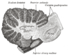
In terms of anatomy, the cerebellum has the appearance of a separate structure attached to the bottom of the brain, tucked underneath the cerebral hemisphere
s. The surface of the cerebellum is covered with finely spaced parallel grooves, in striking contrast to the broad irregular convolutions of the cerebral cortex
. These parallel grooves conceal the fact that the cerebellum is actually a continuous thin layer of tissue (the cerebellar cortex), tightly folded in the style of an accordion
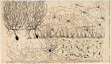
. Within this thin layer are several types of neuron
s with a highly regular arrangement, the most important being Purkinje cell
s and granule cells. This complex neural network gives rise to a massive signal-processing capability, but almost all of its output is directed to a set of small deep cerebellar nuclei
lying in the interior of the cerebellum.
In addition to its direct role in motor control, the cerebellum also is necessary for several types of motor learning
, the most notable one being learning to adjust to changes in sensorimotor relationships. Several theoretical models have been developed to explain sensorimotor calibration in terms of synaptic plasticity
within the cerebellum. Most of them derive from early models formulated by David Marr and James Albus
, which were motivated by the observation that each cerebellar Purkinje cell receives two dramatically different types of input: On one hand, thousands of inputs from parallel fiber
s, each individually very weak; on the other hand, input from one single climbing fiber
, which is, however, so strong that a single climbing fiber action potential will reliably cause a target Purkinje cell to fire a burst of action potentials. The basic concept of the Marr-Albus theory is that the climbing fiber serves as a "teaching signal", which induces a long-lasting change in the strength of synchronously activated parallel fiber inputs. Observations of long-term depression
in parallel fiber inputs have provided support for theories of this type, but their validity remains controversial.
 The cerebellum is located at the bottom of the brain, with the large mass of the cerebral cortex
above it and the portion of the brainstem called the pons
in front of it. It is separated from the overlying cerebrum by a layer of leathery dura mater
; all of its connections with other parts of the brain travel through the pons. Anatomists classify the cerebellum as part of the metencephalon
, which also includes the pons; the metencephalon is the upper part of the rhombencephalon
or "hindbrain". Like the cerebral cortex, the cerebellum is divided into two hemispheres; it also contains a narrow midline zone called the vermis. A set of large folds is, by convention, used to divide the overall structure into 10 smaller "lobules". Because of its large number of tiny granule cell
s, the cerebellum contains more neuron
s than the rest of the brain put together, but it takes up only 10% of total brain volume.
 The unusual surface appearance of the cerebellum conceals the fact that most of its volume is made up of a very tightly folded layer of gray matter
, the cerebellar cortex. It has been estimated that, if the human cerebellar cortex were completely unfolded, it would give rise to a layer of neural tissue about 1 meter long and averaging 5 centimeters wide — a total surface area of about 500 square cm, packed within a volume of dimensions 6 cm × 5 cm × 10 cm. Underneath the gray matter of the cortex lies white matter
, made up largely of myelin
ated nerve fibers running to and from the cortex. Embedded within the white matter — which is sometimes called the arbor vitae
(Tree of Life) because of its branched, tree-like appearance in cross-section — are four deep cerebellar nuclei
, composed of gray matter.
, anterior lobe
(above the primary fissure), and posterior lobe
(below the primary fissure). These lobes divide the cerebellum from rostral to caudal (in humans, top to bottom). In terms of function, however, there is a more important distinction along the medial-to-lateral dimension. Leaving out the flocculonodular part, which has distinct connections and functions, the cerebellum can be parsed functionally into a medial sector called the spinocerebellum and a larger lateral sector called the cerebrocerebellum. A narrow strip of protruding tissue along the midline is called the vermis
(Latin for "worm").
|  >
>

The smallest region, the flocculonodular lobe, is often called the vestibulocerebellum. It is the oldest part in evolutionary terms (archicerebellum) and participates mainly in balance and spatial orientation; its primary connections are with the vestibular nuclei
, although it also receives visual and other sensory input. Damage to it causes disturbances of balance and gait
.
The medial zone of the anterior and posterior lobes constitutes the spinocerebellum, also known as paleocerebellum. This sector of the cerebellum functions mainly to fine-tune body and limb movements. It receives proprioception input from the dorsal columns of the spinal cord
(including the spinocerebellar tract
) and from the trigeminal nerve
, as well as from visual and auditory
systems. It sends fibres to deep cerebellar nuclei that, in turn, project to both the cerebral cortex and the brain stem, thus providing modulation of descending motor systems.
The lateral zone, which in humans is by far the largest part, constitutes the cerebrocerebellum, also known as neocerebellum. It receives input exclusively from the cerebral cortex (especially the parietal lobe
) via the pontine nuclei
(forming cortico-ponto-cerebellar pathways), and sends output mainly to the ventrolateral thalamus
(in turn connected to motor areas of the premotor cortex
and primary motor area of the cerebral cortex) and to the red nucleus
. There is disagreement about the best way to describe the functions of the lateral cerebellum: It is thought to be involved in planning movement that is about to occur, in evaluating sensory information for action, and in a number of purely cognitive functions as well.

 Two types of neuron play dominant roles in the cerebellar circuit: Purkinje cell
s and granule cell
s. Three types of axons also play dominant roles: mossy fibers and climbing fibers (which enter the cerebellum from outside), and parallel fibers (which are the axons of granule cells). There are two main pathways through the cerebellar circuit, originating from mossy fibers and climbing fiber
s, both eventually terminating in the deep cerebellar nuclei.
Mossy fibers project directly to the deep nuclei, but also give rise to the pathway: mossy fiber → granule cells → parallel fibers → Purkinje cells → deep nuclei. Climbing fibers project to Purkinje cells and also send collaterals directly to the deep nuclei. The mossy fiber and climbing fiber inputs each carry fiber-specific information; the cerebellum also receives dopamine
rgic, serotonergic
, noradrenergic
, and cholinergic
inputs that presumably perform global modulation.
The cerebellar cortex is divided into three layers. At the bottom lies the thick granular layer, densely packed with granule cells, along with interneurons, mainly Golgi cell
s but also including Lugaro cells and unipolar brush cells. In the middle lies the Purkinje layer, a narrow zone that contains only the cell bodies of Purkinje cells. At the top lies the molecular layer, which contains the flattened dendritic trees of Purkinje cells, along with the huge array of parallel fibers penetrating the Purkinje cell dendritic trees at right angles. This outermost layer of the cerebellar cortex also contains two types of inhibitory interneuron
s, stellate cell
s, and basket cell
s. Both stellate and basket cells form GABAergic
synapses onto Purkinje cell dendrites.
 Purkinje cell
s are among the most distinctive neurons in the brain, and also among the earliest types to be recognized — they were first described by the Czech anatomist Jan Evangelista Purkyně
in 1837. They are distinguished by the shape of the dendritic tree: The dendrites branch very profusely, but are severely flattened in a plane perpendicular to the cerebellar folds. Thus, the dendrites of a Purkinje cell form a dense planar net, through which parallel fibers pass at right angles. The dendrites are covered with dendritic spine
s, each of which receives synaptic input from a parallel fiber. Purkinje cells receive more synaptic inputs than any other type of cell in the brain — estimates of the number of spines on a single human Purkinje cell run as high as 200,000. The large, spherical cell bodies of Purkinje cells are packed into a narrow layer (one cell thick) of the cerebellar cortex, called the Purkinje layer. After emitting collaterals that innervate nearby parts of the cortex, their axons travel into the deep cerebellar nuclei
, where they make on the order of 1,000 contacts each with several types of nuclear cells, all within a small domain. Purkinje cells use GABA
as their neurotransmitter, and therefore exert inhibitory effects on their targets.
Purkinje cells form the heart of the cerebellar circuit, and their large size and distinctive activity patterns have made it relatively easy to study their response patterns in behaving animals using extracellular
recording techniques. Purkinje cells normally emit action potentials at a high rate even in the absence of synaptic input. In awake, behaving animals, mean rates averaging around 40 Hz are typical. The spike trains show a mixture of what are called simple and complex spikes. A simple spike is a single action potential followed by a refractory period of about 10 msec; a complex spike is a stereotyped sequence of action potentials with very short inter-spike intervals and declining amplitudes. Physiological studies have shown that complex spikes (which occur at baseline rates around 1 Hz and never at rates much higher than 10 Hz) are reliably associated with climbing fiber activation, while simple spikes are produced by a combination of baseline activity and parallel fiber input. Complex spikes are often followed by a pause of several hundred msec during which simple spike activity is suppressed.
s, in contrast to Purkinje cells, are among the smallest neurons in the brain. They are also easily the most numerous neurons in the brain: In humans, estimates of their total number average around 50 billion, which means that about 3/4 of the brain's neurons are cerebellar granule cells. Their cell bodies are packed into a thick layer at the bottom of the cerebellar cortex. A granule cell emits only four to five dendrites, each of which ends in an enlargement called a dendritic claw. These enlargements are sites of excitatory input from mossy fibers and inhibitory input from Golgi cell
s.
The thin, unmyelinated axons of granule cells rise vertically to the upper (molecular) layer of the cortex, where they split in two, with each branch traveling horizontally to form a parallel fiber
; the splitting of the vertical branch into two horizontal branches gives rise to a distinctive "T" shape. A parallel fiber runs for an average of 3 mm in each direction from the split, for a total length of about 6 mm (about 1/10 of the total width of the cortical layer). As they run along, the parallel fibers pass through the dendritic trees of Purkinje cells, contacting one of every 3–5 that they pass, making a total of 80–100 synaptic connections with Purkinje cell dendritic spines. Granule cells use glutamate as their neurotransmitter, and therefore exert excitatory effects on their targets.
Granule cells receive all of their input from mossy fibers, but outnumber them 200 to 1 (in humans). Thus, the information in the granule cell population activity state is the same as the information in the mossy fibers, but recoded in a much more expansive way. Because granule cells are so small and so densely packed, it has been very difficult to record their spike activity in behaving animals, so there is little data to use as a basis of theorizing. The most popular concept of their function was proposed by David Marr, who suggested that they could encode combinations of mossy fiber inputs. The idea is that with each granule cell receiving input from only 4–5 mossy fibers, a granule cell would not respond if only a single one of its inputs were active, but would respond if more than one were active. This combinatorial coding scheme would potentially allow the cerebellum to make much finer distinctions between input patterns than the mossy fibers alone would permit.
s enter the granular layer from their points of origin, many arising from the pontine nuclei, others from the spinal cord, vestibular nuclei, etc. In the human cerebellum, the total number of mossy fibers has been estimated at about 200 million. These fibers form excitatory synapses with the granule cells and the cells of the deep cerebellar nuclei. Within the granular layer, a mossy fiber generates a series of enlargements called rosettes. The contacts between mossy fibers and granule cell dendrites take place within structures called glomeruli. Each glomerulus has a mossy fiber rosette at its center, and up to 20 granule cell dendritic claws contacting it. Terminals from Golgi cell
s infiltrate the structure and make inhibitory synapses onto the granule cell dendrites. The entire assemblage is surrounded by a sheath of glial cells. Each mossy fiber sends collateral branches to several cerebellar folia, generating a total of 20–30 rosettes; thus a single mossy fiber makes contact with an estimated 400–600 granule cells.
(IO) on the contralateral side of the brainstem, via climbing fiber
s. Although the IO lies in the medulla oblongata, and receives input from the spinal cord, brainstem, and cerebral cortex, its output goes entirely to the cerebellum. A climbing fiber gives off collaterals to the deep cerebellar nuclei before entering the cerebellar cortex, where it splits into about 10 terminal branches, each of which innervates a single Purkinje cell. In striking contrast to the 100,000-plus inputs from parallel fibers, each Purkinje cell receives input from exactly one climbing fiber; but this single fiber "climbs" the dendrites of the Purkinje cell, winding around them and making a total of up to 300 synapses as it goes. The net input is so strong that a single action potential
from a climbing fiber is capable of producing an extended complex spike in the Purkinje cell: a burst of several spikes in a row, with diminishing amplitude, followed by a pause during which activity is suppressed. The climbing fiber synapses cover the cell body and proximal dendrites; this zone is devoid of parallel fiber inputs.
Climbing fibers fire at low rates, but a single climbing fiber action potential induces a burst of several action potentials in a target Purkinje cell (a complex spike). The contrast between parallel fiber and climbing fiber inputs to Purkinje cells (over 100,000 of one type versus exactly one of the other type) is perhaps the most provocative feature of cerebellar anatomy, and has motivated much of the theorizing. In fact, the function of climbing fibers is the most controversial topic concerning the cerebellum. There are two schools of thought, one following Marr and Albus in holding that climbing fiber input serves primarily as a teaching signal, the other holding that its function is to shape cerebellar output directly. Both views have been defended in great length in numerous publications. In the words of one review, "In trying to synthesize the various hypotheses on the function of the climbing fibers, one has the sense of looking at a drawing by Escher. Each point of view seems to account for a certain collection of findings, but when one attempts to put the different views together, a coherent picture of what the climbing fibers are doing does not appear. For the majority of researchers, the climbing fibers signal errors in motor performance, either in the usual manner of discharge frequency modulation or as a single announcement of an 'unexpected event'. For other investigators, the message lies in the degree of ensemble synchrony and rhythmicity among a population of climbing fibers."
 The deep nuclei of the cerebellum are clusters of gray matter lying within the white matter at the core of the cerebellum. They are, with the minor exception of the nearby vestibular nuclei
, the sole sources of output from the cerebellum. These nuclei receive collateral projections from mossy fibers and climbing fibers, as well as inhibitory input from the Purkinje cells of the cerebellar cortex. The three nuclei (dentate, interpositus, and fastigial) each communicate with different parts of the brain and cerebellar cortex. The fastigial and interpositus nuclei belong to the spinocerebellum. The dentate nucleus, which in mammals is much larger than the others, is formed as a thin, convoluted layer of gray matter, and communicates exclusively with the lateral parts of the cerebellar cortex. The flocculonodular lobe is the only part of the cerebellar cortex that does not project to the deep nuclei — its output goes to the vestibular nuclei instead.
The majority of neurons in the deep nuclei have large cell bodies and spherical dendritic trees with a radius of about 400 μm, and use glutamate as their neurotransmitter. These cells project to a variety of targets outside the cerebellum. Intermixed with them is a lesser number of small cells, which use GABA
as neurotransmitter and project exclusively to the inferior olivary nucleus
, the source of climbing fiber
s. Thus, the nucleo-olivary projection provides an inhibitory feedback to match the excitatory projection of climbing fibers to the nuclei. There is evidence that each small cluster of nuclear cells projects to the same cluster of olivary cells that send climbing fibers to it; there is strong and matching topography in both directions.
When a Purkinje cell axon enters one of the deep nuclei, it branches to make contact with both large and small nuclear cells, but the total number of cells contacted is only about 35 (in cats). On the converse, a single deep nuclear cell receives input from approximately 860 Purkinje cells (again in cats).
The first indications of compartmental structure came from studies of the receptive fields of cells in various parts of the cerebellum cortex. Each body part maps to specific points in the cerebellum, but there are numerous repetitions of the basic map, forming an arrangement that has been called "fractured somatotopy". A clearer indication of compartmentalization is obtained by immunostaining the cerebellum for certain types of protein. The best-known of these markers are called "zebrins", because staining for them gives rise to a complex pattern reminiscent of the stripes on a zebra. The stripes generated by zebrins and other compartmentalization markers are oriented perpendicular to the cerebellar folds — that is, they are narrow in the mediolateral direction, but much more extended in the longitudinal direction. Different markers generate different sets of stripes, and the widths and lengths vary as a function of location, but they all have the same general shape.
Oscarsson in the late 1970s proposed that these cortical zones can be partitioned into smaller units called microzones. A microzone is defined as a group of Purkinje cells all having the same somatotopic receptive field. Microzones were found to contain on the order of 1000 Purkinje cells each, arranged in a long, narrow strip, oriented perpendicular to the cortical folds. Thus, as the adjoining diagram illustrates, Purkinje cell dendrites are flattened in the same direction as the microzones extend, while parallel fiber
s cross them at right angles.
It is not only receptive fields that define the microzone structure: The climbing fiber
input from the inferior olivary nucleus
is equally important. The branches of a climbing fiber (usually numbering about 10) usually innervate Purkinje cells belonging to the same microzone. Moreover, olivary neurons that send climbing fibers to the same microzone tend to be coupled by gap junction
s, which synchronize their activity, causing Purkinje cells within a microzone to show correlated complex spike activity on a millisecond time scale. Also, the Purkinje cells belonging to a microzone all send their axons to the same small cluster of output cells within the deep cerebellar nuclei
. Finally, the axons of basket cell
s are much longer in the longitudinal direction than in the mediolateral direction, causing them to be confined largely to a single microzone. The consequence of all this structure is that cellular interactions within a microzone are much stronger than interactions between different microzones.
In 2005, Richard Apps and Martin Garwicz summarized evidence that microzones themselves form part of a larger entity they call a multizonal microcomplex. Such a microcomplex includes several spatially separated cortical microzones, all of which project to the same group of deep cerebellar neurons, plus a group of coupled olivary neurons that project to all of the included microzones as well as to the deep nuclear area.
Prior to the 1990s, the function of the cerebellum was almost universally believed to be purely motor-related, but newer findings have brought that view strongly into question. Functional imaging studies have shown cerebellar activation in relation to language, attention, and mental imagery; correlation studies have shown interactions between the cerebellum and non-motoric areas of the cerebral cortex; and a variety of non-motor symptoms have been recognized in people with damage that appears to be confined to the cerebellum.
Kenji Doya has argued that the function of the cerebellum is best understood not in terms of what behaviors it is involved in but rather in terms of what neural computations it performs; the cerebellum consists of a large number of more or less independent modules, all with the same geometrically regular internal structure, and therefore all, it is presumed, performing the same computation. If the input and output connections of a module are with motor areas (as many are), then the module will be involved in motor behavior; but, if the connections are with areas involved in non-motor cognition, the module will show other types of behavioral correlates. The cerebellum, Doya proposes, is best understood as a device for supervised learning
, in contrast to the basal ganglia
, which perform reinforcement learning
, and the cerebral cortex
, which performs unsupervised learning
.
, and Janos Szentágothai. Although a full understanding of cerebellar function has remained elusive, at least four principles have been identified as important: (1) feedforward processing, (2) divergence and convergence, (3) modularity, and (4) plasticity.
1. Feedforward processing: The cerebellum differs from most other parts of the brain (especially the cerebral cortex) in that the signal processing is almost entirely feedforward - that is, signals move unidirectionally through the system from input to output, with very little recurrent internal transmission. The small amount of recurrence that does exist consists of mutual inhibition; there are no mutually excitatory circuits. This feedforward mode of operation means that the cerebellum, in contrast to the cerebral cortex, cannot generate self-sustaining patterns of neural activity. Signals enter the circuit, are processed by each stage in sequential order, and then leave. As Eccles, Ito, and Szentágothai wrote, "This elimination in the design of all possibility of reverberatory chains of neuronal excitation is undoubtedly a great advantage in the performance of the cerebellum as a computer, because what the rest of the nervous system requires from the cerebellum is presumably not some output expressing the operation of complex reverberatory circuits in the cerebellum but rather a quick and clear response to the input of any particular set of information."
2. Divergence and convergence: In the human cerebellum, information from 200 million mossy fiber inputs is expanded to 40 billion granule cell
s, whose parallel fiber
outputs then converge onto 15 million Purkinje cell
s. Because of the way that they are lined up longitudinally, the 1000 or so Purkinje cells belonging to a microzone may receive input from as many as 100 million parallel fibers, and focus their own output down to a group of less than 50 deep nuclear
cells. Thus, the cerebellar network receives a modest number of inputs, processes them very extensively through its rigorously structured internal network, and sends out the results via a very limited number of output cells.
3. Modularity: The cerebellar system is functionally divided into more or less independent modules, which probably number in the hundreds to thousands. All modules have a similar internal structure, but different inputs and outputs. A module (a multizonal microcompartment in the terminology of Apps and Garwicz) consists of a small cluster of neurons in the inferior olivary nucleus, a set of long narrow strips of Purkinje cells in the cerebellar cortex (microzones), and a small cluster of neurons in one of the deep cerebellar nuclei. Different modules share input from mossy fibers and parallel fibers, but in other respects they appear to function independently — the output of one module does not appear to significantly influence the activity of other modules.
4. Plasticity: The synapses between parallel fibers and Purkinje cells, and the synapses between mossy fibers and deep nuclear cells, are both susceptible to modification of their strength. In a single cerebellar module, input from as many as a billion parallel fibers converges onto a group of less than 50 deep nuclear cells, and the influence of each parallel fiber on those nuclear cells is adjustable. This arrangement gives tremendous flexibility for fine-tuning the relationship between cerebellar inputs and outputs.
, who postulated that climbing fiber
s provide a teaching signal that induces synaptic modification in parallel fiber
—Purkinje cell
synapses. Marr assumed that climbing fiber input would cause synchronously activated parallel fiber inputs to be strengthened. Most later cerebellar-learning models, however, have followed Albus in assuming that climbing fiber activity would be an error signal, and would cause synchronously activated parallel fiber inputs to be weakened. Some of these later models, such as the Adaptive Filter model of Fujita made attempts to understand cerebellar function in terms of optimal control theory.
The idea that climbing fiber activity functions as an error signal has been examined in many experimental studies, with some supporting it but others casting doubt. In a pioneering study by Gilbert and Thach from 1977, Purkinje cells from monkeys learning a reaching task showed increased complex spike activity — which is known to reliably indicate activity of the cell's climbing fiber input — during periods when performance was poor. Several studies of motor learning in cats observed complex spike activity when there was a mismatch between an intended movement and the movement that was actually executed. Studies of the vestibulo-ocular reflex
(which stabilizes the visual image on the retina when the head turns) found that climbing fiber activity indicated "retinal slip", although not in a very straightforward way.
One of the most extensively studied cerebellar learning tasks is the eyeblink conditioning
paradigm, in which a neutral conditioned stimulus such as a tone or a light is repeatedly paired with an unconditioned stimulus, such as an air puff, that elicits a blink response. After such repeated presentations of the CS and US, the CS will eventually elicit a blink before the US, a conditioned response or CR. Experiments showed that lesions localized either to a specific part of the interpositus nucleus (one of the deep cerebellar nuclei
) or to a few specific points in the cerebellar cortex would abolish learning of a correctly timed blink response. If cerebellar outputs are pharmacologically inactivated while leaving the inputs and intracellular circuits intact, learning takes place even while the animal fails to show any response, whereas, if intracerebellar circuits are disrupted, no learning takes place — these facts taken together make a strong case that the learning, indeed, occurs inside the cerebellum.
within the cerebellum to account for its role in learning, versus theories that account for aspects of ongoing behavior on the basis of cerebellar signal processing. Several theories of both types have been formulated as mathematical model
s and simulated using computers.
Perhaps the earliest "performance" theory was the "delay line" hypothesis of Valentino Braitenberg. The original theory put forth by Braitenberg and Atwood in 1958 proposed that slow propagation of signals along parallel fibers imposes predictable delays that allow the cerebellum to detect time relationships within a certain window. Experimental data did not support the original form of the theory, but Braitenberg continued to argue for modified versions. The hypothesis that the cerebellum functions essentially as a timing system has also been advocated by Richard Ivry. Another influential "performance" theory is the Tensor Network Theory
of Pellionisz and Llinás, which provided an advanced mathematical formulation of the idea that the fundamental computation performed by the cerebellum is to transform sensory into motor coordinates.
Theories in the "learning" category almost all derive from early publications by David Marr and James Albus
. Marr's 1969 paper proposed that the cerebellum is a device for learning to associate elemental movements encoded by climbing fibers with mossy fiber inputs that encode the sensory context. Albus proposed that a cerebellar Purkinje cell functions as a perceptron
, a neurally inspired abstract learning device. The most basic difference between the Marr and Albus theories is that Marr assumed that climbing fiber activity would cause parallel fiber synapses to be strengthened, whereas Albus proposed that they would be weakened. Albus also formulated his version as a software algorithm he called a CMAC (Cerebellar Model Articulation Controller), which has been tested in a number of applications.
part) may show up as a loss of equilibrium and, in particular, an altered walking gait, with a wide stance that indicates difficulty in balancing. Damage to the lateral zone, or the cerebrocerebellum, results in problems with skilled voluntary and planned movements. This can cause errors in the force, direction, speed and amplitude of movements. Some manifestations include hypotonia
(decreased muscle tone), dysarthria
(problems with speech articulation), dysmetria
(problems judging distances or ranges of movement), dysdiadokinesia (inability to perform rapid alternating movements), impaired check reflex or rebound phenomenon, and tremors (involuntary movement caused by alternating contractions of opposing muscle groups). Damage to the midline portion may disrupt whole-body movements, whereas damage localized more laterally is more likely to disrupt fine movements of the hands or limbs. Damage to the upper part of the cerebellum tends to cause gait impairments and other problems with leg coordination; damage to the lower part is more likely to cause uncoordinated or poorly aimed movements of the arms and hands, as well as difficulties in speed. This complex of motor symptoms is called "ataxia". To identify cerebellar problems, the neurological examination
includes assessment of gait (a broad-based gait being indicative of ataxia), finger-pointing tests and assessment of posture. If cerebellar dysfunction is indicated, a magnetic resonance imaging
scan can be used to obtain a detailed picture of any structural alterations that may exist.
The list of medical problems that can produce cerebellar damage is long: it includes stroke
; hemorrhage; tumors; alcoholism; physical trauma such as gunshot wounds; and chronic degenerative conditions such as olivopontocerebellar atrophy
. Some forms of migraine headache may also produce temporary dysfunction of the cerebellum, of variable severity.
pattern in the human cerebellum shows less age-related alteration than in the cerebral cortex
.
Some studies have reported reductions in numbers of cells or volume of tissue, but the amount of data relating to this question is not very large.
Tumors that commonly arise in the cerebellum include pilocytic astrocytoma
s, medulloblastoma
s (especially in children), ependymoma
s, and hemangioblastoma
s (often in the context of von Hippel-Lindau syndrome).
es of vertebrate
s, including fish, reptiles, birds, and mammals. There is also an analogous brain structure in cephalopod
s with well-developed brains, such as octopus
es. This has been taken as evidence that the cerebellum performs functions important to all animal species
with a brain.
There is considerable variation in the size and shape of the cerebellum in different vertebrate species. In amphibian
s, lamprey
s, and hagfish
, the cerebellum is little developed; in the latter two groups, it is barely distinguishable from the brain-stem. Although the spinocerebellum is present in these groups, the primary structures are small paired nuclei corresponding to the vestibulocerebellum. The cerebellum is a bit larger in reptiles, considerably larger in birds, and larger yet in mammals. The large paired and convoluted lobes found in humans are typical of mammals, but the cerebellum is, in general, a single median lobe in other groups, and is either smooth or only slightly grooved. In mammals, the neocerebellum is the major part of the cerebellum by mass, but, in other vertebrates, it is typically the spinocerebellum.
The cerebellum of cartilaginous and bony fishes is extraordinarily large and complex. In at least one important respect, it differs in internal structure from the mammalian cerebellum: The fish cerebellum does not contain discrete deep cerebellar nuclei
. Instead, the primary targets of Purkinje cells are a distinct type of cell distributed across the cerebellar cortex, a type not seen in mammals. In mormyrid fish
(a family of weakly electrosensitive freshwater fish), the cerebellum is considerably larger than the rest of the brain put together. The largest part of it is a special structure called the valvula, which has an unusually regular architecture and receives much of its input from the electrosensory system.
The hallmark of the mammalian cerebellum is an expansion of the lateral lobes, whose main interactions are with the neocortex. As monkeys evolved into great apes, the expansion of the lateral lobes continued, in tandem with the expansion of the frontal lobes of the neocortex. In ancestral hominids, and in homo sapiens until the middle Pleistocene
period, the cerebellum continued to expand, but the frontal lobes expanded more rapidly. The most recent period of human evolution, however, may actually have been associated with an increase in the relative size of the cerebellum, as the neocortex reduced its size somewhat while the cerebellum expanded.
(DCN), one of the two primary sensory nuclei that receive input directly from the auditory nerve. The DCN is a layered structure, with the bottom layer containing granule cells similar to those of the cerebellum, giving rise to parallel fiber
s that rise to the superficial layer and travel across it horizontally. The superficial layer contains a set of GABAergic neurons called cartwheel cells that resemble Purkinje cells anatomically and chemically — they receive parallel fiber input, but do not have any inputs that resemble climbing fiber
s. The output neurons of the DCN are called fusiform cells. They are glutamatergic, but also resemble Purkinje cells in some respects — they have spiny, flattened superficial dendritic trees that receive parallel fiber input, but they also have basal dendrites that receive input from auditory nerve fibers, which travel across the DCN in a direction at right angles to the parallel fibers. The DCN is most highly developed in rodents and other small animals, and is considerably reduced in primates. Its function is not well understood; the most popular speculations relate it to spatial hearing in one way or another.
Most species of fish and amphibians possess a lateral line
system that senses pressure waves in water. One of the brain areas that receives primary input from the lateral line organ, the medial octavolateral nucleus, has a cerebellum-like structure, with granule cells and parallel fibers. In electrosensitive fish, the input from the electrosensory system goes to the dorsal octavolateral nucleus, which also has a cerebellum-like structure. In ray-finned fishes
(by far the largest group), the optic tectum
has a layer — the marginal layer — that is cerebellum-like.
All of these cerebellum-like structures appear to be primarily sensory-related rather than motor-related. All of them have granule cells that give rise to parallel fibers that connect to Purkinje-like neurons with modifiable synapse
s, but none have climbing fibers comparable to those of the cerebellum — instead they receive direct input from peripheral sensory organs. None has a demonstrated function, but the most influential speculation is that they serve to transform sensory inputs in some sophisticated way, perhaps to compensate for changes in body posture. In fact, James Bower and others have argued, partly on the basis of these structures and partly on the basis of cerebellar studies, that the cerebellum itself is fundamentally a sensory structure, and that it contributes to motor control by moving the body in a way that controls the resulting sensory signals.
 The distinctive appearance of the cerebellum caused even the earliest anatomists to recognize it. Aristotle and Galen, however, did not consider it truly part of the brain: They called it the parencephalon ("same-as-brain"), as opposed to the encephalon or brain proper. Galen was the first to give an extensive description, noting that the cerebellar tissue seemed more solid than the rest of the brain, he speculated that its function is to strengthen the motor nerves.
Further significant developments did not come until the Renaissance. Vesalius discussed the cerebellum briefly, and the anatomy was described more thoroughly by Thomas Willis
in 1664. More anatomical work was done during the 18th century, but it was not until early in the 19th century that the first insights into the function of the cerebellum were obtained. Luigi Rolando
in 1809 established the key insight that damage to the cerebellum results in motor disturbances. Jean Pierre Flourens
in the first half of the 19th century carried out detailed experimental work, which revealed that animals with cerebellar damage can still move, but with a loss of coordination (strange movements, awkward gait, and muscular weakness), and that recovery after the lesion can be nearly complete unless the lesion is very extensive. By the dawn of the 20th century, it was widely accepted that the primary function of the cerebellum relates to motor control; the first half of the 20th century produced several detailed descriptions of the clinical symptoms associated with cerebellar disease in humans.
Brain
The brain is the center of the nervous system in all vertebrate and most invertebrate animals—only a few primitive invertebrates such as sponges, jellyfish, sea squirts and starfishes do not have one. It is located in the head, usually close to primary sensory apparatus such as vision, hearing,...
that plays an important role in motor control
Motor control
Motor control are information processing related activities carried out by the central nervous system that organize the musculoskeletal system to create coordinated movements and skilled actions...
. It may also be involved in some cognitive function
Cognition
In science, cognition refers to mental processes. These processes include attention, remembering, producing and understanding language, solving problems, and making decisions. Cognition is studied in various disciplines such as psychology, philosophy, linguistics, and computer science...
s such as attention
Attention
Attention is the cognitive process of paying attention to one aspect of the environment while ignoring others. Attention is one of the most intensely studied topics within psychology and cognitive neuroscience....
and language
Language
Language may refer either to the specifically human capacity for acquiring and using complex systems of communication, or to a specific instance of such a system of complex communication...
, and in regulating fear
Fear
Fear is a distressing negative sensation induced by a perceived threat. It is a basic survival mechanism occurring in response to a specific stimulus, such as pain or the threat of danger...
and pleasure
Pleasure
Pleasure describes the broad class of mental states that humans and other animals experience as positive, enjoyable, or worth seeking. It includes more specific mental states such as happiness, entertainment, enjoyment, ecstasy, and euphoria...
responses, but its movement-related functions are the most solidly established. The cerebellum does not initiate movement, but it contributes to coordination
Motor coordination
thumb|right|Motor coordination is shown in this animated sequence by [[Eadweard Muybridge]] of himself throwing a diskMotor coordination is the combination of body movements created with the kinematic and kinetic parameters that result in intended actions. Such movements usually smoothly and...
, precision, and accurate timing. It receives input from sensory system
Sensory system
A sensory system is a part of the nervous system responsible for processing sensory information. A sensory system consists of sensory receptors, neural pathways, and parts of the brain involved in sensory perception. Commonly recognized sensory systems are those for vision, hearing, somatic...
s and from other parts of the brain
Brain
The brain is the center of the nervous system in all vertebrate and most invertebrate animals—only a few primitive invertebrates such as sponges, jellyfish, sea squirts and starfishes do not have one. It is located in the head, usually close to primary sensory apparatus such as vision, hearing,...
and spinal cord
Spinal cord
The spinal cord is a long, thin, tubular bundle of nervous tissue and support cells that extends from the brain . The brain and spinal cord together make up the central nervous system...
, and integrates these inputs to fine tune motor activity. Because of this fine-tuning function, damage
Lesion
A lesion is any abnormality in the tissue of an organism , usually caused by disease or trauma. Lesion is derived from the Latin word laesio which means injury.- Types :...
to the cerebellum does not cause paralysis
Paralysis
Paralysis is loss of muscle function for one or more muscles. Paralysis can be accompanied by a loss of feeling in the affected area if there is sensory damage as well as motor. A study conducted by the Christopher & Dana Reeve Foundation, suggests that about 1 in 50 people have been diagnosed...
, but instead produces disorders in fine movement, equilibrium
Equilibrioception
Equilibrioception or sense of balance is one of the physiological senses. It helps prevent humans and animals from falling over when walking or standing still. Balance is the result of a number of body systems working together: the eyes , ears and the body's sense of where it is in space ideally...
, posture, and motor learning
Motor learning
Motor learning is a “relatively permanent” change, resulting from practice or a novel experience, in the capability for responding...
.
In terms of anatomy, the cerebellum has the appearance of a separate structure attached to the bottom of the brain, tucked underneath the cerebral hemisphere
Cerebral hemisphere
A cerebral hemisphere is one of the two regions of the eutherian brain that are delineated by the median plane, . The brain can thus be described as being divided into left and right cerebral hemispheres. Each of these hemispheres has an outer layer of grey matter called the cerebral cortex that is...
s. The surface of the cerebellum is covered with finely spaced parallel grooves, in striking contrast to the broad irregular convolutions of the cerebral cortex
Cerebral cortex
The cerebral cortex is a sheet of neural tissue that is outermost to the cerebrum of the mammalian brain. It plays a key role in memory, attention, perceptual awareness, thought, language, and consciousness. It is constituted of up to six horizontal layers, each of which has a different...
. These parallel grooves conceal the fact that the cerebellum is actually a continuous thin layer of tissue (the cerebellar cortex), tightly folded in the style of an accordion
Accordion
The accordion is a box-shaped musical instrument of the bellows-driven free-reed aerophone family, sometimes referred to as a squeezebox. A person who plays the accordion is called an accordionist....
. Within this thin layer are several types of neuron
Neuron
A neuron is an electrically excitable cell that processes and transmits information by electrical and chemical signaling. Chemical signaling occurs via synapses, specialized connections with other cells. Neurons connect to each other to form networks. Neurons are the core components of the nervous...
s with a highly regular arrangement, the most important being Purkinje cell
Purkinje cell
For the cells of the electrical conduction system of the heart, see Purkinje fibersPurkinje cells, or Purkinje neurons , are a class of GABAergic neurons located in the cerebellar cortex...
s and granule cells. This complex neural network gives rise to a massive signal-processing capability, but almost all of its output is directed to a set of small deep cerebellar nuclei
Deep cerebellar nuclei
The Cerebellum has four deep cerebellar nuclei embedded in the white matter in its center.-Inputs:These nuclei receive inhibitory inputs from Purkinje cells in the cerebellar cortex and excitatory inputs from mossy fiber and climbing fiber pathways. Most output fibers of the cerebellum originate...
lying in the interior of the cerebellum.
In addition to its direct role in motor control, the cerebellum also is necessary for several types of motor learning
Motor learning
Motor learning is a “relatively permanent” change, resulting from practice or a novel experience, in the capability for responding...
, the most notable one being learning to adjust to changes in sensorimotor relationships. Several theoretical models have been developed to explain sensorimotor calibration in terms of synaptic plasticity
Synaptic plasticity
In neuroscience, synaptic plasticity is the ability of the connection, or synapse, between two neurons to change in strength in response to either use or disuse of transmission over synaptic pathways. Plastic change also results from the alteration of the number of receptors located on a synapse...
within the cerebellum. Most of them derive from early models formulated by David Marr and James Albus
James S. Albus
James Sacra Albus was an American engineer, Senior NIST Fellow and founder and former chief of the Intelligent Systems Division of the Manufacturing Engineering Laboratory at the National Institute of Standards and Technology .- Biography :Born in Louisville Ky., Albus received the B.S...
, which were motivated by the observation that each cerebellar Purkinje cell receives two dramatically different types of input: On one hand, thousands of inputs from parallel fiber
Parallel fiber
Parallel fibers arise from granule cells in the cerebellar cortex. They form excitatory synapses onto the dendrites of Purkinje cells ....
s, each individually very weak; on the other hand, input from one single climbing fiber
Climbing fiber
Climbing fibers are the name given to a series of neuronal projections from the inferior olivary nucleus located in the medulla oblongata.These axons pass through the pons and enter the cerebellum via the inferior cerebellar peduncle where they form synapses with the deep cerebellar nuclei and...
, which is, however, so strong that a single climbing fiber action potential will reliably cause a target Purkinje cell to fire a burst of action potentials. The basic concept of the Marr-Albus theory is that the climbing fiber serves as a "teaching signal", which induces a long-lasting change in the strength of synchronously activated parallel fiber inputs. Observations of long-term depression
Long-term depression
Long-term depression , in neurophysiology, is an activity-dependent reduction in the efficacy of neuronal synapses lasting hours or longer. LTD occurs in many areas of the CNS with varying mechanisms depending upon brain region and developmental progress...
in parallel fiber inputs have provided support for theories of this type, but their validity remains controversial.
Structure
At the level of large scale anatomy, the cerebellum consists of a tightly folded and crumpled layer of cortex, with white matter underneath, several deep nuclei embedded in the white matter, and a fluid-filled ventricle at the base. At the microscopic level, each part of the cortex consists of the same small set of neuronal elements, laid out with a highly stereotyped geometry. At an intermediate level, the cerebellum and its auxiliary structures can be decomposed into several hundred or thousand independently functioning modules called "microzones" or "microcompartments".Anatomy
Cerebral cortex
The cerebral cortex is a sheet of neural tissue that is outermost to the cerebrum of the mammalian brain. It plays a key role in memory, attention, perceptual awareness, thought, language, and consciousness. It is constituted of up to six horizontal layers, each of which has a different...
above it and the portion of the brainstem called the pons
Pons
The pons is a structure located on the brain stem, named after the Latin word for "bridge" or the 16th-century Italian anatomist and surgeon Costanzo Varolio . It is superior to the medulla oblongata, inferior to the midbrain, and ventral to the cerebellum. In humans and other bipeds this means it...
in front of it. It is separated from the overlying cerebrum by a layer of leathery dura mater
Dura mater
The dura mater , or dura, is the outermost of the three layers of the meninges surrounding the brain and spinal cord. It is derived from Mesoderm. The other two meningeal layers are the pia mater and the arachnoid mater. The dura surrounds the brain and the spinal cord and is responsible for...
; all of its connections with other parts of the brain travel through the pons. Anatomists classify the cerebellum as part of the metencephalon
Metencephalon
The metencephalon is a developmental categorization of portions of the central nervous system. The metencephalon is composed of the pons and the cerebellum; contains a portion of the fourth ventricle; and the trigeminal nerve , abducens nerve , facial nerve , and a portion of the vestibulocochlear...
, which also includes the pons; the metencephalon is the upper part of the rhombencephalon
Rhombencephalon
The rhombencephalon is a developmental categorization of portions of the central nervous system in vertebrates.The rhombencephalon can be subdivided in a variable number of transversal swellings called rhombomeres...
or "hindbrain". Like the cerebral cortex, the cerebellum is divided into two hemispheres; it also contains a narrow midline zone called the vermis. A set of large folds is, by convention, used to divide the overall structure into 10 smaller "lobules". Because of its large number of tiny granule cell
Granule cell
In neuroscience, granule cells refer to tiny neurons that are around 10 micrometres in diameter. Granule cells are found within the granular layer of the cerebellum , the dentate gyrus of the...
s, the cerebellum contains more neuron
Neuron
A neuron is an electrically excitable cell that processes and transmits information by electrical and chemical signaling. Chemical signaling occurs via synapses, specialized connections with other cells. Neurons connect to each other to form networks. Neurons are the core components of the nervous...
s than the rest of the brain put together, but it takes up only 10% of total brain volume.
Gray Matter
"Gray Matter" is a short story by Stephen King, first published in the October 1973 issue of Cavalier magazine, and later collected in King's 1978 collection Night Shift. It is set in the same area as King's novel Dreamcatcher.-Setting:...
, the cerebellar cortex. It has been estimated that, if the human cerebellar cortex were completely unfolded, it would give rise to a layer of neural tissue about 1 meter long and averaging 5 centimeters wide — a total surface area of about 500 square cm, packed within a volume of dimensions 6 cm × 5 cm × 10 cm. Underneath the gray matter of the cortex lies white matter
White matter
White matter is one of the two components of the central nervous system and consists mostly of myelinated axons. White matter tissue of the freshly cut brain appears pinkish white to the naked eye because myelin is composed largely of lipid tissue veined with capillaries. Its white color is due to...
, made up largely of myelin
Myelin
Myelin is a dielectric material that forms a layer, the myelin sheath, usually around only the axon of a neuron. It is essential for the proper functioning of the nervous system. Myelin is an outgrowth of a type of glial cell. The production of the myelin sheath is called myelination...
ated nerve fibers running to and from the cortex. Embedded within the white matter — which is sometimes called the arbor vitae
Arbor vitae (anatomy)
The arbor vitae is the cerebellar white matter, so called for its branched, tree-like appearance. It brings sensory and motor information to and from the cerebellum....
(Tree of Life) because of its branched, tree-like appearance in cross-section — are four deep cerebellar nuclei
Deep cerebellar nuclei
The Cerebellum has four deep cerebellar nuclei embedded in the white matter in its center.-Inputs:These nuclei receive inhibitory inputs from Purkinje cells in the cerebellar cortex and excitatory inputs from mossy fiber and climbing fiber pathways. Most output fibers of the cerebellum originate...
, composed of gray matter.
Subdivisions
Based on surface appearance, three lobes can be distinguished in the cerebellum, called the flocculonodular lobeFlocculonodular lobe
The flocculonodular lobe is a lobe of the cerebellum consisting of the nodule and the flocculus.It is closely associated with the vestibulocerebellum. This lobe is involved in the maintenance of equilibrium.-External links:...
, anterior lobe
Anterior lobe of cerebellum
The anterior lobe of cerebellum is the portion of the cerebellum responsible for mediating unconscious proprioception.In alcoholics, it can deteriorate.It is sometimes equated to the "paleocerebellum".-External links:...
(above the primary fissure), and posterior lobe
Posterior lobe of cerebellum
The posterior lobe of cerebellum is the portion of the cerebellum caudal to the primary fissure.It is sometimes equated to the "neocerebellum", since phylogenetically it is the newest part of the cerebellum. It plays an important role in fine motor coordination, specifically in the inhibition of...
(below the primary fissure). These lobes divide the cerebellum from rostral to caudal (in humans, top to bottom). In terms of function, however, there is a more important distinction along the medial-to-lateral dimension. Leaving out the flocculonodular part, which has distinct connections and functions, the cerebellum can be parsed functionally into a medial sector called the spinocerebellum and a larger lateral sector called the cerebrocerebellum. A narrow strip of protruding tissue along the midline is called the vermis
Cerebellar vermis
The cerebellar vermis is located in the medial, cortico-nuclear zone of the cerebellum, residing in the posterior fossa of the cranium. The primary fissure in the vermis curves ventrolaterally to the superior surface of the cerebellum, dividing it into anterior and posterior lobes....
(Latin for "worm").
The smallest region, the flocculonodular lobe, is often called the vestibulocerebellum. It is the oldest part in evolutionary terms (archicerebellum) and participates mainly in balance and spatial orientation; its primary connections are with the vestibular nuclei
Vestibular nuclei
The vestibular nuclei are the cranial nuclei for the vestibular nerve.In Terminologia Anatomica they are grouped in both the pons and medulla.-Subnuclei:There are 4 subnuclei; they are situated at the floor of the fourth ventricle....
, although it also receives visual and other sensory input. Damage to it causes disturbances of balance and gait
Gait
Gait is the pattern of movement of the limbs of animals, including humans, during locomotion over a solid substrate. Most animals use a variety of gaits, selecting gait based on speed, terrain, the need to maneuver, and energetic efficiency...
.
The medial zone of the anterior and posterior lobes constitutes the spinocerebellum, also known as paleocerebellum. This sector of the cerebellum functions mainly to fine-tune body and limb movements. It receives proprioception input from the dorsal columns of the spinal cord
Spinal cord
The spinal cord is a long, thin, tubular bundle of nervous tissue and support cells that extends from the brain . The brain and spinal cord together make up the central nervous system...
(including the spinocerebellar tract
Spinocerebellar tract
The spinocerebellar tract is a set of axonal fibers originating in the spinal cord and terminating in the ipsilateral cerebellum. This tract conveys information to the cerebellum about limb and joint position ....
) and from the trigeminal nerve
Trigeminal nerve
The trigeminal nerve contains both sensory and motor fibres. It is responsible for sensation in the face and certain motor functions such as biting, chewing, and swallowing. Sensory information from the face and body is processed by parallel pathways in the central nervous system...
, as well as from visual and auditory
Auditory system
The auditory system is the sensory system for the sense of hearing.- Outer ear :The folds of cartilage surrounding the ear canal are called the pinna...
systems. It sends fibres to deep cerebellar nuclei that, in turn, project to both the cerebral cortex and the brain stem, thus providing modulation of descending motor systems.
The lateral zone, which in humans is by far the largest part, constitutes the cerebrocerebellum, also known as neocerebellum. It receives input exclusively from the cerebral cortex (especially the parietal lobe
Parietal lobe
The parietal lobe is a part of the Brain positioned above the occipital lobe and behind the frontal lobe.The parietal lobe integrates sensory information from different modalities, particularly determining spatial sense and navigation. For example, it comprises somatosensory cortex and the...
) via the pontine nuclei
Pontine nuclei
The pontine nuclei are a part of the pons involved in motor activity. Corticopontine fibres carry information from the primary motor cortex to the ipsilateral pontine nucleus in the ventral pons, and the pontocerebellar projection then carries that information to the contralateral cerebellum via...
(forming cortico-ponto-cerebellar pathways), and sends output mainly to the ventrolateral thalamus
Thalamus
The thalamus is a midline paired symmetrical structure within the brains of vertebrates, including humans. It is situated between the cerebral cortex and midbrain, both in terms of location and neurological connections...
(in turn connected to motor areas of the premotor cortex
Premotor cortex
The premotor cortex is an area of motor cortex lying within the frontal lobe of the brain. It extends 3 mm anterior to the primary motor cortex, near the Sylvian fissure, before narrowing to approximately 1 mm near the medial longitudinal fissure, which serves as the posterior border for...
and primary motor area of the cerebral cortex) and to the red nucleus
Red nucleus
The red nucleus is a structure in the rostral midbrain involved in motor coordination. It comprises a caudal magnocellular and a rostral parvocellular part.-Function:...
. There is disagreement about the best way to describe the functions of the lateral cerebellum: It is thought to be involved in planning movement that is about to occur, in evaluating sensory information for action, and in a number of purely cognitive functions as well.
Cellular components
Purkinje cell
For the cells of the electrical conduction system of the heart, see Purkinje fibersPurkinje cells, or Purkinje neurons , are a class of GABAergic neurons located in the cerebellar cortex...
s and granule cell
Granule cell (cerebellum)
Granule cells of the cerebellum are among the smallest neurons in the brain. Cerebellar granule cells are also easily the most numerous neurons in the brain: in humans, estimates of their total number average around 50 billion, which means that they constitute about 3/4 of the...
s. Three types of axons also play dominant roles: mossy fibers and climbing fibers (which enter the cerebellum from outside), and parallel fibers (which are the axons of granule cells). There are two main pathways through the cerebellar circuit, originating from mossy fibers and climbing fiber
Climbing fiber
Climbing fibers are the name given to a series of neuronal projections from the inferior olivary nucleus located in the medulla oblongata.These axons pass through the pons and enter the cerebellum via the inferior cerebellar peduncle where they form synapses with the deep cerebellar nuclei and...
s, both eventually terminating in the deep cerebellar nuclei.
Mossy fibers project directly to the deep nuclei, but also give rise to the pathway: mossy fiber → granule cells → parallel fibers → Purkinje cells → deep nuclei. Climbing fibers project to Purkinje cells and also send collaterals directly to the deep nuclei. The mossy fiber and climbing fiber inputs each carry fiber-specific information; the cerebellum also receives dopamine
Dopamine
Dopamine is a catecholamine neurotransmitter present in a wide variety of animals, including both vertebrates and invertebrates. In the brain, this substituted phenethylamine functions as a neurotransmitter, activating the five known types of dopamine receptors—D1, D2, D3, D4, and D5—and their...
rgic, serotonergic
Serotonin
Serotonin or 5-hydroxytryptamine is a monoamine neurotransmitter. Biochemically derived from tryptophan, serotonin is primarily found in the gastrointestinal tract, platelets, and in the central nervous system of animals including humans...
, noradrenergic
Norepinephrine
Norepinephrine is the US name for noradrenaline , a catecholamine with multiple roles including as a hormone and a neurotransmitter...
, and cholinergic
Acetylcholine
The chemical compound acetylcholine is a neurotransmitter in both the peripheral nervous system and central nervous system in many organisms including humans...
inputs that presumably perform global modulation.
The cerebellar cortex is divided into three layers. At the bottom lies the thick granular layer, densely packed with granule cells, along with interneurons, mainly Golgi cell
Golgi cell
In neuroscience, Golgi cells are inhibitory interneurons found within the granular layer of the cerebellum. They were first identified as inhibitory by Eccles et al in 1964....
s but also including Lugaro cells and unipolar brush cells. In the middle lies the Purkinje layer, a narrow zone that contains only the cell bodies of Purkinje cells. At the top lies the molecular layer, which contains the flattened dendritic trees of Purkinje cells, along with the huge array of parallel fibers penetrating the Purkinje cell dendritic trees at right angles. This outermost layer of the cerebellar cortex also contains two types of inhibitory interneuron
Interneuron
An interneuron is a multipolar neuron which connects afferent neurons and efferent neurons in neural pathways...
s, stellate cell
Stellate cell
In neuroscience, stellate cells are neurons with several dendrites radiating from the cell body giving them a star shaped appearance. The three most common stellate cells are the inhibitory interneurons found within the molecular layer of the cerebellum, excitatory spiny stellate interneurons and...
s, and basket cell
Basket cell
Basket cells are inhibitory GABAergic interneurons found in several brain regions: the molecular layer of the cerebellum, the hippocampus, and the cortex.-Cerebellum:...
s. Both stellate and basket cells form GABAergic
Gamma-aminobutyric acid
γ-Aminobutyric acid is the chief inhibitory neurotransmitter in the mammalian central nervous system. It plays a role in regulating neuronal excitability throughout the nervous system...
synapses onto Purkinje cell dendrites.
Purkinje cells
Purkinje cell
For the cells of the electrical conduction system of the heart, see Purkinje fibersPurkinje cells, or Purkinje neurons , are a class of GABAergic neurons located in the cerebellar cortex...
s are among the most distinctive neurons in the brain, and also among the earliest types to be recognized — they were first described by the Czech anatomist Jan Evangelista Purkyně
Jan Evangelista Purkyne
Jan Evangelista Purkyně was a Czech anatomist and physiologist. He was one of the best known scientists of his time. His son was the painter Karel Purkyně...
in 1837. They are distinguished by the shape of the dendritic tree: The dendrites branch very profusely, but are severely flattened in a plane perpendicular to the cerebellar folds. Thus, the dendrites of a Purkinje cell form a dense planar net, through which parallel fibers pass at right angles. The dendrites are covered with dendritic spine
Dendritic spine
A dendritic spine is a small membranous protrusion from a neuron's dendrite that typically receives input from a single synapse of an axon. Dendritic spines serve as a storage site for synaptic strength and help transmit electrical signals to the neuron's cell body...
s, each of which receives synaptic input from a parallel fiber. Purkinje cells receive more synaptic inputs than any other type of cell in the brain — estimates of the number of spines on a single human Purkinje cell run as high as 200,000. The large, spherical cell bodies of Purkinje cells are packed into a narrow layer (one cell thick) of the cerebellar cortex, called the Purkinje layer. After emitting collaterals that innervate nearby parts of the cortex, their axons travel into the deep cerebellar nuclei
Deep cerebellar nuclei
The Cerebellum has four deep cerebellar nuclei embedded in the white matter in its center.-Inputs:These nuclei receive inhibitory inputs from Purkinje cells in the cerebellar cortex and excitatory inputs from mossy fiber and climbing fiber pathways. Most output fibers of the cerebellum originate...
, where they make on the order of 1,000 contacts each with several types of nuclear cells, all within a small domain. Purkinje cells use GABA
Gabâ
Gabâ or gabaa, for the people in many parts of the Philippines), is the concept of a non-human and non-divine, imminent retribution. A sort of negative karma, it is generally seen as an evil effect on a person because of their wrongdoings or transgressions...
as their neurotransmitter, and therefore exert inhibitory effects on their targets.
Purkinje cells form the heart of the cerebellar circuit, and their large size and distinctive activity patterns have made it relatively easy to study their response patterns in behaving animals using extracellular
Extracellular field potential
The extracellular field potential is the electrical potential produced by cells, e.g. nerve or muscle cells, outside of the cell. Electrophysiological studies investigate these potentials using extracellular microelectrodes...
recording techniques. Purkinje cells normally emit action potentials at a high rate even in the absence of synaptic input. In awake, behaving animals, mean rates averaging around 40 Hz are typical. The spike trains show a mixture of what are called simple and complex spikes. A simple spike is a single action potential followed by a refractory period of about 10 msec; a complex spike is a stereotyped sequence of action potentials with very short inter-spike intervals and declining amplitudes. Physiological studies have shown that complex spikes (which occur at baseline rates around 1 Hz and never at rates much higher than 10 Hz) are reliably associated with climbing fiber activation, while simple spikes are produced by a combination of baseline activity and parallel fiber input. Complex spikes are often followed by a pause of several hundred msec during which simple spike activity is suppressed.
Granule cells
Cerebellar granule cellGranule cell (cerebellum)
Granule cells of the cerebellum are among the smallest neurons in the brain. Cerebellar granule cells are also easily the most numerous neurons in the brain: in humans, estimates of their total number average around 50 billion, which means that they constitute about 3/4 of the...
s, in contrast to Purkinje cells, are among the smallest neurons in the brain. They are also easily the most numerous neurons in the brain: In humans, estimates of their total number average around 50 billion, which means that about 3/4 of the brain's neurons are cerebellar granule cells. Their cell bodies are packed into a thick layer at the bottom of the cerebellar cortex. A granule cell emits only four to five dendrites, each of which ends in an enlargement called a dendritic claw. These enlargements are sites of excitatory input from mossy fibers and inhibitory input from Golgi cell
Golgi cell
In neuroscience, Golgi cells are inhibitory interneurons found within the granular layer of the cerebellum. They were first identified as inhibitory by Eccles et al in 1964....
s.
The thin, unmyelinated axons of granule cells rise vertically to the upper (molecular) layer of the cortex, where they split in two, with each branch traveling horizontally to form a parallel fiber
Parallel fiber
Parallel fibers arise from granule cells in the cerebellar cortex. They form excitatory synapses onto the dendrites of Purkinje cells ....
; the splitting of the vertical branch into two horizontal branches gives rise to a distinctive "T" shape. A parallel fiber runs for an average of 3 mm in each direction from the split, for a total length of about 6 mm (about 1/10 of the total width of the cortical layer). As they run along, the parallel fibers pass through the dendritic trees of Purkinje cells, contacting one of every 3–5 that they pass, making a total of 80–100 synaptic connections with Purkinje cell dendritic spines. Granule cells use glutamate as their neurotransmitter, and therefore exert excitatory effects on their targets.
Granule cells receive all of their input from mossy fibers, but outnumber them 200 to 1 (in humans). Thus, the information in the granule cell population activity state is the same as the information in the mossy fibers, but recoded in a much more expansive way. Because granule cells are so small and so densely packed, it has been very difficult to record their spike activity in behaving animals, so there is little data to use as a basis of theorizing. The most popular concept of their function was proposed by David Marr, who suggested that they could encode combinations of mossy fiber inputs. The idea is that with each granule cell receiving input from only 4–5 mossy fibers, a granule cell would not respond if only a single one of its inputs were active, but would respond if more than one were active. This combinatorial coding scheme would potentially allow the cerebellum to make much finer distinctions between input patterns than the mossy fibers alone would permit.
Mossy fibers
Mossy fiberMossy fiber (cerebellum)
Mossy fibers are one of the major inputs to cerebellum. There are many sources of this pathway, the largest of which is the cerebral cortex, which sends input to the cerebellum via the pontocerebellar pathway. Other contributors include the vestibular nerve and nuclei, the spinal cord, the...
s enter the granular layer from their points of origin, many arising from the pontine nuclei, others from the spinal cord, vestibular nuclei, etc. In the human cerebellum, the total number of mossy fibers has been estimated at about 200 million. These fibers form excitatory synapses with the granule cells and the cells of the deep cerebellar nuclei. Within the granular layer, a mossy fiber generates a series of enlargements called rosettes. The contacts between mossy fibers and granule cell dendrites take place within structures called glomeruli. Each glomerulus has a mossy fiber rosette at its center, and up to 20 granule cell dendritic claws contacting it. Terminals from Golgi cell
Golgi cell
In neuroscience, Golgi cells are inhibitory interneurons found within the granular layer of the cerebellum. They were first identified as inhibitory by Eccles et al in 1964....
s infiltrate the structure and make inhibitory synapses onto the granule cell dendrites. The entire assemblage is surrounded by a sheath of glial cells. Each mossy fiber sends collateral branches to several cerebellar folia, generating a total of 20–30 rosettes; thus a single mossy fiber makes contact with an estimated 400–600 granule cells.
Climbing fibers
Purkinje cells also receive input from the inferior olivary nucleusInferior olivary nucleus
The inferior olivary nucleus is the largest nucleus situated in the olivary body, part of the medulla oblongata.-Function:It is closely associated with the cerebellum, meaning that it is involved in control and coordination of movements, sensory processing and cognitive tasks likely by encoding the...
(IO) on the contralateral side of the brainstem, via climbing fiber
Climbing fiber
Climbing fibers are the name given to a series of neuronal projections from the inferior olivary nucleus located in the medulla oblongata.These axons pass through the pons and enter the cerebellum via the inferior cerebellar peduncle where they form synapses with the deep cerebellar nuclei and...
s. Although the IO lies in the medulla oblongata, and receives input from the spinal cord, brainstem, and cerebral cortex, its output goes entirely to the cerebellum. A climbing fiber gives off collaterals to the deep cerebellar nuclei before entering the cerebellar cortex, where it splits into about 10 terminal branches, each of which innervates a single Purkinje cell. In striking contrast to the 100,000-plus inputs from parallel fibers, each Purkinje cell receives input from exactly one climbing fiber; but this single fiber "climbs" the dendrites of the Purkinje cell, winding around them and making a total of up to 300 synapses as it goes. The net input is so strong that a single action potential
Action potential
In physiology, an action potential is a short-lasting event in which the electrical membrane potential of a cell rapidly rises and falls, following a consistent trajectory. Action potentials occur in several types of animal cells, called excitable cells, which include neurons, muscle cells, and...
from a climbing fiber is capable of producing an extended complex spike in the Purkinje cell: a burst of several spikes in a row, with diminishing amplitude, followed by a pause during which activity is suppressed. The climbing fiber synapses cover the cell body and proximal dendrites; this zone is devoid of parallel fiber inputs.
Climbing fibers fire at low rates, but a single climbing fiber action potential induces a burst of several action potentials in a target Purkinje cell (a complex spike). The contrast between parallel fiber and climbing fiber inputs to Purkinje cells (over 100,000 of one type versus exactly one of the other type) is perhaps the most provocative feature of cerebellar anatomy, and has motivated much of the theorizing. In fact, the function of climbing fibers is the most controversial topic concerning the cerebellum. There are two schools of thought, one following Marr and Albus in holding that climbing fiber input serves primarily as a teaching signal, the other holding that its function is to shape cerebellar output directly. Both views have been defended in great length in numerous publications. In the words of one review, "In trying to synthesize the various hypotheses on the function of the climbing fibers, one has the sense of looking at a drawing by Escher. Each point of view seems to account for a certain collection of findings, but when one attempts to put the different views together, a coherent picture of what the climbing fibers are doing does not appear. For the majority of researchers, the climbing fibers signal errors in motor performance, either in the usual manner of discharge frequency modulation or as a single announcement of an 'unexpected event'. For other investigators, the message lies in the degree of ensemble synchrony and rhythmicity among a population of climbing fibers."
Deep nuclei
Vestibular nuclei
The vestibular nuclei are the cranial nuclei for the vestibular nerve.In Terminologia Anatomica they are grouped in both the pons and medulla.-Subnuclei:There are 4 subnuclei; they are situated at the floor of the fourth ventricle....
, the sole sources of output from the cerebellum. These nuclei receive collateral projections from mossy fibers and climbing fibers, as well as inhibitory input from the Purkinje cells of the cerebellar cortex. The three nuclei (dentate, interpositus, and fastigial) each communicate with different parts of the brain and cerebellar cortex. The fastigial and interpositus nuclei belong to the spinocerebellum. The dentate nucleus, which in mammals is much larger than the others, is formed as a thin, convoluted layer of gray matter, and communicates exclusively with the lateral parts of the cerebellar cortex. The flocculonodular lobe is the only part of the cerebellar cortex that does not project to the deep nuclei — its output goes to the vestibular nuclei instead.
The majority of neurons in the deep nuclei have large cell bodies and spherical dendritic trees with a radius of about 400 μm, and use glutamate as their neurotransmitter. These cells project to a variety of targets outside the cerebellum. Intermixed with them is a lesser number of small cells, which use GABA
Gabâ
Gabâ or gabaa, for the people in many parts of the Philippines), is the concept of a non-human and non-divine, imminent retribution. A sort of negative karma, it is generally seen as an evil effect on a person because of their wrongdoings or transgressions...
as neurotransmitter and project exclusively to the inferior olivary nucleus
Inferior olivary nucleus
The inferior olivary nucleus is the largest nucleus situated in the olivary body, part of the medulla oblongata.-Function:It is closely associated with the cerebellum, meaning that it is involved in control and coordination of movements, sensory processing and cognitive tasks likely by encoding the...
, the source of climbing fiber
Climbing fiber
Climbing fibers are the name given to a series of neuronal projections from the inferior olivary nucleus located in the medulla oblongata.These axons pass through the pons and enter the cerebellum via the inferior cerebellar peduncle where they form synapses with the deep cerebellar nuclei and...
s. Thus, the nucleo-olivary projection provides an inhibitory feedback to match the excitatory projection of climbing fibers to the nuclei. There is evidence that each small cluster of nuclear cells projects to the same cluster of olivary cells that send climbing fibers to it; there is strong and matching topography in both directions.
When a Purkinje cell axon enters one of the deep nuclei, it branches to make contact with both large and small nuclear cells, but the total number of cells contacted is only about 35 (in cats). On the converse, a single deep nuclear cell receives input from approximately 860 Purkinje cells (again in cats).
Compartmentalization
From the viewpoint of gross anatomy, the cerebellar cortex appears to be a homogeneous sheet of tissue, and, from the viewpoint of microanatomy, all parts of this sheet appear to have the same internal structure. There are, however, a number of respects in which the structure of the cerebellum is compartmentalized. There are large compartments that are generally known as zones; these can be decomposed into smaller compartments known as microzones.The first indications of compartmental structure came from studies of the receptive fields of cells in various parts of the cerebellum cortex. Each body part maps to specific points in the cerebellum, but there are numerous repetitions of the basic map, forming an arrangement that has been called "fractured somatotopy". A clearer indication of compartmentalization is obtained by immunostaining the cerebellum for certain types of protein. The best-known of these markers are called "zebrins", because staining for them gives rise to a complex pattern reminiscent of the stripes on a zebra. The stripes generated by zebrins and other compartmentalization markers are oriented perpendicular to the cerebellar folds — that is, they are narrow in the mediolateral direction, but much more extended in the longitudinal direction. Different markers generate different sets of stripes, and the widths and lengths vary as a function of location, but they all have the same general shape.
Oscarsson in the late 1970s proposed that these cortical zones can be partitioned into smaller units called microzones. A microzone is defined as a group of Purkinje cells all having the same somatotopic receptive field. Microzones were found to contain on the order of 1000 Purkinje cells each, arranged in a long, narrow strip, oriented perpendicular to the cortical folds. Thus, as the adjoining diagram illustrates, Purkinje cell dendrites are flattened in the same direction as the microzones extend, while parallel fiber
Parallel fiber
Parallel fibers arise from granule cells in the cerebellar cortex. They form excitatory synapses onto the dendrites of Purkinje cells ....
s cross them at right angles.
It is not only receptive fields that define the microzone structure: The climbing fiber
Climbing fiber
Climbing fibers are the name given to a series of neuronal projections from the inferior olivary nucleus located in the medulla oblongata.These axons pass through the pons and enter the cerebellum via the inferior cerebellar peduncle where they form synapses with the deep cerebellar nuclei and...
input from the inferior olivary nucleus
Inferior olivary nucleus
The inferior olivary nucleus is the largest nucleus situated in the olivary body, part of the medulla oblongata.-Function:It is closely associated with the cerebellum, meaning that it is involved in control and coordination of movements, sensory processing and cognitive tasks likely by encoding the...
is equally important. The branches of a climbing fiber (usually numbering about 10) usually innervate Purkinje cells belonging to the same microzone. Moreover, olivary neurons that send climbing fibers to the same microzone tend to be coupled by gap junction
Gap junction
A gap junction or nexus is a specialized intercellular connection between a multitude of animal cell-types. It directly connects the cytoplasm of two cells, which allows various molecules and ions to pass freely between cells....
s, which synchronize their activity, causing Purkinje cells within a microzone to show correlated complex spike activity on a millisecond time scale. Also, the Purkinje cells belonging to a microzone all send their axons to the same small cluster of output cells within the deep cerebellar nuclei
Deep cerebellar nuclei
The Cerebellum has four deep cerebellar nuclei embedded in the white matter in its center.-Inputs:These nuclei receive inhibitory inputs from Purkinje cells in the cerebellar cortex and excitatory inputs from mossy fiber and climbing fiber pathways. Most output fibers of the cerebellum originate...
. Finally, the axons of basket cell
Basket cell
Basket cells are inhibitory GABAergic interneurons found in several brain regions: the molecular layer of the cerebellum, the hippocampus, and the cortex.-Cerebellum:...
s are much longer in the longitudinal direction than in the mediolateral direction, causing them to be confined largely to a single microzone. The consequence of all this structure is that cellular interactions within a microzone are much stronger than interactions between different microzones.
In 2005, Richard Apps and Martin Garwicz summarized evidence that microzones themselves form part of a larger entity they call a multizonal microcomplex. Such a microcomplex includes several spatially separated cortical microzones, all of which project to the same group of deep cerebellar neurons, plus a group of coupled olivary neurons that project to all of the included microzones as well as to the deep nuclear area.
Function
The strongest clues to the function of the cerebellum have come from examining the consequences of damage to it. Animals and humans with cerebellar dysfunction show, above all, problems with motor control. They continue to be able to generate motor activity, but it loses precision, producing erratic, uncoordinated, or incorrectly timed movements. A standard test of cerebellar function is to reach with the tip of the finger for a target at arm's length: A healthy person will move the fingertip in a rapid straight trajectory, whereas a person with cerebellar damage will reach slowly and erratically, with many mid-course corrections. Deficits in non-motor functions are more difficult to detect. Thus, the general conclusion reached decades ago is that the basic function of the cerebellum is not to initiate movements, or to decide which movements to execute, but rather to calibrate the detailed form of a movement.Prior to the 1990s, the function of the cerebellum was almost universally believed to be purely motor-related, but newer findings have brought that view strongly into question. Functional imaging studies have shown cerebellar activation in relation to language, attention, and mental imagery; correlation studies have shown interactions between the cerebellum and non-motoric areas of the cerebral cortex; and a variety of non-motor symptoms have been recognized in people with damage that appears to be confined to the cerebellum.
Kenji Doya has argued that the function of the cerebellum is best understood not in terms of what behaviors it is involved in but rather in terms of what neural computations it performs; the cerebellum consists of a large number of more or less independent modules, all with the same geometrically regular internal structure, and therefore all, it is presumed, performing the same computation. If the input and output connections of a module are with motor areas (as many are), then the module will be involved in motor behavior; but, if the connections are with areas involved in non-motor cognition, the module will show other types of behavioral correlates. The cerebellum, Doya proposes, is best understood as a device for supervised learning
Supervised learning
Supervised learning is the machine learning task of inferring a function from supervised training data. The training data consist of a set of training examples. In supervised learning, each example is a pair consisting of an input object and a desired output value...
, in contrast to the basal ganglia
Basal ganglia
The basal ganglia are a group of nuclei of varied origin in the brains of vertebrates that act as a cohesive functional unit. They are situated at the base of the forebrain and are strongly connected with the cerebral cortex, thalamus and other brain areas...
, which perform reinforcement learning
Reinforcement learning
Inspired by behaviorist psychology, reinforcement learning is an area of machine learning in computer science, concerned with how an agent ought to take actions in an environment so as to maximize some notion of cumulative reward...
, and the cerebral cortex
Cerebral cortex
The cerebral cortex is a sheet of neural tissue that is outermost to the cerebrum of the mammalian brain. It plays a key role in memory, attention, perceptual awareness, thought, language, and consciousness. It is constituted of up to six horizontal layers, each of which has a different...
, which performs unsupervised learning
Unsupervised learning
In machine learning, unsupervised learning refers to the problem of trying to find hidden structure in unlabeled data. Since the examples given to the learner are unlabeled, there is no error or reward signal to evaluate a potential solution...
.
Principles
The comparative simplicity and regularity of the cerebellar anatomy led to an early hope that it might imply a similar simplicity of computational function, as expressed in one of the first books on cerebellar electrophysiology, The Cerebellum as a Neuronal Machine by John C. Eccles, Masao ItoMasao Ito
is a Japanese neuroscientist, and director of the Riken Brain Science Institute.He won the 2006 Gruber Neuroscience Prize, and the 1996 Japan Prize.-Life:...
, and Janos Szentágothai. Although a full understanding of cerebellar function has remained elusive, at least four principles have been identified as important: (1) feedforward processing, (2) divergence and convergence, (3) modularity, and (4) plasticity.
1. Feedforward processing: The cerebellum differs from most other parts of the brain (especially the cerebral cortex) in that the signal processing is almost entirely feedforward - that is, signals move unidirectionally through the system from input to output, with very little recurrent internal transmission. The small amount of recurrence that does exist consists of mutual inhibition; there are no mutually excitatory circuits. This feedforward mode of operation means that the cerebellum, in contrast to the cerebral cortex, cannot generate self-sustaining patterns of neural activity. Signals enter the circuit, are processed by each stage in sequential order, and then leave. As Eccles, Ito, and Szentágothai wrote, "This elimination in the design of all possibility of reverberatory chains of neuronal excitation is undoubtedly a great advantage in the performance of the cerebellum as a computer, because what the rest of the nervous system requires from the cerebellum is presumably not some output expressing the operation of complex reverberatory circuits in the cerebellum but rather a quick and clear response to the input of any particular set of information."
2. Divergence and convergence: In the human cerebellum, information from 200 million mossy fiber inputs is expanded to 40 billion granule cell
Granule cell (cerebellum)
Granule cells of the cerebellum are among the smallest neurons in the brain. Cerebellar granule cells are also easily the most numerous neurons in the brain: in humans, estimates of their total number average around 50 billion, which means that they constitute about 3/4 of the...
s, whose parallel fiber
Parallel fiber
Parallel fibers arise from granule cells in the cerebellar cortex. They form excitatory synapses onto the dendrites of Purkinje cells ....
outputs then converge onto 15 million Purkinje cell
Purkinje cell
For the cells of the electrical conduction system of the heart, see Purkinje fibersPurkinje cells, or Purkinje neurons , are a class of GABAergic neurons located in the cerebellar cortex...
s. Because of the way that they are lined up longitudinally, the 1000 or so Purkinje cells belonging to a microzone may receive input from as many as 100 million parallel fibers, and focus their own output down to a group of less than 50 deep nuclear
Deep cerebellar nuclei
The Cerebellum has four deep cerebellar nuclei embedded in the white matter in its center.-Inputs:These nuclei receive inhibitory inputs from Purkinje cells in the cerebellar cortex and excitatory inputs from mossy fiber and climbing fiber pathways. Most output fibers of the cerebellum originate...
cells. Thus, the cerebellar network receives a modest number of inputs, processes them very extensively through its rigorously structured internal network, and sends out the results via a very limited number of output cells.
3. Modularity: The cerebellar system is functionally divided into more or less independent modules, which probably number in the hundreds to thousands. All modules have a similar internal structure, but different inputs and outputs. A module (a multizonal microcompartment in the terminology of Apps and Garwicz) consists of a small cluster of neurons in the inferior olivary nucleus, a set of long narrow strips of Purkinje cells in the cerebellar cortex (microzones), and a small cluster of neurons in one of the deep cerebellar nuclei. Different modules share input from mossy fibers and parallel fibers, but in other respects they appear to function independently — the output of one module does not appear to significantly influence the activity of other modules.
4. Plasticity: The synapses between parallel fibers and Purkinje cells, and the synapses between mossy fibers and deep nuclear cells, are both susceptible to modification of their strength. In a single cerebellar module, input from as many as a billion parallel fibers converges onto a group of less than 50 deep nuclear cells, and the influence of each parallel fiber on those nuclear cells is adjustable. This arrangement gives tremendous flexibility for fine-tuning the relationship between cerebellar inputs and outputs.
Learning
There is considerable evidence that the cerebellum plays an essential role in some types of motor learning. The tasks where the cerebellum most clearly comes into play are those in which it is necessary to make fine adjustments to the way an action is performed. There has, however, been much dispute about whether learning takes place within the cerebellum itself, or whether it merely serves to provide signals that promote learning in other brain structures. Most theories that assign learning to the circuitry of the cerebellum are derived from early ideas of David Marr and James AlbusJames S. Albus
James Sacra Albus was an American engineer, Senior NIST Fellow and founder and former chief of the Intelligent Systems Division of the Manufacturing Engineering Laboratory at the National Institute of Standards and Technology .- Biography :Born in Louisville Ky., Albus received the B.S...
, who postulated that climbing fiber
Climbing fiber
Climbing fibers are the name given to a series of neuronal projections from the inferior olivary nucleus located in the medulla oblongata.These axons pass through the pons and enter the cerebellum via the inferior cerebellar peduncle where they form synapses with the deep cerebellar nuclei and...
s provide a teaching signal that induces synaptic modification in parallel fiber
Parallel fiber
Parallel fibers arise from granule cells in the cerebellar cortex. They form excitatory synapses onto the dendrites of Purkinje cells ....
—Purkinje cell
Purkinje cell
For the cells of the electrical conduction system of the heart, see Purkinje fibersPurkinje cells, or Purkinje neurons , are a class of GABAergic neurons located in the cerebellar cortex...
synapses. Marr assumed that climbing fiber input would cause synchronously activated parallel fiber inputs to be strengthened. Most later cerebellar-learning models, however, have followed Albus in assuming that climbing fiber activity would be an error signal, and would cause synchronously activated parallel fiber inputs to be weakened. Some of these later models, such as the Adaptive Filter model of Fujita made attempts to understand cerebellar function in terms of optimal control theory.
The idea that climbing fiber activity functions as an error signal has been examined in many experimental studies, with some supporting it but others casting doubt. In a pioneering study by Gilbert and Thach from 1977, Purkinje cells from monkeys learning a reaching task showed increased complex spike activity — which is known to reliably indicate activity of the cell's climbing fiber input — during periods when performance was poor. Several studies of motor learning in cats observed complex spike activity when there was a mismatch between an intended movement and the movement that was actually executed. Studies of the vestibulo-ocular reflex
Vestibulo-ocular reflex
The vestibulo-ocular reflex is a reflex eye movement that stabilizes images on the retina during head movement by producing an eye movement in the direction opposite to head movement, thus preserving the image on the center of the visual field. For example, when the head moves to the right, the...
(which stabilizes the visual image on the retina when the head turns) found that climbing fiber activity indicated "retinal slip", although not in a very straightforward way.
One of the most extensively studied cerebellar learning tasks is the eyeblink conditioning
Eyeblink conditioning
Eyeblink conditioning is a form of classical conditioning that has been used extensively to study neural structures and mechanisms that underlie learning and memory. The procedure is relatively simple and usually consists of pairing an auditory or visual stimulus with an eyeblink-eliciting...
paradigm, in which a neutral conditioned stimulus such as a tone or a light is repeatedly paired with an unconditioned stimulus, such as an air puff, that elicits a blink response. After such repeated presentations of the CS and US, the CS will eventually elicit a blink before the US, a conditioned response or CR. Experiments showed that lesions localized either to a specific part of the interpositus nucleus (one of the deep cerebellar nuclei
Deep cerebellar nuclei
The Cerebellum has four deep cerebellar nuclei embedded in the white matter in its center.-Inputs:These nuclei receive inhibitory inputs from Purkinje cells in the cerebellar cortex and excitatory inputs from mossy fiber and climbing fiber pathways. Most output fibers of the cerebellum originate...
) or to a few specific points in the cerebellar cortex would abolish learning of a correctly timed blink response. If cerebellar outputs are pharmacologically inactivated while leaving the inputs and intracellular circuits intact, learning takes place even while the animal fails to show any response, whereas, if intracerebellar circuits are disrupted, no learning takes place — these facts taken together make a strong case that the learning, indeed, occurs inside the cerebellum.
Theories and computational models
The large base of knowledge about the anatomical structure and behavioral functions of the cerebellum have made it a fertile ground for theorizing — there are perhaps more theories of the function of the cerebellum than of any other part of the brain. The most basic distinction among them is between "learning theories" and "performance theories" — that is, theories that make use of synaptic plasticitySynaptic plasticity
In neuroscience, synaptic plasticity is the ability of the connection, or synapse, between two neurons to change in strength in response to either use or disuse of transmission over synaptic pathways. Plastic change also results from the alteration of the number of receptors located on a synapse...
within the cerebellum to account for its role in learning, versus theories that account for aspects of ongoing behavior on the basis of cerebellar signal processing. Several theories of both types have been formulated as mathematical model
Mathematical model
A mathematical model is a description of a system using mathematical concepts and language. The process of developing a mathematical model is termed mathematical modeling. Mathematical models are used not only in the natural sciences and engineering disciplines A mathematical model is a...
s and simulated using computers.
Perhaps the earliest "performance" theory was the "delay line" hypothesis of Valentino Braitenberg. The original theory put forth by Braitenberg and Atwood in 1958 proposed that slow propagation of signals along parallel fibers imposes predictable delays that allow the cerebellum to detect time relationships within a certain window. Experimental data did not support the original form of the theory, but Braitenberg continued to argue for modified versions. The hypothesis that the cerebellum functions essentially as a timing system has also been advocated by Richard Ivry. Another influential "performance" theory is the Tensor Network Theory
Tensor network theory
Tensor network theory is a theory of brain function by Llinas and Pellionisz which provides a mathematical model of transformation of sensory space-time coordinates into motor coordinates by cerebellar neuronal networks....
of Pellionisz and Llinás, which provided an advanced mathematical formulation of the idea that the fundamental computation performed by the cerebellum is to transform sensory into motor coordinates.
Theories in the "learning" category almost all derive from early publications by David Marr and James Albus
James S. Albus
James Sacra Albus was an American engineer, Senior NIST Fellow and founder and former chief of the Intelligent Systems Division of the Manufacturing Engineering Laboratory at the National Institute of Standards and Technology .- Biography :Born in Louisville Ky., Albus received the B.S...
. Marr's 1969 paper proposed that the cerebellum is a device for learning to associate elemental movements encoded by climbing fibers with mossy fiber inputs that encode the sensory context. Albus proposed that a cerebellar Purkinje cell functions as a perceptron
Perceptron
The perceptron is a type of artificial neural network invented in 1957 at the Cornell Aeronautical Laboratory by Frank Rosenblatt. It can be seen as the simplest kind of feedforward neural network: a linear classifier.- Definition :...
, a neurally inspired abstract learning device. The most basic difference between the Marr and Albus theories is that Marr assumed that climbing fiber activity would cause parallel fiber synapses to be strengthened, whereas Albus proposed that they would be weakened. Albus also formulated his version as a software algorithm he called a CMAC (Cerebellar Model Articulation Controller), which has been tested in a number of applications.
Pathology
The most salient symptoms of cerebellar dysfunction are motor-related — the specific symptoms depend on which part of the cerebellum is involved and how it is disrupted. Damage to the flocculonodular lobe (the vestibularVestibular
The Vestibular is a competitive examination and is the primary and widespread system used by Brazilian universities to select their students. The Vestibular usually takes place from November to January, right before the start of school year in February or March, although certain universities hold...
part) may show up as a loss of equilibrium and, in particular, an altered walking gait, with a wide stance that indicates difficulty in balancing. Damage to the lateral zone, or the cerebrocerebellum, results in problems with skilled voluntary and planned movements. This can cause errors in the force, direction, speed and amplitude of movements. Some manifestations include hypotonia
Hypotonia
Hypotonia is a state of low muscle tone , often involving reduced muscle strength. Hypotonia is not a specific medical disorder, but a potential manifestation of many different diseases and disorders that affect motor nerve control by the brain or muscle strength...
(decreased muscle tone), dysarthria
Dysarthria
Dysarthria is a motor speech disorder resulting from neurological injury of the motor component of the motor-speech system and is characterized by poor articulation of phonemes...
(problems with speech articulation), dysmetria
Dysmetria
Dysmetria refers to a lack of coordination of movement typified by the undershoot or overshoot of intended position with the hand, arm, leg, or eye...
(problems judging distances or ranges of movement), dysdiadokinesia (inability to perform rapid alternating movements), impaired check reflex or rebound phenomenon, and tremors (involuntary movement caused by alternating contractions of opposing muscle groups). Damage to the midline portion may disrupt whole-body movements, whereas damage localized more laterally is more likely to disrupt fine movements of the hands or limbs. Damage to the upper part of the cerebellum tends to cause gait impairments and other problems with leg coordination; damage to the lower part is more likely to cause uncoordinated or poorly aimed movements of the arms and hands, as well as difficulties in speed. This complex of motor symptoms is called "ataxia". To identify cerebellar problems, the neurological examination
Neurological examination
A neurological examination is the assessment of sensory neuron and motor responses, especially reflexes, to determine whether the nervous system is impaired...
includes assessment of gait (a broad-based gait being indicative of ataxia), finger-pointing tests and assessment of posture. If cerebellar dysfunction is indicated, a magnetic resonance imaging
Magnetic resonance imaging
Magnetic resonance imaging , nuclear magnetic resonance imaging , or magnetic resonance tomography is a medical imaging technique used in radiology to visualize detailed internal structures...
scan can be used to obtain a detailed picture of any structural alterations that may exist.
The list of medical problems that can produce cerebellar damage is long: it includes stroke
Stroke
A stroke, previously known medically as a cerebrovascular accident , is the rapidly developing loss of brain function due to disturbance in the blood supply to the brain. This can be due to ischemia caused by blockage , or a hemorrhage...
; hemorrhage; tumors; alcoholism; physical trauma such as gunshot wounds; and chronic degenerative conditions such as olivopontocerebellar atrophy
Olivopontocerebellar atrophy
Olivopontocerebellar atrophy is a term used to define neuronal degeneration in the cerebellum, pontine nuclei, and inferior olivary nucleus...
. Some forms of migraine headache may also produce temporary dysfunction of the cerebellum, of variable severity.
Aging
The human cerebellum changes with age. These changes may differ from those of other parts of the brain, for example the gene expressionGene expression
Gene expression is the process by which information from a gene is used in the synthesis of a functional gene product. These products are often proteins, but in non-protein coding genes such as ribosomal RNA , transfer RNA or small nuclear RNA genes, the product is a functional RNA...
pattern in the human cerebellum shows less age-related alteration than in the cerebral cortex
Cerebral cortex
The cerebral cortex is a sheet of neural tissue that is outermost to the cerebrum of the mammalian brain. It plays a key role in memory, attention, perceptual awareness, thought, language, and consciousness. It is constituted of up to six horizontal layers, each of which has a different...
.
Some studies have reported reductions in numbers of cells or volume of tissue, but the amount of data relating to this question is not very large.
Tumors
The cerebellum is occasionally afflicted by tumors. In adults, metastatic tumors are very common.Tumors that commonly arise in the cerebellum include pilocytic astrocytoma
Pilocytic astrocytoma
Pilocytic astrocytoma or juvenile pilocytic astrocytoma or cystic cerebellar astrocytoma is a neoplasm of the brain that occurs more often in children and young adults...
s, medulloblastoma
Medulloblastoma
Medulloblastoma is a highly malignant primary brain tumor that originates in the cerebellum or posterior fossa.Previously, medulloblastomas were thought to represent a subset of primitive neuroectodermal tumor of the posterior fossa...
s (especially in children), ependymoma
Ependymoma
Ependymoma is a tumor that arises from the ependyma, a tissue of the central nervous system. Usually, in pediatric cases the location is intracranial, while in adults it is spinal. The common location of intracranial ependymoma is the fourth ventricle...
s, and hemangioblastoma
Hemangioblastoma
Hemangioblastomas are tumors of the central nervous system that originate from the vascular system usually during middle-age. Sometimes these tumors occur in other sites such as the spinal cord and retina...
s (often in the context of von Hippel-Lindau syndrome).
Comparative anatomy and evolution
The circuits in the cerebellum are similar across all classClass (biology)
In biological classification, class is* a taxonomic rank. Other well-known ranks are life, domain, kingdom, phylum, order, family, genus, and species, with class fitting between phylum and order...
es of vertebrate
Vertebrate
Vertebrates are animals that are members of the subphylum Vertebrata . Vertebrates are the largest group of chordates, with currently about 58,000 species described. Vertebrates include the jawless fishes, bony fishes, sharks and rays, amphibians, reptiles, mammals, and birds...
s, including fish, reptiles, birds, and mammals. There is also an analogous brain structure in cephalopod
Cephalopod
A cephalopod is any member of the molluscan class Cephalopoda . These exclusively marine animals are characterized by bilateral body symmetry, a prominent head, and a set of arms or tentacles modified from the primitive molluscan foot...
s with well-developed brains, such as octopus
Octopus
The octopus is a cephalopod mollusc of the order Octopoda. Octopuses have two eyes and four pairs of arms, and like other cephalopods they are bilaterally symmetric. An octopus has a hard beak, with its mouth at the center point of the arms...
es. This has been taken as evidence that the cerebellum performs functions important to all animal species
Species
In biology, a species is one of the basic units of biological classification and a taxonomic rank. A species is often defined as a group of organisms capable of interbreeding and producing fertile offspring. While in many cases this definition is adequate, more precise or differing measures are...
with a brain.
There is considerable variation in the size and shape of the cerebellum in different vertebrate species. In amphibian
Amphibian
Amphibians , are a class of vertebrate animals including animals such as toads, frogs, caecilians, and salamanders. They are characterized as non-amniote ectothermic tetrapods...
s, lamprey
Lamprey
Lampreys are a family of jawless fish, whose adults are characterized by a toothed, funnel-like sucking mouth. Translated from an admixture of Latin and Greek, lamprey means stone lickers...
s, and hagfish
Hagfish
Hagfish, the clade Myxini , are eel-shaped slime-producing marine animals . They are the only living animals that have a skull but not a vertebral column. Along with lampreys, hagfish are jawless and are living fossils whose next nearest relatives include all vertebrates...
, the cerebellum is little developed; in the latter two groups, it is barely distinguishable from the brain-stem. Although the spinocerebellum is present in these groups, the primary structures are small paired nuclei corresponding to the vestibulocerebellum. The cerebellum is a bit larger in reptiles, considerably larger in birds, and larger yet in mammals. The large paired and convoluted lobes found in humans are typical of mammals, but the cerebellum is, in general, a single median lobe in other groups, and is either smooth or only slightly grooved. In mammals, the neocerebellum is the major part of the cerebellum by mass, but, in other vertebrates, it is typically the spinocerebellum.
The cerebellum of cartilaginous and bony fishes is extraordinarily large and complex. In at least one important respect, it differs in internal structure from the mammalian cerebellum: The fish cerebellum does not contain discrete deep cerebellar nuclei
Deep cerebellar nuclei
The Cerebellum has four deep cerebellar nuclei embedded in the white matter in its center.-Inputs:These nuclei receive inhibitory inputs from Purkinje cells in the cerebellar cortex and excitatory inputs from mossy fiber and climbing fiber pathways. Most output fibers of the cerebellum originate...
. Instead, the primary targets of Purkinje cells are a distinct type of cell distributed across the cerebellar cortex, a type not seen in mammals. In mormyrid fish
Mormyridae
The family Mormyridae, sometimes called "elephantfish" , are freshwater fish in the order Osteoglossiformes native to Africa. It is by far the largest family in the order with around 200 species. Members of the family are popular, if challenging, aquarium species...
(a family of weakly electrosensitive freshwater fish), the cerebellum is considerably larger than the rest of the brain put together. The largest part of it is a special structure called the valvula, which has an unusually regular architecture and receives much of its input from the electrosensory system.
The hallmark of the mammalian cerebellum is an expansion of the lateral lobes, whose main interactions are with the neocortex. As monkeys evolved into great apes, the expansion of the lateral lobes continued, in tandem with the expansion of the frontal lobes of the neocortex. In ancestral hominids, and in homo sapiens until the middle Pleistocene
Pleistocene
The Pleistocene is the epoch from 2,588,000 to 11,700 years BP that spans the world's recent period of repeated glaciations. The name pleistocene is derived from the Greek and ....
period, the cerebellum continued to expand, but the frontal lobes expanded more rapidly. The most recent period of human evolution, however, may actually have been associated with an increase in the relative size of the cerebellum, as the neocortex reduced its size somewhat while the cerebellum expanded.
Cerebellum-like structures
Many vertebrate species have brain areas that resemble the cerebellum in terms of cellular architecture and neurochemistry. The only one found in mammals is the dorsal cochlear nucleusDorsal cochlear nucleus
The dorsal cochlear nucleus , is a cortex-like structure on the dorso-lateral surface of the brainstem...
(DCN), one of the two primary sensory nuclei that receive input directly from the auditory nerve. The DCN is a layered structure, with the bottom layer containing granule cells similar to those of the cerebellum, giving rise to parallel fiber
Parallel fiber
Parallel fibers arise from granule cells in the cerebellar cortex. They form excitatory synapses onto the dendrites of Purkinje cells ....
s that rise to the superficial layer and travel across it horizontally. The superficial layer contains a set of GABAergic neurons called cartwheel cells that resemble Purkinje cells anatomically and chemically — they receive parallel fiber input, but do not have any inputs that resemble climbing fiber
Climbing fiber
Climbing fibers are the name given to a series of neuronal projections from the inferior olivary nucleus located in the medulla oblongata.These axons pass through the pons and enter the cerebellum via the inferior cerebellar peduncle where they form synapses with the deep cerebellar nuclei and...
s. The output neurons of the DCN are called fusiform cells. They are glutamatergic, but also resemble Purkinje cells in some respects — they have spiny, flattened superficial dendritic trees that receive parallel fiber input, but they also have basal dendrites that receive input from auditory nerve fibers, which travel across the DCN in a direction at right angles to the parallel fibers. The DCN is most highly developed in rodents and other small animals, and is considerably reduced in primates. Its function is not well understood; the most popular speculations relate it to spatial hearing in one way or another.
Most species of fish and amphibians possess a lateral line
Lateral line
The lateral line is a sense organ in aquatic organisms , used to detect movement and vibration in the surrounding water. Lateral lines are usually visible as faint lines running lengthwise down each side, from the vicinity of the gill covers to the base of the tail...
system that senses pressure waves in water. One of the brain areas that receives primary input from the lateral line organ, the medial octavolateral nucleus, has a cerebellum-like structure, with granule cells and parallel fibers. In electrosensitive fish, the input from the electrosensory system goes to the dorsal octavolateral nucleus, which also has a cerebellum-like structure. In ray-finned fishes
Actinopterygii
The Actinopterygii or ray-finned fishes constitute a class or sub-class of the bony fishes.The ray-finned fishes are so called because they possess lepidotrichia or "fin rays", their fins being webs of skin supported by bony or horny spines , as opposed to the fleshy, lobed fins that characterize...
(by far the largest group), the optic tectum
Superior colliculus
The optic tectum or simply tectum is a paired structure that forms a major component of the vertebrate midbrain. In mammals this structure is more commonly called the superior colliculus , but, even in mammals, the adjective tectal is commonly used. The tectum is a layered structure, with a...
has a layer — the marginal layer — that is cerebellum-like.
All of these cerebellum-like structures appear to be primarily sensory-related rather than motor-related. All of them have granule cells that give rise to parallel fibers that connect to Purkinje-like neurons with modifiable synapse
Synaptic plasticity
In neuroscience, synaptic plasticity is the ability of the connection, or synapse, between two neurons to change in strength in response to either use or disuse of transmission over synaptic pathways. Plastic change also results from the alteration of the number of receptors located on a synapse...
s, but none have climbing fibers comparable to those of the cerebellum — instead they receive direct input from peripheral sensory organs. None has a demonstrated function, but the most influential speculation is that they serve to transform sensory inputs in some sophisticated way, perhaps to compensate for changes in body posture. In fact, James Bower and others have argued, partly on the basis of these structures and partly on the basis of cerebellar studies, that the cerebellum itself is fundamentally a sensory structure, and that it contributes to motor control by moving the body in a way that controls the resulting sensory signals.
History
Further significant developments did not come until the Renaissance. Vesalius discussed the cerebellum briefly, and the anatomy was described more thoroughly by Thomas Willis
Thomas Willis
Thomas Willis was an English doctor who played an important part in the history of anatomy, neurology and psychiatry. He was a founding member of the Royal Society.-Life:...
in 1664. More anatomical work was done during the 18th century, but it was not until early in the 19th century that the first insights into the function of the cerebellum were obtained. Luigi Rolando
Luigi Rolando
Luigi Rolando was an Italian anatomist.As a University of Turin professor, he devoted his life in studying brain anatomy.The Substantia gelatinosa of Rolando and the fissure of Rolando are named for him.-External links:*...
in 1809 established the key insight that damage to the cerebellum results in motor disturbances. Jean Pierre Flourens
Jean Pierre Flourens
Marie Jean Pierre Flourens , father of Gustave Flourens, was a French physiologist, the founder of experimental brain science and a pioneer in anesthesia....
in the first half of the 19th century carried out detailed experimental work, which revealed that animals with cerebellar damage can still move, but with a loss of coordination (strange movements, awkward gait, and muscular weakness), and that recovery after the lesion can be nearly complete unless the lesion is very extensive. By the dawn of the 20th century, it was widely accepted that the primary function of the cerebellum relates to motor control; the first half of the 20th century produced several detailed descriptions of the clinical symptoms associated with cerebellar disease in humans.
External links
- Cerebellum - Cell Centered Database
- NIF Search - Cerebellum via the Neuroscience Information FrameworkNeuroscience Information FrameworkThe Neuroscience Information Framework is a repository of global neuroscience web resources, including experimental, clinical, and translational neuroscience databases, knowledge bases, atlases, and genetic/genomic resources.-Description:...