

Neuroimaging
Encyclopedia
Medical imaging
Medical imaging is the technique and process used to create images of the human body for clinical purposes or medical science...
the structure
Neuroanatomy
Neuroanatomy is the study of the anatomy and organization of the nervous system. In contrast to animals with radial symmetry, whose nervous system consists of a distributed network of cells, animals with bilateral symmetry have segregated, defined nervous systems, and thus we can begin to speak of...
, function/pharmacology
Pharmacology
Pharmacology is the branch of medicine and biology concerned with the study of drug action. More specifically, it is the study of the interactions that occur between a living organism and chemicals that affect normal or abnormal biochemical function...
of the brain
Brain
The brain is the center of the nervous system in all vertebrate and most invertebrate animals—only a few primitive invertebrates such as sponges, jellyfish, sea squirts and starfishes do not have one. It is located in the head, usually close to primary sensory apparatus such as vision, hearing,...
. It is a relatively new discipline within medicine
Medicine
Medicine is the science and art of healing. It encompasses a variety of health care practices evolved to maintain and restore health by the prevention and treatment of illness....
and neuroscience
Neuroscience
Neuroscience is the scientific study of the nervous system. Traditionally, neuroscience has been seen as a branch of biology. However, it is currently an interdisciplinary science that collaborates with other fields such as chemistry, computer science, engineering, linguistics, mathematics,...
/psychology
Psychology
Psychology is the study of the mind and behavior. Its immediate goal is to understand individuals and groups by both establishing general principles and researching specific cases. For many, the ultimate goal of psychology is to benefit society...
.
Overview
Neuroimaging falls into two broad categories:- Structural imaging, which deals with the structure of the brain and the diagnosis of gross (large scale) intracranial disease (such as tumor), and injury, and
- functional imagingFunctional neuroimagingFunctional neuroimaging is the use of neuroimaging technology to measure an aspect of brain function, often with a view to understanding the relationship between activity in certain brain areas and specific mental functions...
, which is used to diagnose metabolic diseases and lesions on a finer scale (such as Alzheimer's disease) and also for neurological and cognitive psychologyCognitive psychologyCognitive psychology is a subdiscipline of psychology exploring internal mental processes.It is the study of how people perceive, remember, think, speak, and solve problems.Cognitive psychology differs from previous psychological approaches in two key ways....
research and building brain-computer interfaceBrain-computer interfaceA brain–computer interface , sometimes called a direct neural interface or a brain–machine interface , is a direct communication pathway between the brain and an external device...
s.
Functional imaging enables, for example, the processing of information by centers in the brain to be visualized directly. Such processing causes the involved area of the brain to increase metabolism and "light up" on the scan. One of the more controversial uses of neuroimaging has been research into "Thought identification
Thought identification
Thought identification refers to the empirically verified use of technology to, in some sense, read people's minds. Recent research using Neuroimaging has provided some early demonstrations of the technology's potential to recognize high-order patterns in the brain...
" or mind-reading.
History
In 1918 the American neurosurgeon Walter DandyWalter Dandy
Walter Edward Dandy, M.D. was an American neurosurgeon and scientist. He is considered one of the founding fathers of neurosurgery, along with Victor Horsley and Harvey Cushing...
introduced the technique of ventriculography. X-ray
X-ray
X-radiation is a form of electromagnetic radiation. X-rays have a wavelength in the range of 0.01 to 10 nanometers, corresponding to frequencies in the range 30 petahertz to 30 exahertz and energies in the range 120 eV to 120 keV. They are shorter in wavelength than UV rays and longer than gamma...
images of the ventricular system
Ventricular system
The ventricular system is a set of structures containing cerebrospinal fluid in the brain. It is continuous with the central canal of the spinal cord.-Components:The system comprises four ventricles:* right and left lateral ventricles* third ventricle...
within the brain were obtained by injection of filtered air directly into one or both lateral ventricles of the brain. Dandy also observed that air introduced into the subarachnoid space via lumbar spinal puncture could enter the cerebral ventricles and also demonstrate the cerebrospinal fluid compartments around the base of the brain and over its surface. This technique was called pneumoencephalography
Pneumoencephalography
Pneumoencephalography is a medical procedure in which most of the cerebrospinal fluid is drained from around the brain and replaced with air, oxygen, or helium to allow the structure of the brain to show up more clearly on an X-ray image...
.
In 1927 Egas Moniz
Egas Moniz
António Caetano de Abreu Freire Egas Moniz , known as Egas Moniz , was a Portuguese neurologist and the developer of cerebral angiography...
introduced cerebral angiography
Cerebral angiography
Cerebral angiography is a form of angiography which provides images of blood vessels in and around the brain, thereby allowing detection of abnormalities such as arteriovenous malformations and aneurysms....
, whereby both normal and abnormal blood vessels in and around the brain could be visualized with great precision.
In the early 1970s, Allan McLeod Cormack
Allan McLeod Cormack
Allan MacLeod Cormack was a South African-born American physicist who won the 1979 Nobel Prize in Physiology or Medicine for his work on X-ray computed tomography ....
and Godfrey Newbold Hounsfield introduced computerized axial tomography (CAT or CT scanning), and ever more detailed anatomic images of the brain became available for diagnostic and research purposes. Cormack and Hounsfield won the 1979 Nobel Prize for Physiology or Medicine for their work. Soon after the introduction of CAT in the early 1980s, the development of radioligand
Radioligand
A radioligand is a radioactive biochemical substance that is used for diagnosis or for research-oriented study of the receptor systems of the body....
s allowed single photon emission computed tomography
Single photon emission computed tomography
Single-photon emission computed tomography is a nuclear medicine tomographic imaging technique using gamma rays. It is very similar to conventional nuclear medicine planar imaging using a gamma camera. However, it is able to provide true 3D information...
(SPECT) and positron emission tomography
Positron emission tomography
Positron emission tomography is nuclear medicine imaging technique that produces a three-dimensional image or picture of functional processes in the body. The system detects pairs of gamma rays emitted indirectly by a positron-emitting radionuclide , which is introduced into the body on a...
(PET) of the brain.
More or less concurrently, magnetic resonance imaging
Magnetic resonance imaging
Magnetic resonance imaging , nuclear magnetic resonance imaging , or magnetic resonance tomography is a medical imaging technique used in radiology to visualize detailed internal structures...
(MRI or MR scanning) was developed by researchers including Peter Mansfield
Peter Mansfield
Sir Peter Mansfield, FRS, , is a British physicist who was awarded the 2003 Nobel Prize in Physiology or Medicine for his discoveries concerning magnetic resonance imaging . The Nobel Prize was shared with Paul Lauterbur, who also contributed to the development of MRI...
and Paul Lauterbur
Paul Lauterbur
Paul Christian Lauterbur was an American chemist who shared the Nobel Prize in Physiology or Medicine in 2003 with Peter Mansfield for his work which made the development of magnetic resonance imaging possible.Dr...
, who were awarded the Nobel Prize for Physiology or Medicine in 2003. In the early 1980s MRI was introduced clinically, and during the 1980s a veritable explosion of technical refinements and diagnostic MR applications took place. Scientists soon learned that the large blood flow changes measured by PET could also be imaged by the correct type of MRI. Functional magnetic resonance imaging
Functional magnetic resonance imaging
Functional magnetic resonance imaging or functional MRI is a type of specialized MRI scan used to measure the hemodynamic response related to neural activity in the brain or spinal cord of humans or other animals. It is one of the most recently developed forms of neuroimaging...
(fMRI) was born, and
since the 1990s, fMRI has come to dominate the brain mapping field due to its low invasiveness, lack of radiation exposure, and relatively wide availability. As noted above fMRI is also beginning to dominate the field of stroke treatment.
In early 2000s the field of neuroimaging reached the stage where limited practical applications of functional brain imaging have become feasible. The main application area is crude forms of brain-computer interface.
Computed axial tomography
Computed tomographyComputed tomography
X-ray computed tomography or Computer tomography , is a medical imaging method employing tomography created by computer processing...
(CT) or Computed Axial Tomography (CAT) scanning uses a series of x-rays of the head taken from many different directions. Typically used for quickly viewing brain injuries
Acquired brain injury
An acquired brain injury is brain damage caused by events after birth, rather than as part of a genetic or congenital disorder such as fetal alcohol syndrome, perinatal illness or perinatal hypoxia. ABI can result in cognitive, physical, emotional, or behavioural impairments that lead to permanent...
, CT scanning uses a computer program that performs a numerical integral calculation (the inverse Radon transform
Radon transform
thumb|right|Radon transform of the [[indicator function]] of two squares shown in the image below. Lighter regions indicate larger function values. Black indicates zero.thumb|right|Original function is equal to one on the white region and zero on the dark region....
) on the measured x-ray series to estimate how much of an x-ray beam is absorbed in a small volume of the brain. Typically the information is presented as cross sections of the brain.
Diffuse optical imaging
Diffuse optical imaging (DOI) or diffuse optical tomography (DOT) is a medical imagingMedical imaging
Medical imaging is the technique and process used to create images of the human body for clinical purposes or medical science...
modality which uses near infrared
Infrared
Infrared light is electromagnetic radiation with a wavelength longer than that of visible light, measured from the nominal edge of visible red light at 0.74 micrometres , and extending conventionally to 300 µm...
light to generate images of the body. The technique measures the optical absorption of haemoglobin, and relies on the absorption spectrum of haemoglobin varying with its oxygenation status.
Event-related optical signal
Event-related optical signalEvent-related optical signal
Event-related optical signal is a brain-scanning technique which uses infrared light through optical fibers to measure changes in optical properties of active areas of the cerebral cortex...
(EROS) is a brain-scanning technique which uses infrared light through optical fibers to measure changes in optical properties of active areas of the cerebral cortex. Whereas techniques such as diffuse optical imaging (DOT) and near infrared spectroscopy (NIRS) measure optical absorption of haemoglobin, and thus are based on blood flow, EROS takes advantage of the scattering properties of the neurons themselves, and thus provides a much more direct measure of cellular activity. EROS can pinpoint activity in the brain within millimeters (spatially) and within milliseconds (temporally). Its biggest downside is the inability to detect activity more than a few centimeters deep. EROS is a new, relatively inexpensive technique that is non-invasive to the test subject. It was developed at the University of Illinois at Urbana-Champaign where it is now used in the Cognitive Neuroimaging Laboratory of Dr. Gabriele Gratton and Dr. Monica Fabiani.
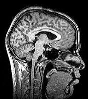
Magnetic resonance imaging
Magnetic resonance imaging
Magnetic resonance imaging , nuclear magnetic resonance imaging , or magnetic resonance tomography is a medical imaging technique used in radiology to visualize detailed internal structures...
(MRI) uses magnetic fields and radio waves to produce high quality two- or three-dimensional images of brain structures without use of ionizing radiation (X-rays) or radioactive tracers.
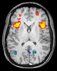
Functional magnetic resonance imaging
Functional magnetic resonance imaging
Functional magnetic resonance imaging or functional MRI is a type of specialized MRI scan used to measure the hemodynamic response related to neural activity in the brain or spinal cord of humans or other animals. It is one of the most recently developed forms of neuroimaging...
(fMRI) relies on the paramagnetic properties of oxygenated and deoxygenated hemoglobin
Hemoglobin
Hemoglobin is the iron-containing oxygen-transport metalloprotein in the red blood cells of all vertebrates, with the exception of the fish family Channichthyidae, as well as the tissues of some invertebrates...
to see images of changing blood flow in the brain associated with neural activity. This allows images to be generated that reflect which brain structures are activated (and how) during performance of different tasks.
Most fMRI scanners allow subjects to be presented with different visual images, sounds and touch stimuli, and to make different actions such as pressing a button or moving a joystick. Consequently, fMRI can be used to reveal brain structures and processes associated with perception, thought and action. The resolution of fMRI is about 2-3 millimeters at present, limited by the spatial spread of the hemodynamic response to neural activity. It has largely superseded PET for the study of brain activation patterns. PET, however, retains the significant advantage of being able to identify specific brain receptors
Receptor (biochemistry)
In biochemistry, a receptor is a molecule found on the surface of a cell, which receives specific chemical signals from neighbouring cells or the wider environment within an organism...
(or transporters
Monoamine transporter
Monoamine transporters are protein structures that function as integral plasma membrane transporters to regulate concentrations of extracellular monoamine neurotransmitters. Three major classes of MATs are responsible for the reuptake of their associated amine neurotransmitters...
) associated with particular neurotransmitters through its ability to image radiolabelled receptor "ligands" (receptor ligands are any chemicals that stick to receptors).
As well as research on healthy subjects, fMRI is increasingly used for the medical diagnosis of disease. Because fMRI is exquisitely sensitive to blood flow, it is extremely sensitive to early changes in the brain resulting from ischemia (abnormally low blood flow), such as the changes which follow stroke
Stroke
A stroke, previously known medically as a cerebrovascular accident , is the rapidly developing loss of brain function due to disturbance in the blood supply to the brain. This can be due to ischemia caused by blockage , or a hemorrhage...
. Early diagnosis of certain types of stroke is increasingly important in neurology, since substances which dissolve blood clots may be used in the first few hours after certain types of stroke occur, but are dangerous to use afterwards. Brain changes seen on fMRI may help to make the decision to treat with these agents.
With between 72% and 90% accuracy where chance would achieve 0.8%, fMRI techniques can decide which of a set of known images the subject is viewing.
Magnetoencephalography
MagnetoencephalographyMagnetoencephalography
Magnetoencephalography is a technique for mapping brain activity by recording magnetic fields produced by electrical currents occurring naturally in the brain, using arrays of SQUIDs...
(MEG) is an imaging technique used to measure the magnetic fields produced by electrical activity in the brain via extremely sensitive devices such as superconducting quantum interference devices
SQUID
A SQUID is a very sensitive magnetometer used to measure extremely weak magnetic fields, based on superconducting loops containing Josephson junctions....
(SQUIDs). MEG offers a very direct measurement of neural electrical activity (compared to fMRI for example) with very high temporal resolution but relatively low spatial resolution. The advantage of measuring the magnetic fields produced by neural activity is that they are likely to be less distorted by surrounding tissue (particularly the skull and scalp) compared to the electric fields measured by EEG. Specifically, it can be shown that magnetic fields produced by electrical activity are not affected by the surrounding head tissue, when the the head is modeled as a set of concentric spherical shells, each being an isotropic homogeneous conductor. Real heads are non-spherical and have largely anisotropic conductivities (particularly white matter and skull). While skull anisotropy has negligible effect on MEG (unlike EEG), white matter anisotropy strongly affects MEG measurements for radial and deep sources. Note, however, that the skull was assumed to be uniformly anisotropic in this study, which is not true for a real head: the absolute and relative thicknesses of diploë
Diploë
Diploë refers to the spongy bone structure of the internal part of short, irregular, and flat bones.In the cranial bones, the layers of compact tissue are familiarly known as the tables of the skull; the outer one is thick and tough; the inner is thin, dense, and brittle, and hence is termed the...
and tables layers vary among and within the skull bones. This makes it likely that MEG is also affected by the skull anisotropy, although probably not to the same degree as EEG.
There are many uses for the MEG, including assisting surgeons in localizing a pathology, assisting researchers in determining the function of various parts of the brain, neurofeedback, and others.
Positron emission tomography
Positron emission tomographyPositron emission tomography
Positron emission tomography is nuclear medicine imaging technique that produces a three-dimensional image or picture of functional processes in the body. The system detects pairs of gamma rays emitted indirectly by a positron-emitting radionuclide , which is introduced into the body on a...
(PET) measures emissions from radioactively labeled metabolically active chemicals that have been injected into the bloodstream. The emission data are computer-processed to produce 2- or 3-dimensional images of the distribution of the chemicals throughout the brain. The positron
Positron
The positron or antielectron is the antiparticle or the antimatter counterpart of the electron. The positron has an electric charge of +1e, a spin of ½, and has the same mass as an electron...
emitting radioisotopes used are produced by a cyclotron
Cyclotron
In technology, a cyclotron is a type of particle accelerator. In physics, the cyclotron frequency or gyrofrequency is the frequency of a charged particle moving perpendicularly to the direction of a uniform magnetic field, i.e. a magnetic field of constant magnitude and direction...
, and chemicals are labeled with these radioactive atoms. The labeled compound, called a radiotracer, is injected into the bloodstream and eventually makes its way to the brain. Sensors in the PET scanner detect the radioactivity as the compound accumulates in various regions of the brain. A computer uses the data gathered by the sensors to create multicolored 2- or 3-dimensional images that show where the compound acts in the brain. Especially useful are a wide array of ligands used to map different aspects of neurotransmitter activity, with by far the most commonly used PET tracer being a labeled form of glucose (see Fludeoxyglucose (18F) (FDG)).
The greatest benefit of PET scanning is that different compounds can show blood flow and oxygen and glucose
Glucose
Glucose is a simple sugar and an important carbohydrate in biology. Cells use it as the primary source of energy and a metabolic intermediate...
metabolism
Metabolism
Metabolism is the set of chemical reactions that happen in the cells of living organisms to sustain life. These processes allow organisms to grow and reproduce, maintain their structures, and respond to their environments. Metabolism is usually divided into two categories...
in the tissues of the working brain. These measurements reflect the amount of brain activity in the various regions of the brain and allow to learn more about how the brain works. PET scans were superior to all other metabolic imaging methods in terms of resolution and speed of completion (as little as 30 seconds), when they first became available. The improved resolution permitted better study to be made as to the area of the brain activated by a particular task. The biggest drawback of PET scanning is that because the radioactivity decays rapidly, it is limited to monitoring short tasks. Before fMRI technology came online, PET scanning was the preferred method of functional (as opposed to structural) brain imaging, and it still continues to make large contributions to neuroscience
Neuroscience
Neuroscience is the scientific study of the nervous system. Traditionally, neuroscience has been seen as a branch of biology. However, it is currently an interdisciplinary science that collaborates with other fields such as chemistry, computer science, engineering, linguistics, mathematics,...
.
PET scanning is also used for diagnosis of brain disease, most notably because brain tumors, strokes, and neuron-damaging diseases which cause dementia (such as Alzheimer's disease) all cause great changes in brain metabolism, which in turn causes easily detectable changes in PET scans. PET is probably most useful in early cases of certain dementias (with classic examples being Alzheimer's disease
Alzheimer's disease
Alzheimer's disease also known in medical literature as Alzheimer disease is the most common form of dementia. There is no cure for the disease, which worsens as it progresses, and eventually leads to death...
and Pick's disease
Pick's disease
Pick's disease, is a rare neurodegenerative disease that causes progressive destruction of nerve cells in the brain. Symptoms include loss of speech , and dementia. While some of the symptoms can initially be alleviated, the disease progresses and patients often die within two to ten years...
) where the early damage is too diffuse and makes too little difference in brain volume and gross structure to change CT and standard MRI images enough to be able to reliably differentiate it from the "normal" range of cortical atrophy which occurs with aging (in many but not all) persons, and which does not cause clinical dementia.
Single photon emission computed tomography
Single photon emission computed tomographySingle photon emission computed tomography
Single-photon emission computed tomography is a nuclear medicine tomographic imaging technique using gamma rays. It is very similar to conventional nuclear medicine planar imaging using a gamma camera. However, it is able to provide true 3D information...
(SPECT) is similar to PET and uses gamma ray
Gamma ray
Gamma radiation, also known as gamma rays or hyphenated as gamma-rays and denoted as γ, is electromagnetic radiation of high frequency . Gamma rays are usually naturally produced on Earth by decay of high energy states in atomic nuclei...
emitting radioisotopes and a gamma camera
Gamma camera
A gamma camera, also called a scintillation camera or Anger camera, is a device used to image gamma radiation emitting radioisotopes, a technique known as scintigraphy...
to record data that a computer uses to construct two- or three-dimensional images of active brain regions SPECT relies on an injection of radioactive tracer, which is rapidly taken up by the brain but does not redistribute. Uptake of SPECT agent is nearly 100% complete within 30 – 60s, reflecting cerebral blood flow (CBF) at the time of injection. These properties of SPECT make it particularly well suited for epilepsy imaging, which is usually made difficult by problems with patient movement and variable seizure types. SPECT provides a "snapshot" of cerebral blood flow since scans can be acquired after seizure termination (so long as the radioactive tracer was injected at the time of the seizure). A significant limitation of SPECT is its poor resolution (about 1 cm) compared to that of MRI.
Like PET, SPECT also can be used to differentiate different kinds of disease processes which produce dementia, and it is increasingly used for this purpose. Neuro-PET has a disadvantage of requiring use of tracers with half-lives of at most 110 minutes, such as FDG. These must be made in a cyclotron, and are expensive or even unavailable if necessary transport times are prolonged more than a few half-lives. SPECT, however, is able to make use of tracers with much longer half-lives, such as technetium-99m, and as a result, is far more widely available.
See also
- Brain mappingBrain mappingBrain mapping is a set of neuroscience techniques predicated on the mapping of quantities or properties onto spatial representations of the brain resulting in maps.- Overview :...
- Functional neuroimagingFunctional neuroimagingFunctional neuroimaging is the use of neuroimaging technology to measure an aspect of brain function, often with a view to understanding the relationship between activity in certain brain areas and specific mental functions...
- functional near-infrared imaging
- History of brain imaging
- Human Cognome ProjectHuman Cognome ProjectThe Human Cognome Project is an effort to reverse engineer the human brain by studying both its structure and function, in order to fully understand mental processes, also known as cognition. The project has many parallels to the Human Genome Project...
- Magnetic resonance imagingMagnetic resonance imagingMagnetic resonance imaging , nuclear magnetic resonance imaging , or magnetic resonance tomography is a medical imaging technique used in radiology to visualize detailed internal structures...
- MagnetoencephalographyMagnetoencephalographyMagnetoencephalography is a technique for mapping brain activity by recording magnetic fields produced by electrical currents occurring naturally in the brain, using arrays of SQUIDs...
- Medical imagingMedical imagingMedical imaging is the technique and process used to create images of the human body for clinical purposes or medical science...
- List of neuroscience databases
- Neuroimaging software
- Statistical parametric mappingStatistical parametric mappingStatistical parametric mapping or SPM is a statistical technique created by Karl Friston for examining differences in brain activity recorded during functional neuroimaging experiments using neuroimaging technologies such as fMRI or PET...
- Transcranial magnetic stimulationTranscranial magnetic stimulationTranscranial magnetic stimulation is a noninvasive method to cause depolarization or hyperpolarization in the neurons of the brain...
- Voxel-based morphometry
External links
- The Whole Brain Atlas @ Harvard
- The McConnell Brain Imaging Center, McGill University
- The American Society of Neuroimaging (ASN).
- UCLA Neuroimaging Training Program.
- Laboratory of Neuro Imaging at UCLA
- A Neuroimaging portal
- BrainMapping.org, a free BrainMapping community information portal
- Lecture notes on mathematical aspects of neuroimaging by Will Penny, University College LondonUniversity College LondonUniversity College London is a public research university located in London, United Kingdom and the oldest and largest constituent college of the federal University of London...
- "Transcranial Magnetic Stimulation". by Michael Leventon in association with MIT AI Lab.
- Foundations of fMRI by Jamie Shorey.
- International Society for Neuroimaging in Psychiatry (ISNIP)
- Journal of Neuroimaging