

Functional neuroimaging
Encyclopedia
Neuroimaging
Neuroimaging includes the use of various techniques to either directly or indirectly image the structure, function/pharmacology of the brain...
technology to measure an aspect of brain function, often with a view to understanding the relationship between activity in certain brain
Brain
The brain is the center of the nervous system in all vertebrate and most invertebrate animals—only a few primitive invertebrates such as sponges, jellyfish, sea squirts and starfishes do not have one. It is located in the head, usually close to primary sensory apparatus such as vision, hearing,...
areas and specific mental functions. It is primarily used as a research tool in cognitive neuroscience
Cognitive neuroscience
Cognitive neuroscience is an academic field concerned with the scientific study of biological substrates underlying cognition, with a specific focus on the neural substrates of mental processes. It addresses the questions of how psychological/cognitive functions are produced by the brain...
, cognitive psychology
Cognitive psychology
Cognitive psychology is a subdiscipline of psychology exploring internal mental processes.It is the study of how people perceive, remember, think, speak, and solve problems.Cognitive psychology differs from previous psychological approaches in two key ways....
, neuropsychology
Neuropsychology
Neuropsychology studies the structure and function of the brain related to specific psychological processes and behaviors. The term neuropsychology has been applied to lesion studies in humans and animals. It has also been applied to efforts to record electrical activity from individual cells in...
, and social neuroscience
Social neuroscience
Social neuroscience is an interdisciplinary field devoted to understanding how biological systems implement social processes and behavior, and to using biological concepts and methods to inform and refine theories of social processes and behavior. Humans are fundamentally a social species, rather...
.
Overview
Common methods of functional neuroimaging include- Positron emission tomographyPositron emission tomographyPositron emission tomography is nuclear medicine imaging technique that produces a three-dimensional image or picture of functional processes in the body. The system detects pairs of gamma rays emitted indirectly by a positron-emitting radionuclide , which is introduced into the body on a...
(PET), - Functional magnetic resonance imagingFunctional magnetic resonance imagingFunctional magnetic resonance imaging or functional MRI is a type of specialized MRI scan used to measure the hemodynamic response related to neural activity in the brain or spinal cord of humans or other animals. It is one of the most recently developed forms of neuroimaging...
(fMRI), - multichannel electroencephalographyElectroencephalographyElectroencephalography is the recording of electrical activity along the scalp. EEG measures voltage fluctuations resulting from ionic current flows within the neurons of the brain...
(EEG), - magnetoencephalographyMagnetoencephalographyMagnetoencephalography is a technique for mapping brain activity by recording magnetic fields produced by electrical currents occurring naturally in the brain, using arrays of SQUIDs...
(MEG), - near infrared spectroscopic imagingNear infrared spectroscopyNear-infrared spectroscopy is a spectroscopic method that uses the near-infrared region of the electromagnetic spectrum...
(NIRSI), and
- Single photon emission computed tomographySingle photon emission computed tomographySingle-photon emission computed tomography is a nuclear medicine tomographic imaging technique using gamma rays. It is very similar to conventional nuclear medicine planar imaging using a gamma camera. However, it is able to provide true 3D information...
(SPECT)
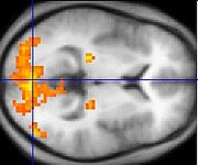
PET, fMRI and NIRSI can measure localized changes in cerebral blood flow related to neural activity. These changes are referred to as activations. Regions of the brain which are activated when a subject performs a particular task may play a role in the neural computations
Computational neuroscience
Computational neuroscience is the study of brain function in terms of the information processing properties of the structures that make up the nervous system...
which contribute to the behaviour. For instance, widespread activation of the occipital lobe
Occipital lobe
The occipital lobe is the visual processing center of the mammalian brain containing most of the anatomical region of the visual cortex. The primary visual cortex is Brodmann area 17, commonly called V1...
is typically seen in tasks which involve visual stimulation (compared with tasks that do not). This part of the brain receives signals from the retina
Retina
The vertebrate retina is a light-sensitive tissue lining the inner surface of the eye. The optics of the eye create an image of the visual world on the retina, which serves much the same function as the film in a camera. Light striking the retina initiates a cascade of chemical and electrical...
and is believed to play a role in visual perception
Visual perception
Visual perception is the ability to interpret information and surroundings from the effects of visible light reaching the eye. The resulting perception is also known as eyesight, sight, or vision...
.
Other methods of neuroimaging involve recording of electrical currents or magnetic fields, for example EEG and MEG. Different methods have different advantages for research; for instance, MEG measures brain activity with high temporal resolution (down to the millsecond level), but is limited in its ability to localize that activity. fMRI does a much better job of localizing brain activity for spatial resolution, but at the cost of speed.
Functional neuroimaging topics
The measure used in a particular study is generally related to the particular question being addressed. Measurement limitations vary amongst the techniques. For instance, MEG and EEG record the magnetic or electrical fluctuations that occur when a population of neurons is active. These methods are excellent for measuring the time-course of neural events (on the order of milliseconds,) but generally bad at measuring where those events happen. PET and fMRI measure changes in the composition of blood near a neural event. Because measurable blood changes are slow (on the order of seconds), these methods are much worse at measuring the time-course of neural events, but are generally better at measuring the location.Traditional "activation studies" focus on determining distributed patterns of brain activity associated with specific tasks. However, scientists are able to more thoroughly understand brain function by studying the interaction of distinct brain regions, as a great deal of neural processing is performed by an integrated network of several regions of the brain. An active area of neuroimaging research involves examining the functional connectivity of spatially remote brain regions. Functional connectivity analyses allow the characterization of interregional neural interactions during particular cognitive or motor tasks or merely from spontaneous activity during rest. FMRI and PET enable creation of functional connectivity maps of distinct spatial distributions of temporally correlated brain regions called functional networks.
A direct method to measure functional connectivity is to observe how stimulation of one part of the brain will affect other areas. This can be done noninvasively in humans by combining transcranial magnetic stimulation
Transcranial magnetic stimulation
Transcranial magnetic stimulation is a noninvasive method to cause depolarization or hyperpolarization in the neurons of the brain...
with one of the neuroimaging tools such as PET, fMRI, or EEG. Massimini et al. (Science, September 30, 2005) used EEG to record how activity spreads from the stimulated site. They reported that in non-REM sleep, although the brain responds vigorously to stimulation, functional connectivity is much attenuated from its level during wakefulness. Thus, during deep sleep, "brain areas do not talk to each other".
Functional neuroimaging draws on data from many areas other than cognitive neuroscience
Cognitive neuroscience
Cognitive neuroscience is an academic field concerned with the scientific study of biological substrates underlying cognition, with a specific focus on the neural substrates of mental processes. It addresses the questions of how psychological/cognitive functions are produced by the brain...
and social neuroscience
Social neuroscience
Social neuroscience is an interdisciplinary field devoted to understanding how biological systems implement social processes and behavior, and to using biological concepts and methods to inform and refine theories of social processes and behavior. Humans are fundamentally a social species, rather...
, including biological sciences (such as neuroanatomy
Neuroanatomy
Neuroanatomy is the study of the anatomy and organization of the nervous system. In contrast to animals with radial symmetry, whose nervous system consists of a distributed network of cells, animals with bilateral symmetry have segregated, defined nervous systems, and thus we can begin to speak of...
and neurophysiology
Neurophysiology
Neurophysiology is a part of physiology. Neurophysiology is the study of nervous system function...
), physics
Physics
Physics is a natural science that involves the study of matter and its motion through spacetime, along with related concepts such as energy and force. More broadly, it is the general analysis of nature, conducted in order to understand how the universe behaves.Physics is one of the oldest academic...
and maths, to further develop and refine the technology.
Critique and careful interpretation
Functional neuroimaging studies have to be carefully designed and interpreted with care.Statistical analysis (often using a technique called statistical parametric mapping
Statistical parametric mapping
Statistical parametric mapping or SPM is a statistical technique created by Karl Friston for examining differences in brain activity recorded during functional neuroimaging experiments using neuroimaging technologies such as fMRI or PET...
) is often needed so that the different sources of activation within the brain can be distinguished from one another.
This can be particularly challenging when considering processes which are difficult to conceptualise or have no easily definable task associated with them (for example belief
Belief
Belief is the psychological state in which an individual holds a proposition or premise to be true.-Belief, knowledge and epistemology:The terms belief and knowledge are used differently in philosophy....
and consciousness
Consciousness
Consciousness is a term that refers to the relationship between the mind and the world with which it interacts. It has been defined as: subjectivity, awareness, the ability to experience or to feel, wakefulness, having a sense of selfhood, and the executive control system of the mind...
).
Functional neuroimaging of interesting phenomena often gets cited in the press.
In one case a group of prominent functional neuroimaging researchers felt compelled to write a letter to New York Times in response to an op-ed
Op-ed
An op-ed, abbreviated from opposite the editorial page , is a newspaper article that expresses the opinions of a named writer who is usually unaffiliated with the newspaper's editorial board...
article about a study of so-called neuropolitics.
They argued that some of the interpretations of the study were "scientifically unfounded".
See also
- Analysis of Functional NeuroImages
- ElectroencephalographyElectroencephalographyElectroencephalography is the recording of electrical activity along the scalp. EEG measures voltage fluctuations resulting from ionic current flows within the neurons of the brain...
- Event related potential
- FMRIB Software LibraryFMRIB Software LibraryThe FMRIB Software Library is a software library containing image analysis and statistical tools for functional, structural and diffusion MRI brain imaging data....
- FreeSurferFreeSurferFreeSurfer is an MRI brain imaging software package developed by the Athinoula A. Martinos Center for Biomedical Imaging at Massachusetts General Hospital. It is an important tool in functional brain mapping and facilitates the visualization of the functional regions of the highly-folded cerebral...
- MagnetoencephalographyMagnetoencephalographyMagnetoencephalography is a technique for mapping brain activity by recording magnetic fields produced by electrical currents occurring naturally in the brain, using arrays of SQUIDs...
- Perfusion ScanningPerfusion scanningPerfusion is defined as the passage of fluid through the lymphatic system or blood vessels to an organ or a tissue. The practice of perfusion scanning, is the process by which this perfusion can be observed, recorded and quantified...
- Single photon emission computed tomographySingle photon emission computed tomographySingle-photon emission computed tomography is a nuclear medicine tomographic imaging technique using gamma rays. It is very similar to conventional nuclear medicine planar imaging using a gamma camera. However, it is able to provide true 3D information...
- Statistical Parametric MappingStatistical parametric mappingStatistical parametric mapping or SPM is a statistical technique created by Karl Friston for examining differences in brain activity recorded during functional neuroimaging experiments using neuroimaging technologies such as fMRI or PET...
- Dynamic causal modellingDynamic causal modellingDynamic causal modelling is a method for the interpretation of functional neuroimaging data . It is implemented in the Statistical parametric mapping software. DCM is a recent technique, developed only in 2003....
Further reading
- Cabeza, R., & Kingstone, K. (eds.) (2006). Handbook of Functional Neuroimaging of Cognition. MIT Press.
- Cacioppo, J.T., Tassinary, L.G., & Berntson, G. G. (2007). Handbook of Psychophysiology. Cambridge University Press.
- Hillary, F.G., & DeLuca, J. (2007). Functional Neuroimaging in Clinical Populations.
- Kanwisher, N., & Duncan, J. (2004). Functional Neuroimaging of Visual Cognition.
- Silbersweig, D., & Stern, E. (2001). Functional Neuroimaging and Neuropsychology Fundamentals and Practice.
- Thatcher, R,W. (1994). Functional Neuroimaging: Technical Foundations.