

Magnetoencephalography
Encyclopedia
Magnetoencephalography is a technique for mapping brain activity by recording magnetic field
s produced by electrical currents occurring naturally in the brain
, using arrays of SQUID
s (superconducting quantum interference devices). Applications of MEG include basic research into perceptual and cognitive brain processes, localizing regions affected by pathology before surgical removal, determining the function of various parts of the brain, and neurofeedback
.
in 1968, before the availability of the SQUID
, using a copper induction coil as the detector. To reduce the magnetic background noise, the measurements were made in a magnetically shielded room. The coil detector was barely sensitive enough, resulting in poor, noisy MEG measurements that were difficult to use. Later, Cohen built a better shielded room at MIT, and used one of the first SQUID detectors, just developed by James E. Zimmerman
, a researcher at Ford Motor Company, to again measure MEG signals. This time the signals were almost as clear as those of EEG
. This stimulated the interest of physicists who had been looking for uses of SQUIDs. Subsequently, various types of spontaneous and evoked MEGs began to be measured.
At first, a single SQUID detector was used to successively measure the magnetic field at a number of points around the subject’s head. This was cumbersome, and in the 1980s, MEG manufacturers began to arrange multiple sensors into arrays to cover a larger area of the head. Present-day MEG arrays are set in helmet-shaped dewar
that typically contain 300 sensors, covering most of the head. In this way, MEGs of a subject or patient can now be accumulated rapidly and efficiently.
(fT) for cortical
activity and 103 fT for the human alpha rhythm, the brain's magnetic field is considerably smaller than the ambient magnetic noise in an urban environment, which is on the order of 108 fT or 0.1 µT. The essential problem of biomagnetism is thus the weakness of the signal relative to the sensitivity of the detectors, and to the competing environmental noise.
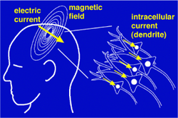 The MEG (and EEG) signals derive from the net effect of ionic currents flowing in the dendrite
s of neurons during synaptic
transmission. In accordance with Maxwell's equations
, any electrical current will produce an orthogonally oriented magnetic field. It is this field which is measured. The net currents can be thought of as electric dipoles
, i.e. currents with a position, orientation, and magnitude, but no spatial extent. According to the right-hand rule
, a current dipole gives rise to a magnetic field that flows around the axis of its vector component.
To generate a signal that is detectable, approximately 50,000 active neurons are needed. Since current dipoles must have similar orientations to generate magnetic fields that reinforce each other, it is often the layer of pyramidal cell
s, which are situated perpendicular to the cortical surface, that give rise to measurable magnetic fields. Bundles of these neurons that are orientated tangentially to the scalp surface project measurable portions of their magnetic fields outside of the head, and these bundles are typically located in the sulci
. Researchers are experimenting with various signal processing
methods in the search for methods that detect deep brain (i.e., non-cortical) signal, but no clinically useful method is currently available.
It is worth noting that action potentials do not usually produce an observable field, mainly because the currents associated with action potentials flow in opposite directions and the magnetic fields cancel out. However, action fields have been measured from peripheral nerves.
, is necessary. Appropriate magnetic shielding can be obtained by constructing rooms made of aluminium
and mu-metal
for reducing high-frequency and low-frequency noise, respectively.

layer, similar in composition to molybdenum permalloy
. The ferromagnetic layer is supplied as 1 mm sheets, while the innermost layer is composed of four sheets in close contact, and the outer two layers are composed of three sheets each. Magnetic continuity is maintained by overlay strips. Insulating washers are used in the screw assemblies to ensure that each main layer is electrically isolated. This helps eliminate radio frequency
radiation, which would degrade SQUID performance. Electrical continuity of the aluminium is also maintained by aluminium overlay strips to ensure AC
eddy-current shielding, which is important at frequencies greater than 1 Hz. The junctions of the inner layer are often electroplated with silver or gold to improve conductivity of the aluminium layers.
s are mounted at the center of each surface and oriented orthogonally to it. This negatively feeds a DC
amplifier through a low-pass network with a slow falloff to minimize positive feedback and oscillation. Built into the system are shaking and degaussing wires. Shaking wires increase the magnetic permeability, while the permanent degaussing wires are applied to all surfaces of the inner main layer to degauss the surfaces. Moreover, noise cancellation algorithms can reduce both low-frequency and high-frequency noise. Modern systems have a noise floor
of around 2–3 fT/Hz0.5 above 1 Hz.
The source models can be either over-determined or under-determined. An over-determined model may consist of a few point-like sources ("equivalent dipoles"), whose locations are then estimated from the data. Under-determined models may be used in cases where many different distributed areas are activated ("distributed source solutions"): there are infinitely many possible current distributions explaining the measurement results, but the most likely is selected. Localization algorithms make use of given source and head models to find a likely location for an underlying focal field generator.
Localization algorithms using overdetermined models operate by expectation-maximization
: the system is initialized with a first guess. A loop is started, in which a forward model is used to simulate the magnetic field that would result from the current guess. The guess is adjusted to reduce the discrepancy between the simulated field and the measured field. This process is iterated until convergence.
The extent to which the constraint-free MEG inverse problem is ill-posed cannot be overemphasized. If one's goal is to estimate the current density within the human brain with say a 5mm resolution then it is well established that the vast majority of the information needed to perform a unique inversion must come not from the magnetic field measurement but rather from the constraints applied to the problem. Furthermore, even when a unique inversion is possible in the presence of such constraints said inversion can be unstable. These conclusions are easily deduced from published works (see for example )
(MRI) images to create magnetic source images (MSI). The two sets of data are combined by measuring the location of a common set of fiducial points
marked during MRI with lipid markers and marked during MEG with electrified coils of wire that give off magnetic fields. The locations of the fiducial points in each data set are then used to define a common coordinate system so that superimposing the functional MEG data onto the structural MRI data ("coregistration") is possible.
A criticism of the use of this technique in clinical practice is that it produces colored areas with definite boundaries superimposed upon an MRI scan: the untrained viewer may not realize that the colors do not represent a physiological certainty, because of the relatively low spatial resolution of MEG, but rather a probability cloud derived from statistical processes. However, when the magnetic source image corroborates other data, it can be of clinical utility.
(MUltiple SIgnal Classification) and MSST (MultiStart Spatial and Temporal) modeling are applied to the analysis of MEG responses. The limitations of dipole models for characterizing neuronal responses are (1) difficulties in localizing extended sources with ECDs, (2) problems with accurately estimating the total number of dipoles in advance, and (3) dependency on dipole location, especially depth in the brain.
(ICA) is another signal processing solution that separates different signals that are statistically independent in time. It is primarily used to remove artifacts such as blinking, eye muscle movement, facial muscle artifacts, cardiac artifacts, etc. from MEG and EEG signals that may be contaminated with outside noise. However, ICA has poor resolution of highly correlated brain sources.
(fMRI), which depends on changes in blood flow, can at best resolve events with a precision of several hundred milliseconds. MEG also accurately pinpoints sources in primary auditory, somatosensory and motor areas. For creating functional maps of human cortex during more complex cognitive tasks, MEG is most often combined with fMRI, as the methods complement each other. Neuronal (MEG) and hemodynamic
(fMRI) data do not necessarily agree, in spite of the tight relationship between local field potentials (LFP) and blood oxygenation level dependent (BOLD) signals. MEG and BOLD signals may originate from the same source (though the BOLD signals are filtered through the hemodynamic response).
Recent studies have reported successful classification of patients with multiple sclerosis
, Alzheimer's disease
, schizophrenia
, Sjögren's syndrome
, chronic alcoholism, and facial pain. MEG can be used to distinguish these patients from healthy control subjects, suggesting a future role of MEG in diagnostics.
, and in localizing eloquent cortex
for surgical planning in patients with brain tumor
s or intractable epilepsy. The goal of epilepsy surgery is to remove the epileptogenic tissue while sparing healthy brain areas. Knowing the exact position of essential brain regions (such as the primary motor cortex
and primary sensory cortex, visual cortex
, and areas involved in speech production and comprehension) helps to avoid surgically induced neurological deficits. Direct cortical stimulation and somatosensory evoked potentials recorded on ECoG are considered the gold standard for localizing essential brain regions. These procedures can be performed either intraoperatively or from chronically indwelling subdural grid electrodes. Both are invasive.
Noninvasive MEG localizations of the central sulcus obtained from somatosensory evoked magnetic fields show strong agreement with these invasive recordings. MEG studies assist in clarification of the functional organization of primary somatosensory cortex and to delineate the spatial extent of hand somatosensory cortex by stimulation of the individual digits. This agreement between invasive localization of cortical tissue and MEG recordings shows the effectiveness of MEG analysis and indicates that MEG may substitute invasive procedures in the future.
, audition
and language processing
in fetuses and newborns.
(better than 1 ms
). Since the MEG signal is a direct measure of neuronal activity, its temporal resolution is comparable with that of intracranial electrodes.
MEG complements other brain activity measurement techniques such as electroencephalography
(EEG), positron emission tomography
(PET), and fMRI. Its strengths consist in independence of head geometry compared to EEG (unless ferromagnetic implant
s are present) and non-invasiveness, as opposed to PET
.
Scalp EEG is sensitive to extracellular volume currents produced by postsynaptic potentials. MEG primarily detects intracellular currents associated with these synaptic potentials because the field components generated by volume currents tend to cancel out in a spherical volume conductor The decay of magnetic fields as a function of distance is more pronounced than for electric fields. MEG is therefore more sensitive to superficial cortical activity, which makes it useful for the study of neocortical epilepsy. Finally, MEG is reference-free, while scalp EEG relies on a reference that, when active, makes interpretation of the data difficult.
Magnetic field
A magnetic field is a mathematical description of the magnetic influence of electric currents and magnetic materials. The magnetic field at any given point is specified by both a direction and a magnitude ; as such it is a vector field.Technically, a magnetic field is a pseudo vector;...
s produced by electrical currents occurring naturally in the brain
Human brain
The human brain has the same general structure as the brains of other mammals, but is over three times larger than the brain of a typical mammal with an equivalent body size. Estimates for the number of neurons in the human brain range from 80 to 120 billion...
, using arrays of SQUID
SQUID
A SQUID is a very sensitive magnetometer used to measure extremely weak magnetic fields, based on superconducting loops containing Josephson junctions....
s (superconducting quantum interference devices). Applications of MEG include basic research into perceptual and cognitive brain processes, localizing regions affected by pathology before surgical removal, determining the function of various parts of the brain, and neurofeedback
Neurofeedback
Neurofeedback , also called neurotherapy, neurobiofeedback or EEG biofeedback is a type of biofeedback that uses realtime displays of electroencephalography or functional magnetic resonance imaging to illustrate brain activity, often with a goal of controlling central nervous system activity...
.
History of MEG
MEG signals were first measured by University of Illinois physicist David CohenDavid Cohen (physicist)
David Cohen is known mostly for his pioneering work in the area of Biomagnetism , where he made many of the first measurements....
in 1968, before the availability of the SQUID
SQUID
A SQUID is a very sensitive magnetometer used to measure extremely weak magnetic fields, based on superconducting loops containing Josephson junctions....
, using a copper induction coil as the detector. To reduce the magnetic background noise, the measurements were made in a magnetically shielded room. The coil detector was barely sensitive enough, resulting in poor, noisy MEG measurements that were difficult to use. Later, Cohen built a better shielded room at MIT, and used one of the first SQUID detectors, just developed by James E. Zimmerman
James Edward Zimmerman
James Edward Zimmerman was born in Lantry, South Dakota. He was a coinventor of the radio-frequency superconducting quantum interference device and he is credited with coining the term.- Career :...
, a researcher at Ford Motor Company, to again measure MEG signals. This time the signals were almost as clear as those of EEG
Electroencephalography
Electroencephalography is the recording of electrical activity along the scalp. EEG measures voltage fluctuations resulting from ionic current flows within the neurons of the brain...
. This stimulated the interest of physicists who had been looking for uses of SQUIDs. Subsequently, various types of spontaneous and evoked MEGs began to be measured.
At first, a single SQUID detector was used to successively measure the magnetic field at a number of points around the subject’s head. This was cumbersome, and in the 1980s, MEG manufacturers began to arrange multiple sensors into arrays to cover a larger area of the head. Present-day MEG arrays are set in helmet-shaped dewar
Vacuum flask
A vacuum flask is an insulating storage vessel which keeps its contents hotter or cooler than its surroundings. Invented by Sir James Dewar in 1892, the vacuum flask consists of two flasks, placed one within the other and joined at the neck...
that typically contain 300 sensors, covering most of the head. In this way, MEGs of a subject or patient can now be accumulated rapidly and efficiently.
The basis of the MEG signal
Synchronized neuronal currents induce weak magnetic fields. At 10 femtoteslaTesla (unit)
The tesla is the SI derived unit of magnetic field B . One tesla is equal to one weber per square meter, and it was defined in 1960 in honour of the inventor, physicist, and electrical engineer Nikola Tesla...
(fT) for cortical
Cerebral cortex
The cerebral cortex is a sheet of neural tissue that is outermost to the cerebrum of the mammalian brain. It plays a key role in memory, attention, perceptual awareness, thought, language, and consciousness. It is constituted of up to six horizontal layers, each of which has a different...
activity and 103 fT for the human alpha rhythm, the brain's magnetic field is considerably smaller than the ambient magnetic noise in an urban environment, which is on the order of 108 fT or 0.1 µT. The essential problem of biomagnetism is thus the weakness of the signal relative to the sensitivity of the detectors, and to the competing environmental noise.
Dendrite
Dendrites are the branched projections of a neuron that act to conduct the electrochemical stimulation received from other neural cells to the cell body, or soma, of the neuron from which the dendrites project...
s of neurons during synaptic
Synapse
In the nervous system, a synapse is a structure that permits a neuron to pass an electrical or chemical signal to another cell...
transmission. In accordance with Maxwell's equations
Maxwell's equations
Maxwell's equations are a set of partial differential equations that, together with the Lorentz force law, form the foundation of classical electrodynamics, classical optics, and electric circuits. These fields in turn underlie modern electrical and communications technologies.Maxwell's equations...
, any electrical current will produce an orthogonally oriented magnetic field. It is this field which is measured. The net currents can be thought of as electric dipoles
Dipole
In physics, there are several kinds of dipoles:*An electric dipole is a separation of positive and negative charges. The simplest example of this is a pair of electric charges of equal magnitude but opposite sign, separated by some distance. A permanent electric dipole is called an electret.*A...
, i.e. currents with a position, orientation, and magnitude, but no spatial extent. According to the right-hand rule
Right-hand rule
In mathematics and physics, the right-hand rule is a common mnemonic for understanding notation conventions for vectors in 3 dimensions. It was invented for use in electromagnetism by British physicist John Ambrose Fleming in the late 19th century....
, a current dipole gives rise to a magnetic field that flows around the axis of its vector component.
To generate a signal that is detectable, approximately 50,000 active neurons are needed. Since current dipoles must have similar orientations to generate magnetic fields that reinforce each other, it is often the layer of pyramidal cell
Pyramidal cell
Pyramidal neurons are a type of neuron found in areas of the brain including cerebral cortex, the hippocampus, and in the amygdala. Pyramidal neurons are the primary excitation units of the mammalian prefrontal cortex and the corticospinal tract. Pyramidal neurons were first discovered and...
s, which are situated perpendicular to the cortical surface, that give rise to measurable magnetic fields. Bundles of these neurons that are orientated tangentially to the scalp surface project measurable portions of their magnetic fields outside of the head, and these bundles are typically located in the sulci
Sulcus (neuroanatomy)
In neuroanatomy, a sulcus is a depression or fissure in the surface of the brain.It surrounds the gyri, creating the characteristic appearance of the brain in humans and other large mammals....
. Researchers are experimenting with various signal processing
Signal processing
Signal processing is an area of systems engineering, electrical engineering and applied mathematics that deals with operations on or analysis of signals, in either discrete or continuous time...
methods in the search for methods that detect deep brain (i.e., non-cortical) signal, but no clinically useful method is currently available.
It is worth noting that action potentials do not usually produce an observable field, mainly because the currents associated with action potentials flow in opposite directions and the magnetic fields cancel out. However, action fields have been measured from peripheral nerves.
Magnetic shielding
Since the magnetic signals emitted by the brain are on the order of a few femtoteslas, shielding from external magnetic signals, including the Earth's magnetic fieldEarth's magnetic field
Earth's magnetic field is the magnetic field that extends from the Earth's inner core to where it meets the solar wind, a stream of energetic particles emanating from the Sun...
, is necessary. Appropriate magnetic shielding can be obtained by constructing rooms made of aluminium
Aluminium
Aluminium or aluminum is a silvery white member of the boron group of chemical elements. It has the symbol Al, and its atomic number is 13. It is not soluble in water under normal circumstances....
and mu-metal
Mu-metal
Mu-metal is a nickel-iron alloy that is notable for its high magnetic permeability. The high permeability makes mu-metal very effective at screening static or low-frequency magnetic fields, which cannot be attenuated by other methods. The name came from the Greek letter mu which represents...
for reducing high-frequency and low-frequency noise, respectively.
Magnetically shielded room (MSR)
A magnetically shielded room (MSR) model consists of three nested main layers. Each of these layers is made of a pure aluminium layer, plus a high permeability ferromagneticFerromagnetism
Ferromagnetism is the basic mechanism by which certain materials form permanent magnets, or are attracted to magnets. In physics, several different types of magnetism are distinguished...
layer, similar in composition to molybdenum permalloy
Permalloy
Permalloy is a nickel-iron magnetic alloy, with about 20% iron and 80% nickel content. It is notable for its very high magnetic permeability, which makes it useful as a magnetic core material in electrical and electronic equipment, and also in magnetic shielding to block magnetic fields...
. The ferromagnetic layer is supplied as 1 mm sheets, while the innermost layer is composed of four sheets in close contact, and the outer two layers are composed of three sheets each. Magnetic continuity is maintained by overlay strips. Insulating washers are used in the screw assemblies to ensure that each main layer is electrically isolated. This helps eliminate radio frequency
Radio frequency
Radio frequency is a rate of oscillation in the range of about 3 kHz to 300 GHz, which corresponds to the frequency of radio waves, and the alternating currents which carry radio signals...
radiation, which would degrade SQUID performance. Electrical continuity of the aluminium is also maintained by aluminium overlay strips to ensure AC
Alternating current
In alternating current the movement of electric charge periodically reverses direction. In direct current , the flow of electric charge is only in one direction....
eddy-current shielding, which is important at frequencies greater than 1 Hz. The junctions of the inner layer are often electroplated with silver or gold to improve conductivity of the aluminium layers.
Active shielding system
Active systems are designed for three dimensional noise cancellation. To implement an active system, low-noise fluxgate magnetometerMagnetometer
A magnetometer is a measuring instrument used to measure the strength or direction of a magnetic field either produced in the laboratory or existing in nature...
s are mounted at the center of each surface and oriented orthogonally to it. This negatively feeds a DC
Direct current
Direct current is the unidirectional flow of electric charge. Direct current is produced by such sources as batteries, thermocouples, solar cells, and commutator-type electric machines of the dynamo type. Direct current may flow in a conductor such as a wire, but can also flow through...
amplifier through a low-pass network with a slow falloff to minimize positive feedback and oscillation. Built into the system are shaking and degaussing wires. Shaking wires increase the magnetic permeability, while the permanent degaussing wires are applied to all surfaces of the inner main layer to degauss the surfaces. Moreover, noise cancellation algorithms can reduce both low-frequency and high-frequency noise. Modern systems have a noise floor
Noise floor
In signal theory, the noise floor is the measure of the signal created from the sum of all the noise sources and unwanted signals within a measurement system, where the noise is defined as any signal other than the one being monitored....
of around 2–3 fT/Hz0.5 above 1 Hz.
The inverse problem
The challenge posed by MEG is to determine the location of electric activity within the brain from the induced magnetic fields outside the head. Problems such as this, where model parameters (the location of the activity) have to be estimated from measured data (the SQUID signals) are referred to as inverse problems (in contrast to forward problems where the model parameters (e.g. source location) are known and the data (e.g. the field at a given distance) is to be estimated.) The primary difficulty is that the inverse problem does not have a unique solution (i.e., there are infinite possible "correct" answers), and the problem of defining the "best" solution is itself the subject of intensive research. Possible solutions can be derived using models involving prior knowledge of brain activity.The source models can be either over-determined or under-determined. An over-determined model may consist of a few point-like sources ("equivalent dipoles"), whose locations are then estimated from the data. Under-determined models may be used in cases where many different distributed areas are activated ("distributed source solutions"): there are infinitely many possible current distributions explaining the measurement results, but the most likely is selected. Localization algorithms make use of given source and head models to find a likely location for an underlying focal field generator.
Localization algorithms using overdetermined models operate by expectation-maximization
Expectation-maximization algorithm
In statistics, an expectation–maximization algorithm is an iterative method for finding maximum likelihood or maximum a posteriori estimates of parameters in statistical models, where the model depends on unobserved latent variables...
: the system is initialized with a first guess. A loop is started, in which a forward model is used to simulate the magnetic field that would result from the current guess. The guess is adjusted to reduce the discrepancy between the simulated field and the measured field. This process is iterated until convergence.
The extent to which the constraint-free MEG inverse problem is ill-posed cannot be overemphasized. If one's goal is to estimate the current density within the human brain with say a 5mm resolution then it is well established that the vast majority of the information needed to perform a unique inversion must come not from the magnetic field measurement but rather from the constraints applied to the problem. Furthermore, even when a unique inversion is possible in the presence of such constraints said inversion can be unstable. These conclusions are easily deduced from published works (see for example )
Magnetic source imaging
The estimated source locations can be combined with magnetic resonance imagingMagnetic resonance imaging
Magnetic resonance imaging , nuclear magnetic resonance imaging , or magnetic resonance tomography is a medical imaging technique used in radiology to visualize detailed internal structures...
(MRI) images to create magnetic source images (MSI). The two sets of data are combined by measuring the location of a common set of fiducial points
Fiduciary marker
A fiducial marker or fiducial is an object used in the field of view of an imaging system which appears in the image produced, for use as a point of reference or a measure...
marked during MRI with lipid markers and marked during MEG with electrified coils of wire that give off magnetic fields. The locations of the fiducial points in each data set are then used to define a common coordinate system so that superimposing the functional MEG data onto the structural MRI data ("coregistration") is possible.
A criticism of the use of this technique in clinical practice is that it produces colored areas with definite boundaries superimposed upon an MRI scan: the untrained viewer may not realize that the colors do not represent a physiological certainty, because of the relatively low spatial resolution of MEG, but rather a probability cloud derived from statistical processes. However, when the magnetic source image corroborates other data, it can be of clinical utility.
Dipole model source localization
A widely accepted source-modeling technique for MEG involves calculating a set of equivalent current dipoles (ECDs), which assumes the underlying neuronal sources to be focal. This dipole fitting procedure is non-linear and over-determined, since the number of unknown dipole parameters is smaller than the number of MEG measurements. Automated multiple dipole model algorithms such as MUSICMusic
Music is an art form whose medium is sound and silence. Its common elements are pitch , rhythm , dynamics, and the sonic qualities of timbre and texture...
(MUltiple SIgnal Classification) and MSST (MultiStart Spatial and Temporal) modeling are applied to the analysis of MEG responses. The limitations of dipole models for characterizing neuronal responses are (1) difficulties in localizing extended sources with ECDs, (2) problems with accurately estimating the total number of dipoles in advance, and (3) dependency on dipole location, especially depth in the brain.
Distributed Source Models
Unlike multiple-dipole modeling, distributed source models divide the source space into a grid containing a large number of dipoles. The inverse problem is to obtain the dipole moments for the grid nodes. As the number of unknown dipole moments is much greater than the number of MEG sensors, the inverse solution is highly underdetermined, so additional constraints are needed to reduce ambiguity of the solution. The primary advantage of this approach is that no prior specification of the source model is necessary. However, the resulting distributions may be difficult to interpret, because they only reflect a "blurred" (or even distorted) image of the true neuronal source distribution. The matter is complicated by the fact that spatial resolution depends strongly on several parameters such as brain area, depth, orientation, number of sensors etc.Independent component analysis (ICA)
Independent component analysisIndependent component analysis
Independent component analysis is a computational method for separating a multivariate signal into additive subcomponents supposing the mutual statistical independence of the non-Gaussian source signals...
(ICA) is another signal processing solution that separates different signals that are statistically independent in time. It is primarily used to remove artifacts such as blinking, eye muscle movement, facial muscle artifacts, cardiac artifacts, etc. from MEG and EEG signals that may be contaminated with outside noise. However, ICA has poor resolution of highly correlated brain sources.
MEG use in the field
In research, MEG's primary use is the measurement of time courses of activity. MEG can resolve events with a precision of 10 milliseconds or faster, while functional MRIFunctional magnetic resonance imaging
Functional magnetic resonance imaging or functional MRI is a type of specialized MRI scan used to measure the hemodynamic response related to neural activity in the brain or spinal cord of humans or other animals. It is one of the most recently developed forms of neuroimaging...
(fMRI), which depends on changes in blood flow, can at best resolve events with a precision of several hundred milliseconds. MEG also accurately pinpoints sources in primary auditory, somatosensory and motor areas. For creating functional maps of human cortex during more complex cognitive tasks, MEG is most often combined with fMRI, as the methods complement each other. Neuronal (MEG) and hemodynamic
Hemodynamics
Hemodynamics, meaning literally "blood movement" is the study of blood flow or the circulation.All animal cells require oxygen for the conversion of carbohydrates, fats and proteins into carbon dioxide , water and energy in a process known as aerobic respiration...
(fMRI) data do not necessarily agree, in spite of the tight relationship between local field potentials (LFP) and blood oxygenation level dependent (BOLD) signals. MEG and BOLD signals may originate from the same source (though the BOLD signals are filtered through the hemodynamic response).
Recent studies have reported successful classification of patients with multiple sclerosis
Multiple sclerosis
Multiple sclerosis is an inflammatory disease in which the fatty myelin sheaths around the axons of the brain and spinal cord are damaged, leading to demyelination and scarring as well as a broad spectrum of signs and symptoms...
, Alzheimer's disease
Alzheimer's disease
Alzheimer's disease also known in medical literature as Alzheimer disease is the most common form of dementia. There is no cure for the disease, which worsens as it progresses, and eventually leads to death...
, schizophrenia
Schizophrenia
Schizophrenia is a mental disorder characterized by a disintegration of thought processes and of emotional responsiveness. It most commonly manifests itself as auditory hallucinations, paranoid or bizarre delusions, or disorganized speech and thinking, and it is accompanied by significant social...
, Sjögren's syndrome
Sjögren's syndrome
Sjögren's syndrome , also known as "Mikulicz disease" and "Sicca syndrome", is a systemic autoimmune disease in which immune cells attack and destroy the exocrine glands that produce tears and saliva....
, chronic alcoholism, and facial pain. MEG can be used to distinguish these patients from healthy control subjects, suggesting a future role of MEG in diagnostics.
Focal epilepsy
The clinical uses of MEG are in detecting and localizing pathological activity in patients with epilepsyEpilepsy
Epilepsy is a common chronic neurological disorder characterized by seizures. These seizures are transient signs and/or symptoms of abnormal, excessive or hypersynchronous neuronal activity in the brain.About 50 million people worldwide have epilepsy, and nearly two out of every three new cases...
, and in localizing eloquent cortex
Eloquent cortex
Eloquent cortex is a name used by neurologists for areas of cortex that—if removed—will result in loss of sensory processing or linguistic ability, minor paralysis, or paralysis...
for surgical planning in patients with brain tumor
Brain tumor
A brain tumor is an intracranial solid neoplasm, a tumor within the brain or the central spinal canal.Brain tumors include all tumors inside the cranium or in the central spinal canal...
s or intractable epilepsy. The goal of epilepsy surgery is to remove the epileptogenic tissue while sparing healthy brain areas. Knowing the exact position of essential brain regions (such as the primary motor cortex
Primary motor cortex
The primary motor cortex is a brain region that in humans is located in the posterior portion of the frontal lobe. Itworks in association with pre-motor areas to plan and execute movements. M1 contains large neurons known as Betz cells, which send long axons down the spinal cord to synapse onto...
and primary sensory cortex, visual cortex
Visual cortex
The visual cortex of the brain is the part of the cerebral cortex responsible for processing visual information. It is located in the occipital lobe, in the back of the brain....
, and areas involved in speech production and comprehension) helps to avoid surgically induced neurological deficits. Direct cortical stimulation and somatosensory evoked potentials recorded on ECoG are considered the gold standard for localizing essential brain regions. These procedures can be performed either intraoperatively or from chronically indwelling subdural grid electrodes. Both are invasive.
Noninvasive MEG localizations of the central sulcus obtained from somatosensory evoked magnetic fields show strong agreement with these invasive recordings. MEG studies assist in clarification of the functional organization of primary somatosensory cortex and to delineate the spatial extent of hand somatosensory cortex by stimulation of the individual digits. This agreement between invasive localization of cortical tissue and MEG recordings shows the effectiveness of MEG analysis and indicates that MEG may substitute invasive procedures in the future.
Fetal MEG
MEG has been used to study cognitive processes such as visionVisual perception
Visual perception is the ability to interpret information and surroundings from the effects of visible light reaching the eye. The resulting perception is also known as eyesight, sight, or vision...
, audition
Hearing (sense)
Hearing is the ability to perceive sound by detecting vibrations through an organ such as the ear. It is one of the traditional five senses...
and language processing
Language processing
Language processing refers to the way human beings process speech or writing and understand it as language. Most recent theories back the idea that this process is made completely by and inside the brain.- Spoken language :...
in fetuses and newborns.
Comparison with related techniques
MEG has been in development since the 1960s but has been greatly aided by recent advances in computing algorithms and hardware, and promises improved spatial resolution coupled with extremely high temporal resolutionTemporal resolution
Temporal resolution refers to the precision of a measurement with respect to time. Often there is a tradeoff between temporal resolution of a measurement and its spatial resolution. This trade-off can be attributed to the finite speed of light and the fact that it takes a certain period of time...
(better than 1 ms
Millisecond
A millisecond is a thousandth of a second.10 milliseconds are called a centisecond....
). Since the MEG signal is a direct measure of neuronal activity, its temporal resolution is comparable with that of intracranial electrodes.
MEG complements other brain activity measurement techniques such as electroencephalography
Electroencephalography
Electroencephalography is the recording of electrical activity along the scalp. EEG measures voltage fluctuations resulting from ionic current flows within the neurons of the brain...
(EEG), positron emission tomography
Positron emission tomography
Positron emission tomography is nuclear medicine imaging technique that produces a three-dimensional image or picture of functional processes in the body. The system detects pairs of gamma rays emitted indirectly by a positron-emitting radionuclide , which is introduced into the body on a...
(PET), and fMRI. Its strengths consist in independence of head geometry compared to EEG (unless ferromagnetic implant
Implant
Implant can refer to:*Implant , or specifically:**Brain implant**Breast implant**Buttock implant**Cochlear implant**Contraceptive implant**Dental implant**Mini dental implant**Extraocular implant**Fetal tissue implant...
s are present) and non-invasiveness, as opposed to PET
Positron emission tomography
Positron emission tomography is nuclear medicine imaging technique that produces a three-dimensional image or picture of functional processes in the body. The system detects pairs of gamma rays emitted indirectly by a positron-emitting radionuclide , which is introduced into the body on a...
.
MEG vs. EEG
Although EEG and MEG signals originate from the same neurophysiological processes, there are important differences. Magnetic fields are less distorted than electric fields by the skull and scalp, which results in a better spatial resolution of the MEG. Whereas scalp EEG is sensitive to both tangential and radial components of a current source in a spherical volume conductor, MEG detects only its tangential components. MEG therefore measures activity in the sulci selectively, whereas scalp EEG measures activity both in the sulci and at the top of the cortical gyri. EEG is therefore sensitive to activity in more brain areas, but activity that is visible in MEG can also be localized with more accuracy.Scalp EEG is sensitive to extracellular volume currents produced by postsynaptic potentials. MEG primarily detects intracellular currents associated with these synaptic potentials because the field components generated by volume currents tend to cancel out in a spherical volume conductor The decay of magnetic fields as a function of distance is more pronounced than for electric fields. MEG is therefore more sensitive to superficial cortical activity, which makes it useful for the study of neocortical epilepsy. Finally, MEG is reference-free, while scalp EEG relies on a reference that, when active, makes interpretation of the data difficult.
See also
- ElectroencephalographyElectroencephalographyElectroencephalography is the recording of electrical activity along the scalp. EEG measures voltage fluctuations resulting from ionic current flows within the neurons of the brain...
- ElectrophysiologyElectrophysiologyElectrophysiology is the study of the electrical properties of biological cells and tissues. It involves measurements of voltage change or electric current on a wide variety of scales from single ion channel proteins to whole organs like the heart...
- Evoked fieldEvoked fieldEvoked fields are part of the magnetoencephalogram. They are brain signals evoked by sensory stimulation, but usually buried by the ongoing brain activity...
- Auditory evoked fieldAuditory evoked fieldAuditory evoked field or AEF is an induced neural activity that is recorded via magnetoencephalography , which is an equivalent of Auditory evoked potential recorded by electroencephalography. The advantage of AEF over AEP is the powerful spatial resolution provided by magnetic field recording,...
- Functional neuroimagingFunctional neuroimagingFunctional neuroimaging is the use of neuroimaging technology to measure an aspect of brain function, often with a view to understanding the relationship between activity in certain brain areas and specific mental functions...
- MagnetocardiographyMagnetocardiographyMagnetocardiography is a technique to measure the magnetic fields produced by electrical activity in the heart using extremely sensitive devices such as the Superconducting Quantum Interference Device...
- MagnetogastrographyMagnetogastrographyMagnetogastrography is the science of recording magnetogastrograms . Magnetogastrograms are recordings of magnetic fields resulting from electrical activity from the stomach and can be considered similar to electrocardiograms. The magnetic fields are typically recorded using SQUIDs.-External links:*...
- MagnetomyographyMagnetomyographyMagnetomyography is a technique for mapping muscle activity by recording magnetic fields produced by electrical currents occurring naturally in the muscles, using arrays of SQUIDs . It has a better capability than electromyography for detecting slow or direct currents.-External links:***...
- MagnetometerMagnetometerA magnetometer is a measuring instrument used to measure the strength or direction of a magnetic field either produced in the laboratory or existing in nature...
- Magnetic source imaging
- Mu-metalMu-metalMu-metal is a nickel-iron alloy that is notable for its high magnetic permeability. The high permeability makes mu-metal very effective at screening static or low-frequency magnetic fields, which cannot be attenuated by other methods. The name came from the Greek letter mu which represents...
- SQUIDSQUIDA SQUID is a very sensitive magnetometer used to measure extremely weak magnetic fields, based on superconducting loops containing Josephson junctions....
- Direct brain interfaces
- Whole brain emulation
Further reading
- Baillet S., Mosher J. C., Leahy R. M.(2001) "Electromagnetic Brain Mapping" in IEEE Signal Processing Magazine, November 2001, 14-30.
- Cohen, D. "Boston and the history of biomagnetism". Neurology and Clinical Neurophysiology 2004; 30: 1.
- Cohen, D., Halgren, E. (2004). "Magnetoencephalography". In: Encyclopedia of Neuroscience, Adelman G., Smith B., editors Elsevier, 1st, 2nd and 3rd (2004) editions.
- Hämäläinen, M., Hari, R., Ilmoniemi, R., Knuutila, J. and Lounasmaa, O. V. (1993) "Magnetoencephalography – theory, instrumentation, and applications to noninvasive studies of signal processing in the human brain" in Reviews of Modern Physics 1993, 65: pp. 413–497
- Hansen, Peter C., Kringelbach, Morten L. and Salmelin, Riita (eds.) (2010) MEG: An Introduction to Methods. New York: Oxford University Press Inc.
- Murakami S, Okada Y. Contributions of principal neocortical neurons to magnetoencephalography and electr, oencephalography signals. J Physiol. 2006 Sep 15;575(Pt 3):925-36.
- Suk, J., Ribary, U., Cappell,J. Yamamoto, T. and Llinas, R. Anatomical localization revealed by MEG recordings of the human somatosensory system. EEG J 78:185-196, 1991.
- Tanzer I.O., (2006) Numerical Modeling in Electro- and Magnetoencephalography, Ph.D. Thesis, Helsinki University of Technology, Finland.
- Gyrus rectus cortical dysplasia revealed by magnetic source imaging with Elekta Neuromag, The Magnetoencephalography Unit, Laboratoire de Cartographie Fonctionnelle du Cerveau, ULB-Hôpital Erasme, Brussels, Belgium