
Functional magnetic resonance imaging
Encyclopedia
Functional magnetic resonance imaging or functional MRI (fMRI) is a type of specialized MRI
scan used to measure the hemodynamic response (change in blood flow) related to neural
activity in the brain
or spinal cord
of human
s or other animals. It is one of the most recently developed forms of neuroimaging
. Since the early 1990s, fMRI has come to dominate the brain mapping field due to its relatively low invasiveness, absence of radiation exposure, and relatively wide availability.
and blood oxygenation in the brain
(collectively known as hemodynamics
) are closely linked to neural activity. When neural cells are active they increase their consumption of energy from glucose
and switch to less energetically effective, but more rapid anaerobic
glycolysis
.
The local response to this energy utilization is to increase blood flow to regions of increased neural activity, which occurs after a delay of approximately 1–2 seconds. This hemodynamic response rises to a peak over 4–6 seconds, before falling back to baseline (and typically undershooting slightly). This leads to changes in local cerebral blood volume
and local changes in the local concentration of oxyhemoglobin that are detectable through their paramagnetic
effects.
at AT&T Bell labs
.
Ogawa and colleagues had recognized the potential importance of BOLD for functional brain imaging with MRI, but the first successful fMRI study was reported by John W. Belliveau and colleagues in 1991 using an intraveneously administered paramagnetic contrast agent (Gadolinium).
Using a visual stimulus paradigm, localized increases in blood volume (32 +/- 10 percent, n = 7 subjects) were detected in the primary visual cortex. In 1991, using the same visual stimulus approach, Hoppel, and her colleagues at the MGH-NMR Center, presented findings at the 1991 conference of the Society for Magnetic Resonance in Medicine demonstrating human brain activity made visible without contrast agents, through the BOLD effect. In 1992, three papers were published using endogenous BOLD contrast MRI. One was submitted by Peter Bandettini at the Medical College of Wisconsin
on February 5, revised March 31, accepted March 31 and published in the June 1992 issue of Magnetic Resonance in Medicine
(MRM). The second by Kenneth Kwong
and colleagues also applied BOLD to image human brain activities with MRI and was submitted on March 26 and published in the June issue of PNAS
in 1992. In the same year, Dr. Ogawa submitted their result on March 31 and published in July issue of PNAS. In the following year, Dr. Ogawa published the biophysics model of BOLD contrast in Biophysical Journal
. Dr. Bandettini also published a further paper in 1993 demonstrating quantitative determination of functional activation maps.
s at a greater rate than in the area of inactive neurons. It results in a surplus of oxyhemoglobin in the veins of the area and distinguishable change of the local ratio of oxyhemoglobin to deoxyhemoglobin, the "marker" of BOLD for MRI.
Hemoglobin
is diamagnetic when oxygenated (oxyhemoglobin) but paramagnetic when deoxygenated (deoxyhemoglobin).
The magnetic resonance
(MR) signal of blood is therefore slightly different depending on the level of oxygenation. Higher BOLD signal intensities arise from increases in the concentration of oxygenated hemoglobin
since the blood magnetic susceptibility
now more closely matches the tissue magnetic susceptibility. By collecting data in an MRI scanner with sequence parameters sensitive to changes in magnetic susceptibility one can assess changes in BOLD contrast. These changes can be either positive or negative depending upon the relative changes in both cerebral blood flow
(CBF) and oxygen consumption. Increases in CBF that outstrip changes in oxygen consumption will lead to increased BOLD signal, conversely decreases in CBF that outstrip changes in oxygen consumption will cause decreased BOLD signal intensity. The signal difference is very small, but given many repetitions of a thought, action or experience, statistical methods can be used to determine the areas of the brain which reliably show more of this difference as a result, and therefore which areas of the brain are active during that thought, action or experience.
Almost all current fMRI research uses BOLD as the method for determining where activity occurs in the brain as the result of various experiences, but because the signals are relative and not individually quantitative, some question its rigor. Other methods which propose to measure neural activity more directly have been attempted (for example measurement of the Oxygen Extraction Fraction (OEF) in regions of the brain, which measures how much of the oxyhemoglobin in the blood has been converted to deoxyhemoglobin
or direct detection of magnetic fields generated by neuronal currents), but because the electromagnetic fields created by an active or firing neuron are so weak, the signal-to-noise ratio
is extremely low and statistical
methods used to extract quantitative data have been largely unsuccessful as of yet.
;
that is, the blood supply is tightly regulated in space and time to provide the nutrients for brain metabolism. However, neuroscientists
have been seeking a more direct relationship between the blood supply and the neural inputs/outputs that can be related to observable electrical activity and circuit models of brain function.
While current data indicate that local field potential
s, an index of integrated electrical activity, form a marginally better correlation with blood flow than the spiking action potential
s that are most directly associated with neural communication
, no simple measure of electrical activity to date has provided an adequate correlation with metabolism and the blood supply across a wide dynamic range. Presumably, this reflects the complex nature of metabolic processes, which form a superset with regards to electrical activity. Some recent results have suggested that the increase in cerebral blood flow (CBF) following neural activity is not causally related to the metabolic demands of the brain region, but rather is driven by the presence of neurotransmitter
s, like glutamate
, serotonin
, nitric oxide
, acetylcholine
, dopamine
and noradrenaline.
Some other recent results suggest that an initial small, negative dip before the main positive BOLD signal is more highly localized and also correlates with measured local decreases in tissue oxygen concentration (perhaps reflecting increased local metabolism
during neuron activation). Use of this more localized negative BOLD signal has enabled imaging of human ocular dominance columns in primary visual cortex
, with resolution of about 0.5 mm. One problem with this technique is that the early negative BOLD signal is small and can only be seen using larger scanners with magnetic fields of at least 3 Tesla
. Further, the signal is much smaller than the normal BOLD signal, making extraction of the signal from noise more difficult. Also, this initial dip occurs within 1–2 seconds of stimulus initiation, which may not be captured when signals are recorded at long repetition (TR). If the TR is sufficiently low, increased speed of the cerebral blood flow response due to consumption of vasoactive drugs (such as caffeine)
or natural differences in vascular responsiveness may further obscure observation of the initial dip.
The BOLD signal is composed of CBF contributions from larger arteries and veins, smaller arterioles and venules, and capillaries. Experimental results indicate that the BOLD signal can be weighted to the smaller vessels, and hence closer to the active neurons, by using larger magnetic fields. For example, whereas about 70% of the BOLD signal arises from larger vessels in a 1.5 tesla scanner, about 70% arises from smaller vessels in a 7 tesla scanner. Furthermore, the size of the BOLD signal increases roughly as the square of the magnetic field strength. Hence there has been a push for larger field scanners to both improve localization and increase the signal. A few 7 tesla commercial scanners have become operational, and experimental 8 and 9 tesla scanners are under development.
s in the resulting image typically represent cubes of tissue about 2–4 millimeters on each side in humans. Recent technical advancements, such as the use of high magnetic fields and multichannel RF reception, have advanced spatial resolution to the millimeter scale. Although responses to stimuli presented as close together as one or two seconds can be distinguished from one another, using a method known as event-related fMRI
, the full time course of a BOLD response to a briefly presented stimulus lasts about 15 seconds for the robust positive response.
For these reasons, Functional imaging
provides insights into neural processing that are complementary to insights of other studies in neurophysiology
.
 Subjects participating in a fMRI experiment are asked to lie still and are usually restrained with soft pads to prevent movement from disturbing measurements. Some labs also employ bite bars to reduce motion, although these are unpopular as they can be uncomfortable. Small head movements can be corrected for in post-processing of the data, but large transient motion cannot be corrected. Motion in excess of around 3 millimeters results in unusable data. Motion is an issue for all populations, but most especially problematic for subjects with certain medical conditions (e.g. Alzheimer's Disease
or schizophrenia
) or with young children. Participants can be habituated to the scanning environment and trained to remain still in an MRI simulator.
An fMRI experiment usually lasts between 15 minutes and an hour. Depending on the purpose of study, subjects may view movies, hear sounds, smell odors, perform cognitive tasks such as n-back
, memorization or imagination, press a few buttons, or perform other tasks. Researchers are required to give detailed instructions and descriptions of the experiment plan to each subject, who must sign a consent form before the experiment.
Safety is an important issue in all experiments involving MRI. Potential subjects must ensure that they are able to enter the MRI environment. The MRI scanner is built around an extremely strong magnet (1.5 teslas
or more), so potential subjects must be thoroughly examined for any ferromagnetic objects (e.g. watches, glasses, hair pins, pacemakers, bone plates and screws, etc.) before entering the scanning environment.
methodology that can detect these changes in neurons. The abrupt onset of increased neuron cell size occurs before the metabolic response commences, is shorter in duration and does not extend significantly beyond the area of the actual cell population involved. This technique is a diffusion weighted technique (DWI). There is some evidence that similar changes in axonal volume in white matter may accompany activity and this has been observed using a DTI (diffusion tensor imaging) technique. The future importance of diffusion-based functional techniques relative to BOLD techniques is not yet clear.
such as an iron oxide
that has been coated by a sugar
or starch
(to hide from the body's defense system), causes a local disturbance in the magnetic field
that is measurable by the MRI scanner. The signals associated with these kinds of contrast agents are proportional to the cerebral blood volume. While this semi-invasive method presents a considerable disadvantage in terms of studying brain function in normal subjects, it enables far greater detection sensitivity than BOLD signal, which may increase the viability of fMRI in clinical populations. Other methods of investigating blood volume that do not require an injection are a subject of current research, although no alternative technique in theory can match the high sensitivity provided by injection of contrast agent.
ASL fMRI is less popular than BOLD, as it suffers from a lower signal to noise ratio, can be less sensitive to weak stimuli and its temporal resolution is poorer than in BOLD studies. On the plus side, it can provide quantitative measures of a single well-defined parameter, CBF, whose baseline value can also be determined in the same experiment. It has also been found to outperform BOLD in terms of stability to slow signal drifts and localization of the activation area. The ASL activation signal is believed to be dominated by changes in the capillary bed of the activated area of the cortex, wheareas the BOLD signal is likely to be dominated by changes in the oxygenation of nearby veins.
The ASL Experiment
The mains steps of a standard pulsed ASL experiment are summarized below. It starts with the application of an inversion pulse to part of the neck (the inversion slab). This step aims to invert (i.e. label) the magnetization of arterial blood. A time TI later, when the labelled blood is expected to have reached the brain, an excitation pulse is applied to the area of the brain where CBF is to be measured (the imaging slice). Some of the signal in the acquired image will have come from the inverted blood that has reached the imaging slice (this is typically 1% of the total signal), but the overwhelming majority will have come from static brain tissue. This image is called the labelled (or tag) image.
To remove the contribution from static tissue, an identical image (the control image) is acquired in the absence of the blood labeling step. To mimic magnetization transfer (MT) effects of the blood labeling pulse, however, the control image acquisition needs to be preceded by a pulse with a similar RF deposition to the one used to invert blood.
The tag image can be subtracted from the control to yield an image whose signal comes exclusively from labelled blood – this is the perfusion weighted image (PWI). Fast imaging techniques such as EPI are usually employed to allow the perfusion state of the brain to be captured in as short a time as possible.
To improve the SNR of the PWI, several pairs of similar control and tag images are usually acquired and then averaged. Finally, an absolute CBF map can be generated from PWIs acquired at one or more inversion times by the application of a suitable model.
There is an immense number of variations to this basic experiment, according to the way labeling is performed, the method used to control MT effects and the types of image readout employed. Some of these are discussed below.
Labeling in ASL
Two basic methods exist to perform labeling in ASL: continuous ASL (CASL) and pulsed ASL (PASL). In CASL, a 2-4 second long flow-induced adiabatic inversion pulse is applied at the level of the internal carotid arteries. The CASL labeling scheme has a relatively low inversion efficiency and causes high levels of power deposition. Some of these problems can be minimized using a train of RF pulses to perform a flow-induced adiabatic inversion, a technique called pseudo-continuous arterial spin labeling (PCASL).
Blood inversion can also be accomplished using a short inversion pulse applied to a larger (100-150 mm wide) region of the neck. This method, pulsed arterial spin labeling (PASL), shows higher inversion efficiencies (very close to 1), particularly when adiabatic inversion pulses are used. However, imperfect slice selection profiles imply that a gap of 10-30 mm must be left between the inversion slab and the brain area to be imaged. This introduces a delay in the arrival of the inverted blood to the imaging slice, during which it experiences T1 decay, leading to a decreased sensitivity of the experiment when compared to CASL.
Another recently proposed labeling technique is velocity-selective arterial spin labeling (VS-ASL), where labeling is performed as a function of spin velocity rather than position. Only spins whose velocity is initially above a given threshold (i.e. arterial blood water spins), but subsequently drops (as it is delivered to the capillary bed) are tagged. This way, all spins traveling in small arteries and arterioles are expected to be labelled, with no constraints on spatial position.
Control of Magnetization Transfer Effects
In PASL and CASL, the labeling pulses are in off-resonance with respect to the imaging slice, as they are applied to the neck region. They can therefore give rise to MT effects, which cause a decrease in the signal of the water protons in the imaging slice and must be cancelled to avoid confounding ASL perfusion measurements.
One way of avoiding MT effects is to use a small dedicated coil placed in the neck region for the labeling pulses. No MT effects will take place in the imaging slice due to the localized transmit sensitivity of the labeling coil. This method is most often used for CASL, where MT effects are particularly important due to the high power of the labeling pulses used.
In most situations, methods that do not involve non-standard hardware configurations are preferred. These usually mimic the MT effects produced in the labeling process during the control image generation, so that no MT-related signal loss will be present in the subtracted images. These techniques are used both in CASL and PASL, but the remainder of this discussion will focus on PASL, as this is the ASL technique used in all the experimental work described in this thesis.
One of the earliest methods devised to balance magnetization transfer effects is EPISTAR, Echo Planar Imaging and Signal Targeting with Alternating Radiofrequency. To acquire the EPISTAR control image, an inversion slab identical to the one used for the tag image acquisition is placed at the same distance from the imaging slice, but distally to it. Assuming that the power spectrum of fat is symmetric around the Larmor frequency, MT effects will be identical in both runs of the experiment. A method similar to EPISTAR where the control inversion pulse is applied non-selectively, called PICORE, Proximal Inversion with a Control for Off-Resonance Effects, has also been proposed.
In another MT control scheme, the control experiment is performed applying two contiguous 180º pulses, each with half the power of the labeling pulse to the labeling slab. This method, PULSAR, Pulsed STAR Labeling of Arterial Regions, was described above under the subheading "The ASL Experiment". It is similar in rationale to TILT, Transfer Insensitive Labeling Technique, where two 90º pulses are applied in or out of phase during the tag or control image generation, respectively.
Another very popular PASL technique, which is intrinsically MT balanced, is FAIR, Flow-sensitive Alternating Inversion Recovery. In FAIR, a PWI is obtained by subtracting two images acquired following an inversion recovery. One of the images is acquired following a slice-selective inversion, whereas the other follows a non-selective inversion pulse (at the same TI). Even though FAIR images have a very different contrast from the control/tag pairs in other types of ASL, the subtraction image also shows the inverted arterial blood that has reached the imaging slice and is similar to the PWI obtained using other methods.
The Buxton Model
The perfusion weighted images obtained in ASL are not CBF maps in themselves, as they are arbitrarily scaled and the intensity of the signal in each voxel is determined by a complicated interplay of CBF and arrival and dwelling times of the blood water bolus in each voxel.
To obtain a CBF map from one or more PWIs, the signal in each voxel must be processed using a suitable mathematical model. These models can be derived either by modifying Bloch’s equations to account for the perfusion phenomenon or using kinetic theory. Using the kinetic methodology developed by Buxton and colleagues, the longitudinal magnetization difference, ΔM(t)=Mzcontrol(t)-Mztag(t), in a voxel in a
PWI can be interpreted as the convolution (conv) of an arterial input function (AIF),which will be denoted by c(t), and a residue
function, R(t), such that:
M(t)= 2*IE*M0b*f conv(c(t),R(t)) (1)
In this equation, M0b refers to the longitudinal magnetization of arterial blood, IE to the inversion efficiency of the inversion pulse and f is the temporal rate at which the inverted magnetization reaches the voxel of interest, i.e. f is the cerebral blood flow in units of inverse time. The AIF represents the amount of labelled blood that reaches the tissue, whereas the residue function describes the washout of the bolus due to T1 decay and venous outflow.
In PASL, according to Buxton’s model, the arterial input function, which is created by the labeling pulse, has the ideal shape of a hat function. This perfectly well-defined bolus travels upwards in the arterial tree, “decaying” with arterial blood’s relaxation time constant, T1b, until it reaches the perfused tissue at time Δt, the bolus arrival time. The AIF can then be written as:
c(t) = exp (-t/T1b) for Δt < t < \Δt + τ (2)
and c(t) = 0 at all other times. τ is the duration of the bolus, which is related to the spatial width of the inversion pulse and the mean blood velocity at the inversion site and from there to the imaging plane. In Buxton's model, the perfused voxels are modeled by a single compartment, implying that upon reaching the perfused voxel, all labelled water spins immediately exchange with the tissue (i.e. the exchange time, Tex, is null). It is also assumed that the water spins immediately reach equilibrium with the venous system, so that the rate at which they leave the voxel due to venous outflow is f/λ, where λ is the blood-brain partition coefficient of the tissue.
Under these assumptions, the residue function in Equation 1 can be written as:
R(t) = exp (-t/T1t) exp (-f t/λ) (3)
where T1t is the relaxation time constant of brain tissue (WM or GM). The bolus decays due to T1 relaxation and, usually to a lesser extent, venous outflow. The latter can be interpreted as an apparent change in the T1 of the tissue, so that R(t) = exp(-t/T1tapp), with 1/T1tapp = 1/T1t + f/λ.
Inserting Equations 2 and 3 into Equation 6.1, ΔM can be written as:
For 0 < t < Δt: ΔM (t) = 0
For Δt < t < Δt + τ: ΔM (t) = 2 M0b IE f exp(-t/T1app)*(exp(-k*Δt)-exp(-k*t))
For t > Δt + τ: ΔM (t) = 2 M0b IE f exp(-t/T1app)*exp(-k*Δt)*(1-exp(-k*τ)) (4)
where k = 1/T1b – 1/T1tapp and t=0 is the time at which the inversion pulse was applied.
It was shown that despite all the simplifying assumptions made, calculating absolute CBF values from an ASL experiment involves using a complicated model requiring the estimate of 7 parameters independent from perfusion: M0b, Δt, τ, IE, T1b, T1t and λ. These can be estimated from literature values or else separately measured. If perfusion weighted images are acquired at different delays following inversion, further increasing the duration of the ASL experiment, some of the unknowns other than CBF can also be estimated from a multi-parameter fit. In practice, this is only possible for one (usually Δt) or two (Δt and τ) additional variables.
QUIPSS-II
To allow for better CBF estimation using PWIs acquired at a single time point following the labeling, Wong and colleagues proposed in 1998 applying a saturation pulse to the distal end of the inversion pulse, at time TIsat, to sharply define the end of the bolus. If TIsat<τand TI> TIsat + Δt, it can be shown that:
ΔM(TI) = 2 M0b IE f exp(-TI/T1b) (5)
This method, Quantitative Imaging of Perfusion using a Single Subtraction (version II), QUIPSS-II, greatly reduces the number of unknowns needed to estimate perfusion. In theory, QUIPSS-II allows CBF to be measured accurately using a single TI. In practice, however, it is difficult to ensure that the two conditions are met for all areas of the brain, particularly in patients with abnormal hemodynamics.
Look-Locker ASL
As in an inversion recovery experiment, acquiring signal in an ASL experiment at different times following labeling involves repeating the entire experiment for each delay time, making it extremely time consuming. A variant of the ASL experiment that involves acquiring PWIs at several times following a single inversion, in an analogy with the Look-Locker method, has been proposed.
In this method, the Buxton model is modified to account for the effect of previous excitation pulses on the tissue’s magnetization. By assuming that the excitation pulses only affect water molecules after they have exchanged into tissue, the tissue longitudinal relaxation time constant can be modified as in a Look-Locker experiment. In the Buxton model (Equation 4), T1tapp should be replaced by:
T1tappeff = 1/T1t + f/λ– ln (cos FA)/ΔTI (6)
and the shape of the PWI time curve shape remains similar to that of the standard Buxton model. ΔTI, the gap between excitation pulses, is usually a well-defined experimental parameter. However, FA, the excitation flip angle, can vary spatially to a great extent, affecting the uncertainty of the experimental measurement of CBF.
Look-Locker ASL has been implemented using the FAIR scheme, taking the name ITS-FAIR, Inflow Turbo Sampling FAIR or LL-EPI, Look-Locker EPI, and also using the PULSAR method, where it was renamed QUASAR, Quantitative STAR Labelling of Arterial Regions. As Look-Locker ASL allows the perfusion curve to be sampled at several time points following a single inversion, scan time can be considerably reduced, at the expense of SNR and the simplicity of the model used.
Other ASL Models
The Buxton model can be complicated further by making more realistic assumptions about the shape of the AIF and placing restrictions and a temporal delay on the exchange of water molecules between the capillaries and tissue. Two-compartment models (tissue + capillaries) are often used to model these effects, usually by including an additional parameter such as the permeability of the capillary wall to water and/or a non negligible exchange time. These models are often analytically quite complex.
Finally, a model-free approach similar to the one used in DSC-MRI was recently proposed for ASL by Petersen and colleagues in 2006. In this method, the AIF is estimated from the signal in neighboring arterial voxels and R(t) is calculated directly from a deconvolution of ΔM (Equation 1). CBF can be estimated taking into account that R(0)=1. This method suffers, however, from the problems associated with the deconvolution of noisy data and can only be used if the perfusion signal is sampled at several time points following labeling.
White Matter Perfusion
As mentioned in Chapter 3, white matter is much less metabolically active than grey matter and, in healthy adults, its CBF is 3 to 4 times lower than in the cortex. This places WM perfusion at the limit of detection in a typical ASL experiment. The capillary transit time of white matter is also much longer than that of GM, implying that by the time the bolus reaches white matter tissue, its longitudinal magnetization will have almost fully recovered. This implies that measuring white matter perfusion using ASL is particularly challenging.
Modifications to the Standard ASL Experiment
Some modifications have been proposed to the ASL experiment to minimize its sensitivity and improve its robustness. Some of these modifications will now be discussed.
Vascular Crushers
Perfusion measurements in ASL can be confounded by intra-arterial blood signal, which is hyperintense at early time points. As most of this intravascular signal flows through the imaging slice to perfuse more distal brain areas, it should not be taken into account for CBF estimates of the imaging slice. In Look-Locker ASL, arteries can appear even more hyperintense than static tissue, due to flow related enhancement.
Although macroscopic vessels can potentially be discarded with the use of an arteriogram, this approach does not take into account the existence of partial volume effects between the arteries and brain tissue or the contribution of the arteries to the tissue signal of neighbouring voxels, due to the definition of the point spread function in MRI.
A better strategy is to dephase arterial blood signal using bipolar gradients, similar to the ones employed in phase contrast angiography. These gradients, which in ASL are usually called crushers, are added between the excitation and readout modules with an encoding velocity (vENC) of about 3 cm/s. At this value, it is expected that the blood in capillaries remains virtually undisturbed, whereas signal from macroscopic flow is eliminated. Bipolar gradients are in practice only used in a single direction (usually the foot-head, FH, direction) to avoid a sizable increase in TE and the consequent drop in SNR. Signal from macroscopic in-plane flow will therefore not be eliminated.
Multi-Slice Imaging
Most ASL schemes permit magnetization transfer effects to be balanced across the brain, allowing the acquisition of more than once slice. The effective number of acquired slices is limited by the temporal duration of the bolus. Multi-slice ASL usually provides incomplete brain coverage, as gaps must be left between different slices to avoid cross-talk.
Ascending slice schemes are often used, as the acquisition order accompanies the bolus arrival order, allowing the perfusion curve to be sampled at approximately the same temporal position across the brain. The tagged blood reaching the upper slices may, however, be affected by the excitation of downstream slices.
Bolus Length Definition
As explained above, the bolus temporal length, τ, is defined by the inversion slab's width, but cannot in practice be determined accurately as the velocity of blood at the inversion site is not known. A QUIPSS-II saturation slab applied at time TIsat can be used to define the temporal width of the bolus. If TIsat < , can be approximated by the time at which the saturation pulse is applied following the inversion, i.e. τ is now equal to TIsat.
Signal Pre-Saturation
ASL is based on image subtraction, making it extremely sensitive to differences between control and tag images which are unrelated to perfusion. These may include subject motion (discussed in more detail later in this chapter), magnetization transfer effects and imperfect slice profiles of the control and tagged pulses. It has been found that applying a pre-saturation pulse to the imaging slice in both control and tag experiments can improve perfusion quantification, as it minimises any potential imbalance between the slice profiles and increases the fraction of the measured signal that can be attributed to perfusion.
Non-EPI Readout
ASL can be performed with fast imaging techniques other than echo planar imaging. T1-FFE has been used both in the brain and perfusion studies of other organs, such as the kidney. Some studies have also reported CBF measurements using 3D-GRASE (Gradient and Spin Echo), a 3D fast imaging technique that can be regarded as a hybrid of EPI and a turbo spin echo. 3D-GRASE has the potential to provide better brain coverage than multi-slice EPI, which would in turn permit the implementation of more efficient motion correction schemes, as will be discussed below.
Vessel Selective ASL
One of the most exciting possibilities of ASL is its potential to determine the perfusion territories of individual arteries, by selectively labelling the desired arteries only. An analysis of the methods used in these applications can be found in a review article by Paiva and colleagues written in 2007.
-based process for assessing function within the living brain. MRS takes advantage of the fact that proton
s (hydrogen
atoms) residing in differing chemical environments depending upon the molecule they inhabit (H2O vs. protein
, for example) possess slightly different resonant properties (chemical shift
). For a given volume of brain (typically > 1 cubic cm), the distribution of these H resonances can be displayed as a spectrum
.
The area under the peak for each resonance provides a quantitative measure of the relative abundance of that compound. The largest peak is composed of H2O. However, there are also discernible peaks for choline
, creatine
, N-acetylaspartate (NAA) and lactate
. Fortuitously, NAA is mostly inactive within the neuron, serving as a precursor to glutamate and as storage for acetyl groups (to be used in fatty acid
synthesis) — but its relative levels are a reasonable approximation of neuronal integrity and functional status. Brain diseases (schizophrenia
, stroke
, certain tumor
s, multiple sclerosis
) can be characterized by the regional alteration in NAA levels when compared to healthy subjects. Creatine is used as a relative control value since its levels remain fairly constant, while choline and lactate levels have been used to evaluate brain tumor
s.
function provided by BOLD fMRI. White matter
bundles carry functional information between brain regions. The diffusion of water molecules is hindered across the axes of these bundles, such that measurements of water diffusion can reveal information about the location of large white matter pathways. Illnesses that disrupt the normal organization or integrity of cerebral white matter (such as multiple sclerosis) have a quantitative impact on DTI measures.
(EEG) directly measures the brain's electrical activity, giving high temporal resolution (~milliseconds) but low spatial resolution. The two techniques are therefore complementary and may be used simultaneously to record brain activity.
Recording an EEG signal inside an MRI system is technically challenging. The MRI system introduces artifacts into the EEG recording by inducing currents in the EEG leads via Faraday induction
. This can happen through several different mechanisms. An imaging sequence applies a series of short radiofrequency pulses which induce a signal in the EEG system. The pulses are short and relatively infrequent, so interference may be avoided by blanking (switching off) the EEG system during their transmission. Magnetic field gradients used during imaging also induce a signal, which is harder to remove as it is in a similar frequency range to the EEG signal. Current is also induced when EEG leads move inside the magnet bore (i.e. when the patient moves during the exam). Finally, pulsed blood flow in the patient in the static magnetic field also induces a signal (called a ballistocardiograph
ic artifact), which is also within the frequency range of interest. The EEG system also affects the MRI scan. Metal in the EEG leads and electrodes can introduce susceptibility
artifacts into MR images. Care must also be taken to limit currents induced in the EEG leads via the MRI RF system, which could heat the leads sufficiently to burn the subject.
Having simultaneously recorded EEG and fMRI data, the final hurdle is to co-register
the two datasets, as each is reconstructed using a different algorithm, subject to different distortions (see EEG-fMRI
).
(PET) scanners or more rarely with SPECT scanners.
Niels A. Lassen
and his coworkers lead the earliest efforts of functional neuroimaging, using radioactive gases to construct images of the working brain.
These nuclear imaging techniques do not use the nuclear magnetic resonance property and employ entirely different scanners.
For a typical fMRI scan, the 3D volume of the subject's head is imaged every one or two seconds, producing a few hundred to a few thousand complete images per scanning session. The nature of MRI is such that these images are acquired in Fourier transform
space, so they must be transformed back to image space to be useful. Because of practical limitations of the scanner the Fourier samples are not acquired on a grid, and scanner imperfections like thermal drift and spike noise introduce additional distortions. Small motions on the part of the subject and the subject's pulse and respiration will also affect the images.
The most common situation is that the researcher uses a pulse sequence supplied by the scanner vendor, such as an echo-planar imaging (EPI) sequence that allows for relatively rapid acquisition of many images. Software in the scanner platform itself then performs the reconstruction of images from Fourier transform space. During this stage some information is lost (specifically the complex phase of the reconstructed signal). Some types of artifacts, for example spike noise, become more difficult to remove after reconstruction, but if the scanner is working well these artifacts are thought to be relatively unimportant. For pulse sequences not provided by the vendor, for example spiral EPI, reconstruction may have to be done by software running on a separate platform.
After reconstruction the output of the scanning session consists of a series of 3D images of the brain. The most common corrections performed on these images are motion correction and correction for physiological effects. Outlier correction and spatial and/or temporal filtering may also be performed. If the task performed by the subject is thought to produce bursts of activation which are short compared to the BOLD response time (on the order of 6 seconds), temporal filtering may be performed at this stage to attempt to deconvolve
out the BOLD response and recover the temporal pattern of activation.
At this point the data provides a time series of samples for each voxel in the scanned volume. A variety of methods are used to correlate these voxel time series with the task in order to produce maps of task-dependent activation.
There are many software packages available for analysing fMRI data.
Reconstruction of MRI data needs to be tested, calibrated and confirmed. MRI can suffer from numerous artifacts that include, geometric distortions, Nyquist ghosting, and signal dropout. Medical Imaging Phantoms are used to provide a consistent geometrical source for calibration and testing purposes. Minute tumor changes can require recalibration by use of a phantom to quantify the change.
MRI procedures themselves can vary considerably in cost but generally fall somewhere between $400 and $3,500, depending on the facility and which region of the body is being scanned. Extremity scans (feet, hands, etc.) tend to be lower in price while body scans (including the brain) tend to be higher.
At least two companies have been set up to use fMRI in lie detection
: No Lie MRI
and the Cephos Corporation).
In using fMRI techniques for use in lie detection, activated areas of the brain are observed while the subject is making a statement. Depending on what regions are the most active, the technician might determine whether a subject is telling the truth or not. Since a specific combination of brain functions are needed in order to tell a lie, the simultaneous activation of these regions often indicates deception. This technology is in its early stages of development, and many of its proponents hope to replace older lie detection techniques.
In clinical trials, the usage of fMRI as a method of lie detection has appeared reliable, with studies from 2005 by Kozel et al. indicating a 90% to 93% success rate.
However, there is still a fair amount of controversy over whether these techniques are reliable enough to be used in a legal setting. Some studies indicate that while there is an overall positive correlation, there is a great deal of variation between findings and in some cases considerable difficulty in replicating the findings.
Magnetic resonance imaging
Magnetic resonance imaging , nuclear magnetic resonance imaging , or magnetic resonance tomography is a medical imaging technique used in radiology to visualize detailed internal structures...
scan used to measure the hemodynamic response (change in blood flow) related to neural
Neuron
A neuron is an electrically excitable cell that processes and transmits information by electrical and chemical signaling. Chemical signaling occurs via synapses, specialized connections with other cells. Neurons connect to each other to form networks. Neurons are the core components of the nervous...
activity in the brain
Brain
The brain is the center of the nervous system in all vertebrate and most invertebrate animals—only a few primitive invertebrates such as sponges, jellyfish, sea squirts and starfishes do not have one. It is located in the head, usually close to primary sensory apparatus such as vision, hearing,...
or spinal cord
Spinal cord
The spinal cord is a long, thin, tubular bundle of nervous tissue and support cells that extends from the brain . The brain and spinal cord together make up the central nervous system...
of human
Human
Humans are the only living species in the Homo genus...
s or other animals. It is one of the most recently developed forms of neuroimaging
Neuroimaging
Neuroimaging includes the use of various techniques to either directly or indirectly image the structure, function/pharmacology of the brain...
. Since the early 1990s, fMRI has come to dominate the brain mapping field due to its relatively low invasiveness, absence of radiation exposure, and relatively wide availability.
Background
Since the 1890s it has been known that changes in blood flowBlood flow
Blood flow is the continuous running of blood in the cardiovascular system.The human body is made up of several processes all carrying out various functions. We have the gastrointestinal system which aids the digestion and the absorption of food...
and blood oxygenation in the brain
Brain
The brain is the center of the nervous system in all vertebrate and most invertebrate animals—only a few primitive invertebrates such as sponges, jellyfish, sea squirts and starfishes do not have one. It is located in the head, usually close to primary sensory apparatus such as vision, hearing,...
(collectively known as hemodynamics
Hemodynamics
Hemodynamics, meaning literally "blood movement" is the study of blood flow or the circulation.All animal cells require oxygen for the conversion of carbohydrates, fats and proteins into carbon dioxide , water and energy in a process known as aerobic respiration...
) are closely linked to neural activity. When neural cells are active they increase their consumption of energy from glucose
Glucose
Glucose is a simple sugar and an important carbohydrate in biology. Cells use it as the primary source of energy and a metabolic intermediate...
and switch to less energetically effective, but more rapid anaerobic
Anaerobic respiration
Anaerobic respiration is a form of respiration using electron acceptors other than oxygen. Although oxygen is not used as the final electron acceptor, the process still uses a respiratory electron transport chain; it is respiration without oxygen...
glycolysis
Glycolysis
Glycolysis is the metabolic pathway that converts glucose C6H12O6, into pyruvate, CH3COCOO− + H+...
.
The local response to this energy utilization is to increase blood flow to regions of increased neural activity, which occurs after a delay of approximately 1–2 seconds. This hemodynamic response rises to a peak over 4–6 seconds, before falling back to baseline (and typically undershooting slightly). This leads to changes in local cerebral blood volume
Blood volume
Blood volume is the volume of blood in the circulatory system of an individual.-Humans:A typical adult has a blood volume of approximately between 4.7 and 5 liters, with females generally having less blood volume than males....
and local changes in the local concentration of oxyhemoglobin that are detectable through their paramagnetic
Paramagnetism
Paramagnetism is a form of magnetism whereby the paramagnetic material is only attracted when in the presence of an externally applied magnetic field. In contrast with this, diamagnetic materials are repulsive when placed in a magnetic field...
effects.
History
Blood-oxygen-level dependence (BOLD) is the MRI contrast of blood deoxyhemoglobin, first discovered in 1990 by Seiji OgawaSeiji Ogawa
Seiji Ogawa is a Japanese researcher best known for discovering the technique that underlies Functional Magnetic Resonance Imaging . He determined that the contrast in blood oxygen levels can be mapped in magnetic resonance imaging, thus showing which areas of the brain respond to the brain's...
at AT&T Bell labs
Bell Labs
Bell Laboratories is the research and development subsidiary of the French-owned Alcatel-Lucent and previously of the American Telephone & Telegraph Company , half-owned through its Western Electric manufacturing subsidiary.Bell Laboratories operates its...
.
Ogawa and colleagues had recognized the potential importance of BOLD for functional brain imaging with MRI, but the first successful fMRI study was reported by John W. Belliveau and colleagues in 1991 using an intraveneously administered paramagnetic contrast agent (Gadolinium).
Using a visual stimulus paradigm, localized increases in blood volume (32 +/- 10 percent, n = 7 subjects) were detected in the primary visual cortex. In 1991, using the same visual stimulus approach, Hoppel, and her colleagues at the MGH-NMR Center, presented findings at the 1991 conference of the Society for Magnetic Resonance in Medicine demonstrating human brain activity made visible without contrast agents, through the BOLD effect. In 1992, three papers were published using endogenous BOLD contrast MRI. One was submitted by Peter Bandettini at the Medical College of Wisconsin
Medical College of Wisconsin
Medical College of Wisconsin is a private, freestanding medical school and graduate school located in Milwaukee, Wisconsin, United States. It was formerly affiliated with Marquette University....
on February 5, revised March 31, accepted March 31 and published in the June 1992 issue of Magnetic Resonance in Medicine
Magnetic Resonance in Medicine
Magnetic Resonance in Medicine is a peer-reviewed journal devoted to the publication of original investigations concerned with all aspects of the development and use of nuclear magnetic resonance and electron paramagnetic resonance techniques for medical applications.The journal is aimed at...
(MRM). The second by Kenneth Kwong
Kenneth Kwong
Kenneth Kwong is an Associate Professor in radiology at Harvard Medical School. His work, along with that of Seiji Ogawa, was significant in the development of fMRI....
and colleagues also applied BOLD to image human brain activities with MRI and was submitted on March 26 and published in the June issue of PNAS
Proceedings of the National Academy of Sciences
The Proceedings of the National Academy of Sciences of the United States of America, usually referred to as PNAS, is the official journal of the United States National Academy of Sciences...
in 1992. In the same year, Dr. Ogawa submitted their result on March 31 and published in July issue of PNAS. In the following year, Dr. Ogawa published the biophysics model of BOLD contrast in Biophysical Journal
Biophysical Journal
The Biophysical Journal is the journal of the Biophysical Society published by Cell Press. The society comprises chemists, biochemists, and physical chemists, biologists, neuroscientists, plant and animal physiologists, engineers, mathematicians, and physicists...
. Dr. Bandettini also published a further paper in 1993 demonstrating quantitative determination of functional activation maps.
Physiology
As neurons do not have internal reserves for glucose and oxygen, more neuronal activity requires more glucose and oxygen to be delivered rapidly through the blood stream. Through a process called the hemodynamic response, blood releases glucose to neurons and astrocyteAstrocyte
Astrocytes , also known collectively as astroglia, are characteristic star-shaped glial cells in the brain and spinal cord...
s at a greater rate than in the area of inactive neurons. It results in a surplus of oxyhemoglobin in the veins of the area and distinguishable change of the local ratio of oxyhemoglobin to deoxyhemoglobin, the "marker" of BOLD for MRI.
Hemoglobin
Hemoglobin
Hemoglobin is the iron-containing oxygen-transport metalloprotein in the red blood cells of all vertebrates, with the exception of the fish family Channichthyidae, as well as the tissues of some invertebrates...
is diamagnetic when oxygenated (oxyhemoglobin) but paramagnetic when deoxygenated (deoxyhemoglobin).
The magnetic resonance
Nuclear magnetic resonance
Nuclear magnetic resonance is a physical phenomenon in which magnetic nuclei in a magnetic field absorb and re-emit electromagnetic radiation...
(MR) signal of blood is therefore slightly different depending on the level of oxygenation. Higher BOLD signal intensities arise from increases in the concentration of oxygenated hemoglobin
Hemoglobin
Hemoglobin is the iron-containing oxygen-transport metalloprotein in the red blood cells of all vertebrates, with the exception of the fish family Channichthyidae, as well as the tissues of some invertebrates...
since the blood magnetic susceptibility
Magnetic susceptibility
In electromagnetism, the magnetic susceptibility \chi_m is a dimensionless proportionality constant that indicates the degree of magnetization of a material in response to an applied magnetic field...
now more closely matches the tissue magnetic susceptibility. By collecting data in an MRI scanner with sequence parameters sensitive to changes in magnetic susceptibility one can assess changes in BOLD contrast. These changes can be either positive or negative depending upon the relative changes in both cerebral blood flow
Cerebral blood flow
Cerebral blood flow, or CBF, is the blood supply to the brain in a given time. In an adult, CBF is typically 750 millitres per minute or 15% of the cardiac output. This equates to 50 to 54 millilitres of blood per 100 grams of brain tissue per minute. CBF is tightly regulated to meet the brain's...
(CBF) and oxygen consumption. Increases in CBF that outstrip changes in oxygen consumption will lead to increased BOLD signal, conversely decreases in CBF that outstrip changes in oxygen consumption will cause decreased BOLD signal intensity. The signal difference is very small, but given many repetitions of a thought, action or experience, statistical methods can be used to determine the areas of the brain which reliably show more of this difference as a result, and therefore which areas of the brain are active during that thought, action or experience.
Almost all current fMRI research uses BOLD as the method for determining where activity occurs in the brain as the result of various experiences, but because the signals are relative and not individually quantitative, some question its rigor. Other methods which propose to measure neural activity more directly have been attempted (for example measurement of the Oxygen Extraction Fraction (OEF) in regions of the brain, which measures how much of the oxyhemoglobin in the blood has been converted to deoxyhemoglobin
or direct detection of magnetic fields generated by neuronal currents), but because the electromagnetic fields created by an active or firing neuron are so weak, the signal-to-noise ratio
Signal-to-noise ratio
Signal-to-noise ratio is a measure used in science and engineering that compares the level of a desired signal to the level of background noise. It is defined as the ratio of signal power to the noise power. A ratio higher than 1:1 indicates more signal than noise...
is extremely low and statistical
Statistics
Statistics is the study of the collection, organization, analysis, and interpretation of data. It deals with all aspects of this, including the planning of data collection in terms of the design of surveys and experiments....
methods used to extract quantitative data have been largely unsuccessful as of yet.
Neural correlates of BOLD
The precise relationship between neural signals and BOLD is under active research. In general, changes in BOLD signal are well correlated with changes in blood flow. Numerous studies during the past several decades have identified a coupling between blood flow and metabolic rateMetabolism
Metabolism is the set of chemical reactions that happen in the cells of living organisms to sustain life. These processes allow organisms to grow and reproduce, maintain their structures, and respond to their environments. Metabolism is usually divided into two categories...
;
that is, the blood supply is tightly regulated in space and time to provide the nutrients for brain metabolism. However, neuroscientists
Neuroscience
Neuroscience is the scientific study of the nervous system. Traditionally, neuroscience has been seen as a branch of biology. However, it is currently an interdisciplinary science that collaborates with other fields such as chemistry, computer science, engineering, linguistics, mathematics,...
have been seeking a more direct relationship between the blood supply and the neural inputs/outputs that can be related to observable electrical activity and circuit models of brain function.
While current data indicate that local field potential
Local field potential
A local field potential is a particular class of electrophysiological signals, which is dominated by the electrical current flowing from all nearby dendritic synaptic activity within a volume of tissue. A voltage is produced by the summed synaptic current flowing across the resistance of the local...
s, an index of integrated electrical activity, form a marginally better correlation with blood flow than the spiking action potential
Action potential
In physiology, an action potential is a short-lasting event in which the electrical membrane potential of a cell rapidly rises and falls, following a consistent trajectory. Action potentials occur in several types of animal cells, called excitable cells, which include neurons, muscle cells, and...
s that are most directly associated with neural communication
, no simple measure of electrical activity to date has provided an adequate correlation with metabolism and the blood supply across a wide dynamic range. Presumably, this reflects the complex nature of metabolic processes, which form a superset with regards to electrical activity. Some recent results have suggested that the increase in cerebral blood flow (CBF) following neural activity is not causally related to the metabolic demands of the brain region, but rather is driven by the presence of neurotransmitter
Neurotransmitter
Neurotransmitters are endogenous chemicals that transmit signals from a neuron to a target cell across a synapse. Neurotransmitters are packaged into synaptic vesicles clustered beneath the membrane on the presynaptic side of a synapse, and are released into the synaptic cleft, where they bind to...
s, like glutamate
Glutamic acid
Glutamic acid is one of the 20 proteinogenic amino acids, and its codons are GAA and GAG. It is a non-essential amino acid. The carboxylate anions and salts of glutamic acid are known as glutamates...
, serotonin
Serotonin
Serotonin or 5-hydroxytryptamine is a monoamine neurotransmitter. Biochemically derived from tryptophan, serotonin is primarily found in the gastrointestinal tract, platelets, and in the central nervous system of animals including humans...
, nitric oxide
Nitric oxide
Nitric oxide, also known as nitrogen monoxide, is a diatomic molecule with chemical formula NO. It is a free radical and is an important intermediate in the chemical industry...
, acetylcholine
Acetylcholine
The chemical compound acetylcholine is a neurotransmitter in both the peripheral nervous system and central nervous system in many organisms including humans...
, dopamine
Dopamine
Dopamine is a catecholamine neurotransmitter present in a wide variety of animals, including both vertebrates and invertebrates. In the brain, this substituted phenethylamine functions as a neurotransmitter, activating the five known types of dopamine receptors—D1, D2, D3, D4, and D5—and their...
and noradrenaline.
Some other recent results suggest that an initial small, negative dip before the main positive BOLD signal is more highly localized and also correlates with measured local decreases in tissue oxygen concentration (perhaps reflecting increased local metabolism
Metabolism
Metabolism is the set of chemical reactions that happen in the cells of living organisms to sustain life. These processes allow organisms to grow and reproduce, maintain their structures, and respond to their environments. Metabolism is usually divided into two categories...
during neuron activation). Use of this more localized negative BOLD signal has enabled imaging of human ocular dominance columns in primary visual cortex
Visual cortex
The visual cortex of the brain is the part of the cerebral cortex responsible for processing visual information. It is located in the occipital lobe, in the back of the brain....
, with resolution of about 0.5 mm. One problem with this technique is that the early negative BOLD signal is small and can only be seen using larger scanners with magnetic fields of at least 3 Tesla
Tesla (unit)
The tesla is the SI derived unit of magnetic field B . One tesla is equal to one weber per square meter, and it was defined in 1960 in honour of the inventor, physicist, and electrical engineer Nikola Tesla...
. Further, the signal is much smaller than the normal BOLD signal, making extraction of the signal from noise more difficult. Also, this initial dip occurs within 1–2 seconds of stimulus initiation, which may not be captured when signals are recorded at long repetition (TR). If the TR is sufficiently low, increased speed of the cerebral blood flow response due to consumption of vasoactive drugs (such as caffeine)
or natural differences in vascular responsiveness may further obscure observation of the initial dip.
The BOLD signal is composed of CBF contributions from larger arteries and veins, smaller arterioles and venules, and capillaries. Experimental results indicate that the BOLD signal can be weighted to the smaller vessels, and hence closer to the active neurons, by using larger magnetic fields. For example, whereas about 70% of the BOLD signal arises from larger vessels in a 1.5 tesla scanner, about 70% arises from smaller vessels in a 7 tesla scanner. Furthermore, the size of the BOLD signal increases roughly as the square of the magnetic field strength. Hence there has been a push for larger field scanners to both improve localization and increase the signal. A few 7 tesla commercial scanners have become operational, and experimental 8 and 9 tesla scanners are under development.
Technique
BOLD effects are measured using rapid volumetric acquisition of images with contrast weighed by T1 or T2*. Such images can be acquired with moderately good spatial and temporal resolution; images are usually taken every 1–4 seconds, and the voxelVoxel
A voxel is a volume element, representing a value on a regular grid in three dimensional space. This is analogous to a pixel, which represents 2D image data in a bitmap...
s in the resulting image typically represent cubes of tissue about 2–4 millimeters on each side in humans. Recent technical advancements, such as the use of high magnetic fields and multichannel RF reception, have advanced spatial resolution to the millimeter scale. Although responses to stimuli presented as close together as one or two seconds can be distinguished from one another, using a method known as event-related fMRI
Event-related functional magnetic resonance imaging
Event-related functional magnetic resonance imaging is a form of functional Magnetic Resonance Imaging in which a series of fMRI images are time-locked to a stimulus presentation and averaged together over many trials...
, the full time course of a BOLD response to a briefly presented stimulus lasts about 15 seconds for the robust positive response.
fMRI studies draw from many disciplines
fMRI is a highly interdisciplinary research area and many studies draw on knowledge in several fields:- PhysicsPhysicsPhysics is a natural science that involves the study of matter and its motion through spacetime, along with related concepts such as energy and force. More broadly, it is the general analysis of nature, conducted in order to understand how the universe behaves.Physics is one of the oldest academic...
: Physical principles underlie fMRI signals and many studies require an understanding of these underlying principles. - PsychologyPsychologyPsychology is the study of the mind and behavior. Its immediate goal is to understand individuals and groups by both establishing general principles and researching specific cases. For many, the ultimate goal of psychology is to benefit society...
: Almost all fMRI studies are essentially cognitive psychologicalCognitive psychologyCognitive psychology is a subdiscipline of psychology exploring internal mental processes.It is the study of how people perceive, remember, think, speak, and solve problems.Cognitive psychology differs from previous psychological approaches in two key ways....
, cognitive psychophysiologicalPhysiological psychologyPhysiological psychology is a subdivision of behavioral neuroscience that studies the neural mechanisms of perception and behavior through direct manipulation of the brains of nonhuman animal subjects in controlled experiments...
, and/or psychophysicalPsychophysicsPsychophysics quantitatively investigates the relationship between physical stimuli and the sensations and perceptions they effect. Psychophysics has been described as "the scientific study of the relation between stimulus and sensation" or, more completely, as "the analysis of perceptual...
experiments in which the MRI scanner is used to obtain an extra set of measurements in addition to behavioral or electroencephalographic measurements. - NeuroanatomyNeuroanatomyNeuroanatomy is the study of the anatomy and organization of the nervous system. In contrast to animals with radial symmetry, whose nervous system consists of a distributed network of cells, animals with bilateral symmetry have segregated, defined nervous systems, and thus we can begin to speak of...
: The fMRI signals can be put into the context of previous knowledge only with an understanding of the neuroanatomy. - StatisticsStatisticsStatistics is the study of the collection, organization, analysis, and interpretation of data. It deals with all aspects of this, including the planning of data collection in terms of the design of surveys and experiments....
: Correct application of statistics is essential to "tease out" observations and avoid false-positiveType I and type II errorsIn statistical test theory the notion of statistical error is an integral part of hypothesis testing. The test requires an unambiguous statement of a null hypothesis, which usually corresponds to a default "state of nature", for example "this person is healthy", "this accused is not guilty" or...
results. - ElectrophysiologyElectrophysiologyElectrophysiology is the study of the electrical properties of biological cells and tissues. It involves measurements of voltage change or electric current on a wide variety of scales from single ion channel proteins to whole organs like the heart...
: Familiarity with neuronal behavior at the electrophysiological level can help investigators design a useful fMRI study.
Advantages and Disadvantages of fMRI
Like any technique, fMRI has advantages and disadvantages, and in order to be useful, the experiments that employ it must be carefully designed and conducted to maximize its strengths and minimize its weaknesses.Advantages of fMRI
- It can noninvasively record brain signals without risks of ionising radiation inherent in other scanning methods, such as CTComputed tomographyX-ray computed tomography or Computer tomography , is a medical imaging method employing tomography created by computer processing...
or PETPositron emission tomographyPositron emission tomography is nuclear medicine imaging technique that produces a three-dimensional image or picture of functional processes in the body. The system detects pairs of gamma rays emitted indirectly by a positron-emitting radionuclide , which is introduced into the body on a...
scans. - It has high spatial resolution. 2–3 mm is typical but resolution can be as good as 1mm.
- It can record signal from all regions of the brain, unlike EEG/MEG which are biased towards the cortical surface.
- fMRI is widely used and standard data-analysis approaches have been developed which allow researchers to compare results across labs.
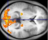
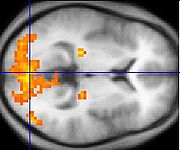
- fMRI produces compelling images of brain "activation".
Disadvantages of fMRI
- The images produced must be interpreted carefully, since correlation does not imply causality, and brain processes are complex and often non-localized.
- Statistical methods must be used carefully because they can produce false positives. One team of researchers studying reactions to pictures of human emotional expressions reported a few activated voxels in the brain of a dead salmon when no correction for multiple comparisonsMultiple comparisonsIn statistics, the multiple comparisons or multiple testing problem occurs when one considers a set of statistical inferences simultaneously. Errors in inference, including confidence intervals that fail to include their corresponding population parameters or hypothesis tests that incorrectly...
was applied, illustrating the need for rigorous statistical analyses. - The BOLD signal is only an indirect measure of neural activity, and is therefore susceptible to influence by non-neural changes in the body. This also means that it is difficult to interpret positive and negative BOLD responses
- BOLD signals are most strongly associated with the input to a given area rather than with the output. It is therefore possible (although unlikely) that a BOLD signal could be present in a given area even if there is no single unit activity.
- fMRI has poor temporal resolution. The BOLD response peaks approximately 5 seconds after neuronal firing begins in an area. This means that it is hard to distinguish BOLD responses to different events which occur within a short time window. Careful experimental design can reduce this problem. Also, some research groups are attempting to combine fMRI signals that have relatively high spatial resolution with signals recorded with other techniques, electroencephalographyElectroencephalographyElectroencephalography is the recording of electrical activity along the scalp. EEG measures voltage fluctuations resulting from ionic current flows within the neurons of the brain...
(EEG) or magnetoencephalographyMagnetoencephalographyMagnetoencephalography is a technique for mapping brain activity by recording magnetic fields produced by electrical currents occurring naturally in the brain, using arrays of SQUIDs...
(MEG), which have higher temporal resolution but worse spatial resolution. - fMRI has often been used to show activation localized to specific regions, thus minimizing the distributed nature of processing in neural networksBiological neural networkIn neuroscience, a biological neural network describes a population of physically interconnected neurons or a group of disparate neurons whose inputs or signalling targets define a recognizable circuit. Communication between neurons often involves an electrochemical process...
. Several recent multivariate statisticalMultivariate statisticsMultivariate statistics is a form of statistics encompassing the simultaneous observation and analysis of more than one statistical variable. The application of multivariate statistics is multivariate analysis...
techniques work around this issue by characterizing interactions between "active" regions found via traditional univariateUnivariateIn mathematics, univariate refers to an expression, equation, function or polynomial of only one variable. Objects of any of these types but involving more than one variable may be called multivariate...
techniques. - The BOLD response can be affected by a variety of factors, including: drugs/substances; age, brain pathology; local differences in neurovascular coupling; attention; amount of carbon dioxide in the blood; etc.
For these reasons, Functional imaging
Functional imaging
Functional imaging , is a method of detecting or measuring changes in metabolism, blood flow, regional chemical composition, and absorption....
provides insights into neural processing that are complementary to insights of other studies in neurophysiology
Neurophysiology
Neurophysiology is a part of physiology. Neurophysiology is the study of nervous system function...
.
Scanning in practice
Alzheimer's disease
Alzheimer's disease also known in medical literature as Alzheimer disease is the most common form of dementia. There is no cure for the disease, which worsens as it progresses, and eventually leads to death...
or schizophrenia
Schizophrenia
Schizophrenia is a mental disorder characterized by a disintegration of thought processes and of emotional responsiveness. It most commonly manifests itself as auditory hallucinations, paranoid or bizarre delusions, or disorganized speech and thinking, and it is accompanied by significant social...
) or with young children. Participants can be habituated to the scanning environment and trained to remain still in an MRI simulator.
An fMRI experiment usually lasts between 15 minutes and an hour. Depending on the purpose of study, subjects may view movies, hear sounds, smell odors, perform cognitive tasks such as n-back
N-back
The n-back task is a Continuous Performance Task that is commonly used in neuroimaging to stimulate brain activity in test subjects. It was introduced by Wayne Kirchner in 1958.-The task:...
, memorization or imagination, press a few buttons, or perform other tasks. Researchers are required to give detailed instructions and descriptions of the experiment plan to each subject, who must sign a consent form before the experiment.
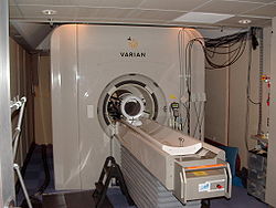
Safety is an important issue in all experiments involving MRI. Potential subjects must ensure that they are able to enter the MRI environment. The MRI scanner is built around an extremely strong magnet (1.5 teslas
Tesla (unit)
The tesla is the SI derived unit of magnetic field B . One tesla is equal to one weber per square meter, and it was defined in 1960 in honour of the inventor, physicist, and electrical engineer Nikola Tesla...
or more), so potential subjects must be thoroughly examined for any ferromagnetic objects (e.g. watches, glasses, hair pins, pacemakers, bone plates and screws, etc.) before entering the scanning environment.
Related techniques
Aside from BOLD fMRI, there are other related ways to probe brain activity using magnetic resonance properties:Diffusion based functional MRI
Neuronal activity produces some immediate physical changes in cell shape that can be detected because they affect the compartment shape and size for water diffusion. A much improved spatial and temporal resolution for fMRI data collection has now been achieved by using diffusion MRIDiffusion MRI
Diffusion MRI is a magnetic resonance imaging method that produces in vivo images of biological tissues weighted with the local microstructural characteristics of water diffusion, which is capable of showing connections between brain regions...
methodology that can detect these changes in neurons. The abrupt onset of increased neuron cell size occurs before the metabolic response commences, is shorter in duration and does not extend significantly beyond the area of the actual cell population involved. This technique is a diffusion weighted technique (DWI). There is some evidence that similar changes in axonal volume in white matter may accompany activity and this has been observed using a DTI (diffusion tensor imaging) technique. The future importance of diffusion-based functional techniques relative to BOLD techniques is not yet clear.
Contrast MR
An injected contrast agentMRI contrast agent
MRI contrast agents are a group of contrast media used to improve the visibility of internal body structures in magnetic resonance imaging . The most commonly used compounds for contrast enhancement are gadolinium-based. MRI contrast agents alter the relaxation times of tissues and body cavities...
such as an iron oxide
Iron oxide
Iron oxides are chemical compounds composed of iron and oxygen. All together, there are sixteen known iron oxides and oxyhydroxides.Iron oxides and oxide-hydroxides are widespread in nature, play an important role in many geological and biological processes, and are widely utilized by humans, e.g.,...
that has been coated by a sugar
Sugar
Sugar is a class of edible crystalline carbohydrates, mainly sucrose, lactose, and fructose, characterized by a sweet flavor.Sucrose in its refined form primarily comes from sugar cane and sugar beet...
or starch
Starch
Starch or amylum is a carbohydrate consisting of a large number of glucose units joined together by glycosidic bonds. This polysaccharide is produced by all green plants as an energy store...
(to hide from the body's defense system), causes a local disturbance in the magnetic field
Magnetic field
A magnetic field is a mathematical description of the magnetic influence of electric currents and magnetic materials. The magnetic field at any given point is specified by both a direction and a magnitude ; as such it is a vector field.Technically, a magnetic field is a pseudo vector;...
that is measurable by the MRI scanner. The signals associated with these kinds of contrast agents are proportional to the cerebral blood volume. While this semi-invasive method presents a considerable disadvantage in terms of studying brain function in normal subjects, it enables far greater detection sensitivity than BOLD signal, which may increase the viability of fMRI in clinical populations. Other methods of investigating blood volume that do not require an injection are a subject of current research, although no alternative technique in theory can match the high sensitivity provided by injection of contrast agent.
Arterial spin Labeling (ASL)
Arterial Spin Labelling (ASL), also known as arterial spin tagging, is an MRI technique capable of measuring cerebral blood flow (CBF) in vivo. ASL is capable of providing cerebral perfusion maps, without requiring the administration of a contrast agent or the use of ionising radiation, as it uses magnetically-labelled endogenous blood water as a freely-diffusible tracer. It was first proposed in 1992 and has since benefited from a number of modifications aimed at improving its robustness. ASL can monitor changes in CBF with activation and fMRI studies can therefore be conducted using ASL instead of relying on the BOLD effect.ASL fMRI is less popular than BOLD, as it suffers from a lower signal to noise ratio, can be less sensitive to weak stimuli and its temporal resolution is poorer than in BOLD studies. On the plus side, it can provide quantitative measures of a single well-defined parameter, CBF, whose baseline value can also be determined in the same experiment. It has also been found to outperform BOLD in terms of stability to slow signal drifts and localization of the activation area. The ASL activation signal is believed to be dominated by changes in the capillary bed of the activated area of the cortex, wheareas the BOLD signal is likely to be dominated by changes in the oxygenation of nearby veins.
The ASL Experiment
The mains steps of a standard pulsed ASL experiment are summarized below. It starts with the application of an inversion pulse to part of the neck (the inversion slab). This step aims to invert (i.e. label) the magnetization of arterial blood. A time TI later, when the labelled blood is expected to have reached the brain, an excitation pulse is applied to the area of the brain where CBF is to be measured (the imaging slice). Some of the signal in the acquired image will have come from the inverted blood that has reached the imaging slice (this is typically 1% of the total signal), but the overwhelming majority will have come from static brain tissue. This image is called the labelled (or tag) image.
To remove the contribution from static tissue, an identical image (the control image) is acquired in the absence of the blood labeling step. To mimic magnetization transfer (MT) effects of the blood labeling pulse, however, the control image acquisition needs to be preceded by a pulse with a similar RF deposition to the one used to invert blood.
The tag image can be subtracted from the control to yield an image whose signal comes exclusively from labelled blood – this is the perfusion weighted image (PWI). Fast imaging techniques such as EPI are usually employed to allow the perfusion state of the brain to be captured in as short a time as possible.
To improve the SNR of the PWI, several pairs of similar control and tag images are usually acquired and then averaged. Finally, an absolute CBF map can be generated from PWIs acquired at one or more inversion times by the application of a suitable model.
There is an immense number of variations to this basic experiment, according to the way labeling is performed, the method used to control MT effects and the types of image readout employed. Some of these are discussed below.
Labeling in ASL
Two basic methods exist to perform labeling in ASL: continuous ASL (CASL) and pulsed ASL (PASL). In CASL, a 2-4 second long flow-induced adiabatic inversion pulse is applied at the level of the internal carotid arteries. The CASL labeling scheme has a relatively low inversion efficiency and causes high levels of power deposition. Some of these problems can be minimized using a train of RF pulses to perform a flow-induced adiabatic inversion, a technique called pseudo-continuous arterial spin labeling (PCASL).
Blood inversion can also be accomplished using a short inversion pulse applied to a larger (100-150 mm wide) region of the neck. This method, pulsed arterial spin labeling (PASL), shows higher inversion efficiencies (very close to 1), particularly when adiabatic inversion pulses are used. However, imperfect slice selection profiles imply that a gap of 10-30 mm must be left between the inversion slab and the brain area to be imaged. This introduces a delay in the arrival of the inverted blood to the imaging slice, during which it experiences T1 decay, leading to a decreased sensitivity of the experiment when compared to CASL.
Another recently proposed labeling technique is velocity-selective arterial spin labeling (VS-ASL), where labeling is performed as a function of spin velocity rather than position. Only spins whose velocity is initially above a given threshold (i.e. arterial blood water spins), but subsequently drops (as it is delivered to the capillary bed) are tagged. This way, all spins traveling in small arteries and arterioles are expected to be labelled, with no constraints on spatial position.
Control of Magnetization Transfer Effects
In PASL and CASL, the labeling pulses are in off-resonance with respect to the imaging slice, as they are applied to the neck region. They can therefore give rise to MT effects, which cause a decrease in the signal of the water protons in the imaging slice and must be cancelled to avoid confounding ASL perfusion measurements.
One way of avoiding MT effects is to use a small dedicated coil placed in the neck region for the labeling pulses. No MT effects will take place in the imaging slice due to the localized transmit sensitivity of the labeling coil. This method is most often used for CASL, where MT effects are particularly important due to the high power of the labeling pulses used.
In most situations, methods that do not involve non-standard hardware configurations are preferred. These usually mimic the MT effects produced in the labeling process during the control image generation, so that no MT-related signal loss will be present in the subtracted images. These techniques are used both in CASL and PASL, but the remainder of this discussion will focus on PASL, as this is the ASL technique used in all the experimental work described in this thesis.
One of the earliest methods devised to balance magnetization transfer effects is EPISTAR, Echo Planar Imaging and Signal Targeting with Alternating Radiofrequency. To acquire the EPISTAR control image, an inversion slab identical to the one used for the tag image acquisition is placed at the same distance from the imaging slice, but distally to it. Assuming that the power spectrum of fat is symmetric around the Larmor frequency, MT effects will be identical in both runs of the experiment. A method similar to EPISTAR where the control inversion pulse is applied non-selectively, called PICORE, Proximal Inversion with a Control for Off-Resonance Effects, has also been proposed.
In another MT control scheme, the control experiment is performed applying two contiguous 180º pulses, each with half the power of the labeling pulse to the labeling slab. This method, PULSAR, Pulsed STAR Labeling of Arterial Regions, was described above under the subheading "The ASL Experiment". It is similar in rationale to TILT, Transfer Insensitive Labeling Technique, where two 90º pulses are applied in or out of phase during the tag or control image generation, respectively.
Another very popular PASL technique, which is intrinsically MT balanced, is FAIR, Flow-sensitive Alternating Inversion Recovery. In FAIR, a PWI is obtained by subtracting two images acquired following an inversion recovery. One of the images is acquired following a slice-selective inversion, whereas the other follows a non-selective inversion pulse (at the same TI). Even though FAIR images have a very different contrast from the control/tag pairs in other types of ASL, the subtraction image also shows the inverted arterial blood that has reached the imaging slice and is similar to the PWI obtained using other methods.
The Buxton Model
The perfusion weighted images obtained in ASL are not CBF maps in themselves, as they are arbitrarily scaled and the intensity of the signal in each voxel is determined by a complicated interplay of CBF and arrival and dwelling times of the blood water bolus in each voxel.
To obtain a CBF map from one or more PWIs, the signal in each voxel must be processed using a suitable mathematical model. These models can be derived either by modifying Bloch’s equations to account for the perfusion phenomenon or using kinetic theory. Using the kinetic methodology developed by Buxton and colleagues, the longitudinal magnetization difference, ΔM(t)=Mzcontrol(t)-Mztag(t), in a voxel in a
PWI can be interpreted as the convolution (conv) of an arterial input function (AIF),which will be denoted by c(t), and a residue
function, R(t), such that:
M(t)= 2*IE*M0b*f conv(c(t),R(t)) (1)
In this equation, M0b refers to the longitudinal magnetization of arterial blood, IE to the inversion efficiency of the inversion pulse and f is the temporal rate at which the inverted magnetization reaches the voxel of interest, i.e. f is the cerebral blood flow in units of inverse time. The AIF represents the amount of labelled blood that reaches the tissue, whereas the residue function describes the washout of the bolus due to T1 decay and venous outflow.
In PASL, according to Buxton’s model, the arterial input function, which is created by the labeling pulse, has the ideal shape of a hat function. This perfectly well-defined bolus travels upwards in the arterial tree, “decaying” with arterial blood’s relaxation time constant, T1b, until it reaches the perfused tissue at time Δt, the bolus arrival time. The AIF can then be written as:
c(t) = exp (-t/T1b) for Δt < t < \Δt + τ (2)
and c(t) = 0 at all other times. τ is the duration of the bolus, which is related to the spatial width of the inversion pulse and the mean blood velocity at the inversion site and from there to the imaging plane. In Buxton's model, the perfused voxels are modeled by a single compartment, implying that upon reaching the perfused voxel, all labelled water spins immediately exchange with the tissue (i.e. the exchange time, Tex, is null). It is also assumed that the water spins immediately reach equilibrium with the venous system, so that the rate at which they leave the voxel due to venous outflow is f/λ, where λ is the blood-brain partition coefficient of the tissue.
Under these assumptions, the residue function in Equation 1 can be written as:
R(t) = exp (-t/T1t) exp (-f t/λ) (3)
where T1t is the relaxation time constant of brain tissue (WM or GM). The bolus decays due to T1 relaxation and, usually to a lesser extent, venous outflow. The latter can be interpreted as an apparent change in the T1 of the tissue, so that R(t) = exp(-t/T1tapp), with 1/T1tapp = 1/T1t + f/λ.
Inserting Equations 2 and 3 into Equation 6.1, ΔM can be written as:
For 0 < t < Δt: ΔM (t) = 0
For Δt < t < Δt + τ: ΔM (t) = 2 M0b IE f exp(-t/T1app)*(exp(-k*Δt)-exp(-k*t))
For t > Δt + τ: ΔM (t) = 2 M0b IE f exp(-t/T1app)*exp(-k*Δt)*(1-exp(-k*τ)) (4)
where k = 1/T1b – 1/T1tapp and t=0 is the time at which the inversion pulse was applied.
It was shown that despite all the simplifying assumptions made, calculating absolute CBF values from an ASL experiment involves using a complicated model requiring the estimate of 7 parameters independent from perfusion: M0b, Δt, τ, IE, T1b, T1t and λ. These can be estimated from literature values or else separately measured. If perfusion weighted images are acquired at different delays following inversion, further increasing the duration of the ASL experiment, some of the unknowns other than CBF can also be estimated from a multi-parameter fit. In practice, this is only possible for one (usually Δt) or two (Δt and τ) additional variables.
QUIPSS-II
To allow for better CBF estimation using PWIs acquired at a single time point following the labeling, Wong and colleagues proposed in 1998 applying a saturation pulse to the distal end of the inversion pulse, at time TIsat, to sharply define the end of the bolus. If TIsat<τand TI> TIsat + Δt, it can be shown that:
ΔM(TI) = 2 M0b IE f exp(-TI/T1b) (5)
This method, Quantitative Imaging of Perfusion using a Single Subtraction (version II), QUIPSS-II, greatly reduces the number of unknowns needed to estimate perfusion. In theory, QUIPSS-II allows CBF to be measured accurately using a single TI. In practice, however, it is difficult to ensure that the two conditions are met for all areas of the brain, particularly in patients with abnormal hemodynamics.
Look-Locker ASL
As in an inversion recovery experiment, acquiring signal in an ASL experiment at different times following labeling involves repeating the entire experiment for each delay time, making it extremely time consuming. A variant of the ASL experiment that involves acquiring PWIs at several times following a single inversion, in an analogy with the Look-Locker method, has been proposed.
In this method, the Buxton model is modified to account for the effect of previous excitation pulses on the tissue’s magnetization. By assuming that the excitation pulses only affect water molecules after they have exchanged into tissue, the tissue longitudinal relaxation time constant can be modified as in a Look-Locker experiment. In the Buxton model (Equation 4), T1tapp should be replaced by:
T1tappeff = 1/T1t + f/λ– ln (cos FA)/ΔTI (6)
and the shape of the PWI time curve shape remains similar to that of the standard Buxton model. ΔTI, the gap between excitation pulses, is usually a well-defined experimental parameter. However, FA, the excitation flip angle, can vary spatially to a great extent, affecting the uncertainty of the experimental measurement of CBF.
Look-Locker ASL has been implemented using the FAIR scheme, taking the name ITS-FAIR, Inflow Turbo Sampling FAIR or LL-EPI, Look-Locker EPI, and also using the PULSAR method, where it was renamed QUASAR, Quantitative STAR Labelling of Arterial Regions. As Look-Locker ASL allows the perfusion curve to be sampled at several time points following a single inversion, scan time can be considerably reduced, at the expense of SNR and the simplicity of the model used.
Other ASL Models
The Buxton model can be complicated further by making more realistic assumptions about the shape of the AIF and placing restrictions and a temporal delay on the exchange of water molecules between the capillaries and tissue. Two-compartment models (tissue + capillaries) are often used to model these effects, usually by including an additional parameter such as the permeability of the capillary wall to water and/or a non negligible exchange time. These models are often analytically quite complex.
Finally, a model-free approach similar to the one used in DSC-MRI was recently proposed for ASL by Petersen and colleagues in 2006. In this method, the AIF is estimated from the signal in neighboring arterial voxels and R(t) is calculated directly from a deconvolution of ΔM (Equation 1). CBF can be estimated taking into account that R(0)=1. This method suffers, however, from the problems associated with the deconvolution of noisy data and can only be used if the perfusion signal is sampled at several time points following labeling.
White Matter Perfusion
As mentioned in Chapter 3, white matter is much less metabolically active than grey matter and, in healthy adults, its CBF is 3 to 4 times lower than in the cortex. This places WM perfusion at the limit of detection in a typical ASL experiment. The capillary transit time of white matter is also much longer than that of GM, implying that by the time the bolus reaches white matter tissue, its longitudinal magnetization will have almost fully recovered. This implies that measuring white matter perfusion using ASL is particularly challenging.
Modifications to the Standard ASL Experiment
Some modifications have been proposed to the ASL experiment to minimize its sensitivity and improve its robustness. Some of these modifications will now be discussed.
Vascular Crushers
Perfusion measurements in ASL can be confounded by intra-arterial blood signal, which is hyperintense at early time points. As most of this intravascular signal flows through the imaging slice to perfuse more distal brain areas, it should not be taken into account for CBF estimates of the imaging slice. In Look-Locker ASL, arteries can appear even more hyperintense than static tissue, due to flow related enhancement.
Although macroscopic vessels can potentially be discarded with the use of an arteriogram, this approach does not take into account the existence of partial volume effects between the arteries and brain tissue or the contribution of the arteries to the tissue signal of neighbouring voxels, due to the definition of the point spread function in MRI.
A better strategy is to dephase arterial blood signal using bipolar gradients, similar to the ones employed in phase contrast angiography. These gradients, which in ASL are usually called crushers, are added between the excitation and readout modules with an encoding velocity (vENC) of about 3 cm/s. At this value, it is expected that the blood in capillaries remains virtually undisturbed, whereas signal from macroscopic flow is eliminated. Bipolar gradients are in practice only used in a single direction (usually the foot-head, FH, direction) to avoid a sizable increase in TE and the consequent drop in SNR. Signal from macroscopic in-plane flow will therefore not be eliminated.
Multi-Slice Imaging
Most ASL schemes permit magnetization transfer effects to be balanced across the brain, allowing the acquisition of more than once slice. The effective number of acquired slices is limited by the temporal duration of the bolus. Multi-slice ASL usually provides incomplete brain coverage, as gaps must be left between different slices to avoid cross-talk.
Ascending slice schemes are often used, as the acquisition order accompanies the bolus arrival order, allowing the perfusion curve to be sampled at approximately the same temporal position across the brain. The tagged blood reaching the upper slices may, however, be affected by the excitation of downstream slices.
Bolus Length Definition
As explained above, the bolus temporal length, τ, is defined by the inversion slab's width, but cannot in practice be determined accurately as the velocity of blood at the inversion site is not known. A QUIPSS-II saturation slab applied at time TIsat can be used to define the temporal width of the bolus. If TIsat < , can be approximated by the time at which the saturation pulse is applied following the inversion, i.e. τ is now equal to TIsat.
Signal Pre-Saturation
ASL is based on image subtraction, making it extremely sensitive to differences between control and tag images which are unrelated to perfusion. These may include subject motion (discussed in more detail later in this chapter), magnetization transfer effects and imperfect slice profiles of the control and tagged pulses. It has been found that applying a pre-saturation pulse to the imaging slice in both control and tag experiments can improve perfusion quantification, as it minimises any potential imbalance between the slice profiles and increases the fraction of the measured signal that can be attributed to perfusion.
Non-EPI Readout
ASL can be performed with fast imaging techniques other than echo planar imaging. T1-FFE has been used both in the brain and perfusion studies of other organs, such as the kidney. Some studies have also reported CBF measurements using 3D-GRASE (Gradient and Spin Echo), a 3D fast imaging technique that can be regarded as a hybrid of EPI and a turbo spin echo. 3D-GRASE has the potential to provide better brain coverage than multi-slice EPI, which would in turn permit the implementation of more efficient motion correction schemes, as will be discussed below.
Vessel Selective ASL
One of the most exciting possibilities of ASL is its potential to determine the perfusion territories of individual arteries, by selectively labelling the desired arteries only. An analysis of the methods used in these applications can be found in a review article by Paiva and colleagues written in 2007.
Magnetic resonance spectroscopic imaging
Magnetic resonance spectroscopic imaging (MRS) is another, NMRNuclear magnetic resonance
Nuclear magnetic resonance is a physical phenomenon in which magnetic nuclei in a magnetic field absorb and re-emit electromagnetic radiation...
-based process for assessing function within the living brain. MRS takes advantage of the fact that proton
Proton
The proton is a subatomic particle with the symbol or and a positive electric charge of 1 elementary charge. One or more protons are present in the nucleus of each atom, along with neutrons. The number of protons in each atom is its atomic number....
s (hydrogen
Hydrogen
Hydrogen is the chemical element with atomic number 1. It is represented by the symbol H. With an average atomic weight of , hydrogen is the lightest and most abundant chemical element, constituting roughly 75% of the Universe's chemical elemental mass. Stars in the main sequence are mainly...
atoms) residing in differing chemical environments depending upon the molecule they inhabit (H2O vs. protein
Protein
Proteins are biochemical compounds consisting of one or more polypeptides typically folded into a globular or fibrous form, facilitating a biological function. A polypeptide is a single linear polymer chain of amino acids bonded together by peptide bonds between the carboxyl and amino groups of...
, for example) possess slightly different resonant properties (chemical shift
Chemical shift
In nuclear magnetic resonance spectroscopy, the chemical shift is the resonant frequency of a nucleus relative to a standard. Often the position and number of chemical shifts are diagnostic of the structure of a molecule...
). For a given volume of brain (typically > 1 cubic cm), the distribution of these H resonances can be displayed as a spectrum
Spectroscopy
Spectroscopy is the study of the interaction between matter and radiated energy. Historically, spectroscopy originated through the study of visible light dispersed according to its wavelength, e.g., by a prism. Later the concept was expanded greatly to comprise any interaction with radiative...
.
The area under the peak for each resonance provides a quantitative measure of the relative abundance of that compound. The largest peak is composed of H2O. However, there are also discernible peaks for choline
Choline
Choline is a water-soluble essential nutrient. It is usually grouped within the B-complex vitamins. Choline generally refers to the various quaternary ammonium salts containing the N,N,N-trimethylethanolammonium cation....
, creatine
Creatine
Creatine is a nitrogenous organic acid that occurs naturally in vertebrates and helps to supply energy to all cells in the body, primarily muscle. This is achieved by increasing the formation of Adenosine triphosphate...
, N-acetylaspartate (NAA) and lactate
Lactic acid
Lactic acid, also known as milk acid, is a chemical compound that plays a role in various biochemical processes and was first isolated in 1780 by the Swedish chemist Carl Wilhelm Scheele. Lactic acid is a carboxylic acid with the chemical formula C3H6O3...
. Fortuitously, NAA is mostly inactive within the neuron, serving as a precursor to glutamate and as storage for acetyl groups (to be used in fatty acid
Fatty acid
In chemistry, especially biochemistry, a fatty acid is a carboxylic acid with a long unbranched aliphatic tail , which is either saturated or unsaturated. Most naturally occurring fatty acids have a chain of an even number of carbon atoms, from 4 to 28. Fatty acids are usually derived from...
synthesis) — but its relative levels are a reasonable approximation of neuronal integrity and functional status. Brain diseases (schizophrenia
Schizophrenia
Schizophrenia is a mental disorder characterized by a disintegration of thought processes and of emotional responsiveness. It most commonly manifests itself as auditory hallucinations, paranoid or bizarre delusions, or disorganized speech and thinking, and it is accompanied by significant social...
, stroke
Stroke
A stroke, previously known medically as a cerebrovascular accident , is the rapidly developing loss of brain function due to disturbance in the blood supply to the brain. This can be due to ischemia caused by blockage , or a hemorrhage...
, certain tumor
Tumor
A tumor or tumour is commonly used as a synonym for a neoplasm that appears enlarged in size. Tumor is not synonymous with cancer...
s, multiple sclerosis
Multiple sclerosis
Multiple sclerosis is an inflammatory disease in which the fatty myelin sheaths around the axons of the brain and spinal cord are damaged, leading to demyelination and scarring as well as a broad spectrum of signs and symptoms...
) can be characterized by the regional alteration in NAA levels when compared to healthy subjects. Creatine is used as a relative control value since its levels remain fairly constant, while choline and lactate levels have been used to evaluate brain tumor
Brain tumor
A brain tumor is an intracranial solid neoplasm, a tumor within the brain or the central spinal canal.Brain tumors include all tumors inside the cranium or in the central spinal canal...
s.
Diffusion tensor imaging
Diffusion tensor imaging (DTI) is a related use of MR to measure anatomical connectivity between areas. Although it is not strictly a functional imaging technique because it does not measure dynamic changes in brain function, the measures of inter-area connectivity it provides are complementary to images of corticalCerebral cortex
The cerebral cortex is a sheet of neural tissue that is outermost to the cerebrum of the mammalian brain. It plays a key role in memory, attention, perceptual awareness, thought, language, and consciousness. It is constituted of up to six horizontal layers, each of which has a different...
function provided by BOLD fMRI. White matter
White matter
White matter is one of the two components of the central nervous system and consists mostly of myelinated axons. White matter tissue of the freshly cut brain appears pinkish white to the naked eye because myelin is composed largely of lipid tissue veined with capillaries. Its white color is due to...
bundles carry functional information between brain regions. The diffusion of water molecules is hindered across the axes of these bundles, such that measurements of water diffusion can reveal information about the location of large white matter pathways. Illnesses that disrupt the normal organization or integrity of cerebral white matter (such as multiple sclerosis) have a quantitative impact on DTI measures.
fMRI and EEG
Functional MRI has high spatial resolution but relatively poor temporal resolution (of the order of several seconds). ElectroencephalographyEEG
EEG commonly refers to electroencephalography, a measurement of the electrical activity of the brain.EEG may also refer to:* Emperor Entertainment Group, a Hong Kong-based entertainment company...
(EEG) directly measures the brain's electrical activity, giving high temporal resolution (~milliseconds) but low spatial resolution. The two techniques are therefore complementary and may be used simultaneously to record brain activity.
Recording an EEG signal inside an MRI system is technically challenging. The MRI system introduces artifacts into the EEG recording by inducing currents in the EEG leads via Faraday induction
Faraday's law of induction
Faraday's law of induction dates from the 1830s, and is a basic law of electromagnetism relating to the operating principles of transformers, inductors, and many types of electrical motors and generators...
. This can happen through several different mechanisms. An imaging sequence applies a series of short radiofrequency pulses which induce a signal in the EEG system. The pulses are short and relatively infrequent, so interference may be avoided by blanking (switching off) the EEG system during their transmission. Magnetic field gradients used during imaging also induce a signal, which is harder to remove as it is in a similar frequency range to the EEG signal. Current is also induced when EEG leads move inside the magnet bore (i.e. when the patient moves during the exam). Finally, pulsed blood flow in the patient in the static magnetic field also induces a signal (called a ballistocardiograph
Ballistocardiograph
Ballistocardiograph or BCG detects and measures recoil of the human body due to the momentum of the blood that the heart is currently pumping....
ic artifact), which is also within the frequency range of interest. The EEG system also affects the MRI scan. Metal in the EEG leads and electrodes can introduce susceptibility
Magnetic susceptibility
In electromagnetism, the magnetic susceptibility \chi_m is a dimensionless proportionality constant that indicates the degree of magnetization of a material in response to an applied magnetic field...
artifacts into MR images. Care must also be taken to limit currents induced in the EEG leads via the MRI RF system, which could heat the leads sufficiently to burn the subject.
Having simultaneously recorded EEG and fMRI data, the final hurdle is to co-register
Image registration
Image registration is the process of transforming different sets of data into one coordinate system. Data may be multiple photographs, data from different sensors, from different times, or from different viewpoints. It is used in computer vision, medical imaging, military automatic target...
the two datasets, as each is reconstructed using a different algorithm, subject to different distortions (see EEG-fMRI
EEG-fMRI
EEG-fMRI is a multimodal neuroimaging technique whereby EEG and fMRI data are recorded synchronously for the study of electrical brain activity in correlation with haemodynamic changes in brain during the electrical activity, be it normal function or associated with...
).
Nuclear neuroimaging
Before the advent of fMRI functional neuroimaging was typically performed with positron emission tomographyPositron emission tomography
Positron emission tomography is nuclear medicine imaging technique that produces a three-dimensional image or picture of functional processes in the body. The system detects pairs of gamma rays emitted indirectly by a positron-emitting radionuclide , which is introduced into the body on a...
(PET) scanners or more rarely with SPECT scanners.
Niels A. Lassen
Niels A. Lassen
Niels Alexander Lassen was a Danish medical doctor and pioneer of neuroimaging.His father, H.C.A...
and his coworkers lead the earliest efforts of functional neuroimaging, using radioactive gases to construct images of the working brain.
These nuclear imaging techniques do not use the nuclear magnetic resonance property and employ entirely different scanners.
Approaches to fMRI data analysis
The ultimate goal of fMRI data analysis is to detect correlations between brain activation and the task the subject performs during the scan. The BOLD signature of activation is relatively weak, however, so other sources of noise in the acquired data must be carefully controlled. This means that a series of processing steps must be performed on the acquired images before the actual statistical search for task-related activation can begin.For a typical fMRI scan, the 3D volume of the subject's head is imaged every one or two seconds, producing a few hundred to a few thousand complete images per scanning session. The nature of MRI is such that these images are acquired in Fourier transform
Fourier transform
In mathematics, Fourier analysis is a subject area which grew from the study of Fourier series. The subject began with the study of the way general functions may be represented by sums of simpler trigonometric functions...
space, so they must be transformed back to image space to be useful. Because of practical limitations of the scanner the Fourier samples are not acquired on a grid, and scanner imperfections like thermal drift and spike noise introduce additional distortions. Small motions on the part of the subject and the subject's pulse and respiration will also affect the images.
The most common situation is that the researcher uses a pulse sequence supplied by the scanner vendor, such as an echo-planar imaging (EPI) sequence that allows for relatively rapid acquisition of many images. Software in the scanner platform itself then performs the reconstruction of images from Fourier transform space. During this stage some information is lost (specifically the complex phase of the reconstructed signal). Some types of artifacts, for example spike noise, become more difficult to remove after reconstruction, but if the scanner is working well these artifacts are thought to be relatively unimportant. For pulse sequences not provided by the vendor, for example spiral EPI, reconstruction may have to be done by software running on a separate platform.
After reconstruction the output of the scanning session consists of a series of 3D images of the brain. The most common corrections performed on these images are motion correction and correction for physiological effects. Outlier correction and spatial and/or temporal filtering may also be performed. If the task performed by the subject is thought to produce bursts of activation which are short compared to the BOLD response time (on the order of 6 seconds), temporal filtering may be performed at this stage to attempt to deconvolve
Deconvolution
In mathematics, deconvolution is an algorithm-based process used to reverse the effects of convolution on recorded data. The concept of deconvolution is widely used in the techniques of signal processing and image processing...
out the BOLD response and recover the temporal pattern of activation.
At this point the data provides a time series of samples for each voxel in the scanned volume. A variety of methods are used to correlate these voxel time series with the task in order to produce maps of task-dependent activation.
There are many software packages available for analysing fMRI data.
Reconstruction of MRI data needs to be tested, calibrated and confirmed. MRI can suffer from numerous artifacts that include, geometric distortions, Nyquist ghosting, and signal dropout. Medical Imaging Phantoms are used to provide a consistent geometrical source for calibration and testing purposes. Minute tumor changes can require recalibration by use of a phantom to quantify the change.
Cost of fMRI
The major cost of an fMRI experiment is the MR scanner itself. New 1.5 tesla scanners often cost between $1,000,000 USD and $1,500,000 USD. New 3.0 tesla scanners often cost between $2,000,000 and $2,300,000 USD. Construction of MRI suites can cost $500,000 USD.MRI procedures themselves can vary considerably in cost but generally fall somewhere between $400 and $3,500, depending on the facility and which region of the body is being scanned. Extremity scans (feet, hands, etc.) tend to be lower in price while body scans (including the brain) tend to be higher.
Commercial use
Most fMRI scans are for research or clinical use. Commercial use is limited. However, a few companies have been set up that attempt to sell fMRI specific hardware or services for research or clinical use.At least two companies have been set up to use fMRI in lie detection
Lie detection
Lie detection is the practice of attempting to determine whether someone is lying. Activities of the body not easily controlled by the conscious mind are compared under different circumstances. Usually this involves asking the subject control questions where the answers are known to the examiner...
: No Lie MRI
No Lie MRI
No Lie MRI is a San Diego-based company which has been offering brain-scan lie detection services using functional magnetic resonance imaging since 2006. Its president is Joel Huizenga...
and the Cephos Corporation).
In using fMRI techniques for use in lie detection, activated areas of the brain are observed while the subject is making a statement. Depending on what regions are the most active, the technician might determine whether a subject is telling the truth or not. Since a specific combination of brain functions are needed in order to tell a lie, the simultaneous activation of these regions often indicates deception. This technology is in its early stages of development, and many of its proponents hope to replace older lie detection techniques.
In clinical trials, the usage of fMRI as a method of lie detection has appeared reliable, with studies from 2005 by Kozel et al. indicating a 90% to 93% success rate.
However, there is still a fair amount of controversy over whether these techniques are reliable enough to be used in a legal setting. Some studies indicate that while there is an overall positive correlation, there is a great deal of variation between findings and in some cases considerable difficulty in replicating the findings.
See also
- Brain MappingBrain mappingBrain mapping is a set of neuroscience techniques predicated on the mapping of quantities or properties onto spatial representations of the brain resulting in maps.- Overview :...
- Issues in fMRIIssues in fMRIFunctional Magnetic Resonance Imaging, also known as ƒMRI, is a neuroimaging technique that measures the blood oxygenation, also called BOLD, that accompanies neuronal activity. The blood oxygenation level dependent fMRI has relatively high spatial resolution , but lower temporal resolution , in...
- Brain function
- Event related fMRI
- SEEP fMRISignal enhancement by extravascular water protonsSignal enhancement by extravascular water protons, or SEEP, is a contrast mechanism for functional magnetic resonance imaging , which is an alternative to the more commonly employed BOLD contrast. This mechanism for image contrast changes corresponding to changes in neuronal activity was first...
- List of neuroscience databases
- Functional neuroimagingFunctional neuroimagingFunctional neuroimaging is the use of neuroimaging technology to measure an aspect of brain function, often with a view to understanding the relationship between activity in certain brain areas and specific mental functions...
Textbooks
- Scott A. Huettel, Allen W. Song, Gregory McCarthy, Functional Magnetic Resonance Imaging, Sinauer Associates, 2004, ISBN 0-87893-288-7
- Richard B. Buxton, An Introduction to Functional Magnetic Resonance Imaging: Principles and Techniques, Cambridge Univ Press, 2002, ISBN 0-521-58113-3
- Roberto Cabeza and Alan Kingstone, Editors, Handbook of Functional Neuroimaging of Cognition, Second Edition, MIT Press, 2006, ISBN 0-262-03344-5
Journal articles
External links
- www.mri-tutorial.com- MRI-TUTORIAL.COM | A free learning repository about neuroimaging
- RadiologyInfo- The radiology information resource for patients: Functional Magnetic Resonance Imaging of the Brain
- Introduction to FMRI from the Oxford Centre for Functional Magnetic Resonance Imaging of the Brain, Oxford University
- BrainMapping.ORG project Community web site for information Brain Mapping and methods
- fMRI Videos RadiologyTube.com - a collection of fMRI videos
- fMRI Methods Wiki - with advice on how to report on fMRI studies
- fMRI entry at Scholarpedia