
X-ray
Overview
Electromagnetic radiation
Electromagnetic radiation is a form of energy that exhibits wave-like behavior as it travels through space...
. X-rays have a wavelength
Wavelength
In physics, the wavelength of a sinusoidal wave is the spatial period of the wave—the distance over which the wave's shape repeats.It is usually determined by considering the distance between consecutive corresponding points of the same phase, such as crests, troughs, or zero crossings, and is a...
in the range of 0.01 to 10 nanometers, corresponding to frequencies
Frequency
Frequency is the number of occurrences of a repeating event per unit time. It is also referred to as temporal frequency.The period is the duration of one cycle in a repeating event, so the period is the reciprocal of the frequency...
in the range 30 petahertz
Hertz
The hertz is the SI unit of frequency defined as the number of cycles per second of a periodic phenomenon. One of its most common uses is the description of the sine wave, particularly those used in radio and audio applications....
to 30 exahertz
Hertz
The hertz is the SI unit of frequency defined as the number of cycles per second of a periodic phenomenon. One of its most common uses is the description of the sine wave, particularly those used in radio and audio applications....
(3×10¹⁶ Hz to 3×10¹⁹ Hz) and energies in the range 120 eV
Electronvolt
In physics, the electron volt is a unit of energy equal to approximately joule . By definition, it is equal to the amount of kinetic energy gained by a single unbound electron when it accelerates through an electric potential difference of one volt...
to 120 keV
Kev
Kev can refer to:*Kev Hawkins, a fictional character.*Kevin, a given name occasionally shortened to "Kev".*Kiloelectronvolt, a unit of energy who symbol is "KeV".* Krefelder Eislauf-VereinKEV can refer to:...
. They are shorter in wavelength than UV
Ultraviolet
Ultraviolet light is electromagnetic radiation with a wavelength shorter than that of visible light, but longer than X-rays, in the range 10 nm to 400 nm, and energies from 3 eV to 124 eV...
rays and longer than gamma ray
Gamma ray
Gamma radiation, also known as gamma rays or hyphenated as gamma-rays and denoted as γ, is electromagnetic radiation of high frequency . Gamma rays are usually naturally produced on Earth by decay of high energy states in atomic nuclei...
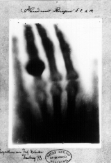
s. In many languages, X-radiation is called Röntgen radiation, after Wilhelm Conrad Röntgen, who is usually credited as its discoverer, and who had named it X-radiation to signify an unknown type of radiation.