
Mechanical ventilation
Encyclopedia
In medicine
, mechanical ventilation is a method to mechanically assist or replace spontaneous breathing
. This may involve a machine called a ventilator or the breathing may be assisted by a physician
, respiratory therapist or other suitable person compressing a bag
or set of bellows. Traditionally divided into negative-pressure ventilation
, where air is essentially sucked into the lungs, or positive pressure ventilation, where air (or another gas mix) is pushed into the trachea
. There are two main divisions of mechanical ventilation; Invasive ventilation, and non-invasive ventilation.. There are two main modes of mechanical ventilation
within the two divisions; positive pressure ventilation and negative pressure ventilation.
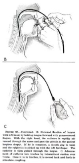
may have been the first to describe mechanical ventilation: "If you take a dead animal and blow air through its larynx [through a reed], you will fill its bronchi and watch its lungs attain the greatest distention." Vesalius
too describes ventilation by inserting a reed or cane into the trachea
of animals. In 1908 George Poe
demonstrated his mechanical respirator by asphyxiating dogs and seemingly bringing them back to life.
, airway injury, alveolar damage, and ventilator-associated pneumonia.
In many healthcare systems prolonged ventilation as part of intensive care is a limited resource (in that there are only so many patients that can receive care at any given moment). It is used to support a single failing organ system (the lungs) and cannot reverse any underlying disease process (such as terminal cancer). For this reason there can be (occasionally difficult) decisions to be made about whether it is suitable to commence someone on mechanical ventilation. Equally many ethical issues surround the decision to discontinue mechanical ventilation.
). It may be used at home or in a nursing or rehabilitation institution if patients have chronic illnesses that require long-term ventilatory assistance. Owing to the anatomy of the human pharynx
, larynx
, and esophagus
and the circumstances for which ventilation is required then additional measures are often required to secure the airway
during positive pressure ventilation to allow unimpeded passage of air into the trachea and avoid air passing into the esophagus and stomach. Commonly this is by insertion of a tube into the trachea
which provides a clear route for the air. This can be either an endotracheal tube
, inserted through the natural openings of mouth or nose or a tracheostomy inserted through an artificial opening in the neck. In other circumstances simple airway maneuvres, an oropharyngeal airway
or laryngeal mask airway
may be employed. If the patient is able to protect their own airway and non-invasive ventilation or negative-pressure ventilation
is used then a airway adjunct
may not be needed.
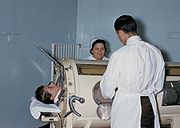
The iron lung
, also known as the Drinker and Shaw tank, was developed in 1929 and was one of the first negative-pressure machines used for long-term ventilation. It was refined and used in the 20th century largely as a result of the polio epidemic
that struck the world in the 1940s. The machine is effectively a large elongated tank
, which encases the patient up to the neck. The neck is sealed with a rubber gasket
so that the patient's face (and airway) are exposed to the room air.
While the exchange of oxygen
and carbon dioxide
between the bloodstream and the pulmonary airspace works by diffusion
and requires no external work, air must be moved into and out of the lungs
to make it available to the gas exchange
process. In spontaneous breathing, a negative pressure is created in the pleural cavity
by the muscles of respiration, and the resulting gradient between the atmospheric pressure
and the pressure inside the thorax generates a flow of air.
In the iron lung by means of a pump, the air is withdrawn mechanically to produce a vacuum inside the tank, thus creating negative pressure. This negative pressure leads to expansion of the chest, which causes a decrease in intrapulmonary pressure, and increases flow of ambient air into the lungs. As the vacuum is released, the pressure inside the tank equalizes to that of the ambient pressure, and the elastic coil of the chest and lungs leads to passive exhalation. However, when the vacuum is created, the abdomen also expands along with the lung, cutting off venous flow back to the heart, leading to pooling of venous blood in the lower extremities. There are large portholes for nurse or home assistant access. The patients can talk and eat normally, and can see the world through a well-placed series of mirrors. Some could remain in these iron lungs for years at a time quite successfully.
Today, negative pressure mechanical ventilators are still in use, notably with the polio wing hospitals in England
such as St Thomas' Hospital
in London and the John Radcliffe
in Oxford
. The prominent device used is a smaller device known as the cuirass
. The cuirass is a shell-like unit, creating negative pressure only to the chest using a combination of a fitting shell and a soft bladder. Its main use is in patients with neuromuscular disorders who have some residual muscular function. However, it was prone to falling off and caused severe chafing and skin damage and was not used as a long term device. In recent years this device has re-surfaced as a modern polycarbonate
shell with multiple seals and a high pressure oscillation pump in order to carry out biphasic cuirass ventilation
.
 The design of the modern positive-pressure ventilators were mainly based on technical developments by the military during World War II to supply oxygen to fighter pilots in high altitude. Such ventilators replaced the iron lungs as safe endotracheal tubes with high volume/low pressure cuffs were developed. The popularity of positive-pressure ventilators rose during the polio epidemic in the 1950s in Scandinavia and the United States and was the beginning of modern ventilation therapy. Positive pressure through manual supply of 50% oxygen through a tracheostomy tube led to a reduced mortality rate among patients with polio and respiratory paralysis. However, because of the sheer amount of man-power required for such manual intervention, mechanical positive-pressure ventilators became increasingly popular.
Positive-pressure ventilators work by increasing the patient's airway pressure through an endotracheal or tracheostomy tube. The positive pressure allows air to flow into the airway until the ventilator breath is terminated. Subsequently, the airway pressure drops to zero, and the elastic recoil of the chest wall and lungs push the tidal volume
— the breath—out through passive exhalation.
Common medical indications for use include:
, subcutaneous emphysema
, pneumomediastinum
, and pneumoperitoneum
.
Ventilator-associated lung injury — Ventilator-associated lung injury
(VALI) refers to acute lung injury that occurs during mechanical ventilation. It is clinically indistinguishable from acute lung injury
or acute respiratory distress syndrome
(ALI/ARDS).
Diaphragm — Controlled mechanical ventilation may lead to a rapid type of disuse atrophy
involving the diaphragmatic muscle fibers, which can develop within the first day of mechanical ventilation. This cause of atrophy in the diaphragm is also a cause of atrophy in all respiratory related muscles during controlled mechanical ventilation.
Motility of mucocilia in the airways — Positive pressure ventilation appears to impair mucociliary motility in the airways. Bronchial mucus transport was frequently impaired and associated with retention of secretions and pneumonia
.
 Ventilators come in many different styles and method of giving a breath to sustain life.
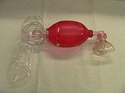
Hand controlled — Manual ventilators such as Bag valve mask
s and anesthesia bags require the user to hold the ventilator to the face or to an artificial airway
and maintain breaths with their hands.
Transport ventilators — These ventilators are small, more rugged, and can be powered pneumatically or via AC or DC power sources.
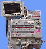
Intensive-care ventilators — These ventilators are larger and usually run on AC power (though virtually all contain a battery to facilitate intra-facility transport and as a back-up in the event of a power failure). This style of ventilator often provides greater control of a wide variety of ventilation parameters (such as inspiratory rise time). Many ICU ventilators also incorporate graphics to provide visual feedback of each breath.
Neonatal ventilators — Designed with the preterm neonate in mind, these are a specialized subset of ICU ventilators which are designed to deliver the smaller, more precise volumes and pressures required to ventilate these patients.
Positive airway pressure ventilators (PAP) — These ventilators are specifically designed for non-invasive ventilation. this includes ventilators for use at home for treatment of chronic conditions such as sleep apnea
or COPD.
has consumed it into its definition and overall has combined any CMV mode for mechanical ventilation into the more accepted term in nomenclature for mechanical ventilation.
Volume controlled continuous mandatory ventilation
— In this mode the ventilator provides a mechanical breath with either a pre-set tidal volume or peak pressure every time the patient initiates a breath. Traditional assist-control used only a pre-set tidal volume—when a preset peak pressure is used this is also sometimes termed intermittent positive pressure ventilation (IPPV). However, the initiation timing is the same—both provide a ventilator breath with every patient effort. In most ventilators a back-up minimum breath rate can be set in the event that the patient becomes apnoeic. Although a maximum rate is not usually set, an alarm can be set if the ventilator cycles too frequently. This can alert that the patient is tachypneic or that the ventilator may be auto-cycling (a problem that results when the ventilator interprets fluctuations in the circuit due to the last breath termination as a new breath initiation attempt).
(PC-CMV) — mechanical ventilation with preset inspiratory pressure (PIP) and inspiratory time (Ti). Every breath is machine initiated and mandatory.
.
(PSV). When a patient attempts to breathe spontaneously through an endotracheal tube, the narrowed diameter of the airway results in higher resistance to airflow, and thus a higher work of breathing. PSV was developed as a method to decrease the work of breathing in-between ventilator mandated breaths by providing an elevated pressure triggered by spontaneous breathing that "supports" ventilation during inspiration. Thus, for example, SIMV might be combined with PSV so that additional breaths beyond the SIMV programmed breaths are supported. However, while the SIMV mandated breaths have a preset volume or peak pressure, the PSV breaths are designed to cut short when the inspiratory flow reaches a percentage of the peak inspiratory flow (e.g. 10–25%). New generation of ventilators provides user-adjustable inspiration cycling off threshold, and some even are equipped with automatic inspiration cycling off threshold function. This helps the patient ventilator synchrony. The peak pressure set for the PSV breaths is usually a lower pressure than that set for the full ventilator mandated breath. PSV can be also be used as an independent mode.
(CPAP). A continuous level of elevated pressure is provided through the patient circuit to maintain adequate oxygenation, decrease the work of breathing, and decrease the work of the heart (such as in left-sided heart failure CHF). Note that no cycling of ventilator pressures occurs and the patient must initiate all breaths. In addition, no additional pressure above the CPAP pressure is provided during those breaths. CPAP may be used invasively through an endotracheal tube or tracheostomy or non-invasively with a face mask or nasal prongs. Non-invasive ventilation has become more common for treatment of acute respiratory failure.
, in which the patient has an increased respiratory drive. Such hyperventilation and hypocapnia
(decreased systemic carbon dioxide due to hyperventilation) usually occurs in patients with end-stage liver disease, hyperventilatory sepsis, and head trauma. Respiratory alkalosis
will be evident from the initial arterial blood gas
obtained, and the mode of ventilation can then be changed if so desired.
Positive End Expiratory Pressure may or may not be employed to prevent atelectasis
in adult patients. It is almost always used for pediatric and neonatal patients due to their increased tendency for atelectasis.
High frequency oscillation is used most frequently in neonates, but is also used as an always alternative mode in adults with severe ARDS.
Pressure regulated volume control
is another option.
As a general rule, whenever possible, spontaneous breathing must be maintained or supported, to avoid muscular atrophy of the diaphragm (Ventilator Induced Dysfunction of Diaphragm, VIDD) . To limit VALI and VILI, protective ventilation pattern should be applied to the patient. If this results in severe hypercapnia, exceeding accepted levels for permissive hypercapnia (pH below 7.2), measures for extracorporeal removal (iLA Membranventilator, Novalung) should be installed at an early stage of mechanical ventilation, to terminate cascades of inflammatory response from the lung tissue, resulting in multiorgan failure respective.
As the amount of tidal volume increases, the pressure required to administer that volume is increased. This pressure is known as the peak airway pressure. If the peak airway pressure is persistently above 45 cm (4.4 kPa) for adults, the risk of barotrauma is increased (see below) and efforts should be made to try to reduce the peak airway pressure (such as acceptable alarm limits). In infants and children it is unclear what level of peak pressure may cause damage. In general, keeping peak pressures below 30 cm (2.9 kPa) is desirable.
Monitoring for barotrauma can also involve measuring the plateau pressure, which is the pressure after the delivery of the tidal volume but before the patient is allowed to exhale. Normal breathing pattern involves inspiration, then expiration. The ventilator is programmed so that after delivery of the tidal volume (inspiration), the patient is not allowed to exhale for a half a second. Therefore, pressure must be maintained in order to prevent exhalation, and this pressure is the plateau pressure. Barotrauma is minimized when the plateau pressure is maintained < 30–35 cm.
, and this has led some to propose that ventilators should deliver 1½–2 times the amount of the preset tidal volume 6–8 times per hour to account for the sighs. However, such high quantity of volume delivery requires very high peak pressure that predisposes to barotrauma. Currently, accounting for sighs is not recommended if the patient is receiving 10–12 mL/kg or is on PEEP. If the tidal volume used is lower, the sigh adjustment can be used, as long as the peak and plateau pressures are acceptable.
Sighs are not generally used with ventilation of infants and young children.
(FRC). At the end of expiration, the PEEP exerts pressure to oppose passive emptying of the lung and to keep the airway pressure above the atmospheric pressure. The presence of PEEP opens up collapsed or unstable alveoli and increases the FRC and surface area for gas exchange, thus reducing the size of the shunt. For example, if a large shunt is found to exist based on the estimation from 100% Fi (see above), then PEEP can be considered and the Fi can be lowered (< 60%) in order to maintain an adequate Pa, thus reducing the risk of oxygen toxicity.
In addition to treating a shunt, PEEP may also be useful to decrease the work of breathing. In pulmonary physiology, compliance
is a measure of the "stiffness" of the lung and chest wall. The mathematical formula for compliance (C) equals change in volume divided by change in pressure. The higher the compliance, the more easily the lungs will inflate in response to positive pressure. An underinflated lung will have low compliance and PEEP will improve this initially by increasing the FRC, since the partially inflated lung takes less energy to inflate further. Excessive PEEP can however produce overinflation, which will again decrease compliance. Therefore it is important to maintain an adequate, but not excessive FRC.
Indications. PEEP can cause significant haemodynamic consequences through decreasing venous return to the right heart and decreasing right ventricular function. As such, it should be judiciously used and is indicated for adults in two circumstances.
If used, PEEP is usually set with the minimal positive pressure to maintain an adequate Pa with a safe Fi. As PEEP increases intrathoracic pressure, there can be a resulting decrease in venous return and decrease in cardiac output. A PEEP of less than 10 cm (1 kPa) is usually safe in adults if intravascular volume depletion is absent. Lower levels are used for pediatric patients.
Older literature recommended routine placement of a Swan-Ganz catheter if the amount of PEEP used is greater than 10 cm for hemodynamic monitoring. More recent literature has failed to find outcome benefits with routine PA catheterisation when compared to simple central venous pressure
monitoring.
If cardiac output measurement is required, minimally invasive techniques, such as oesophageal doppler monitoring or arterial waveform contour monitoring may be sufficient alternatives.
When withdrawing, it is decreased through 1–2 cm decrements while monitoring haemoglobin-oxygen saturations. Any unacceptable haemoglobin-oxygen saturation should prompt reinstitution of the last PEEP level that maintained good saturation.
and severe hypoxemia
. It improves FRC, drainage of secretions, and ventilation-perfusion
matching (efficiency of gas exchange). It may improve oxygenation in > 50% of patients, but no survival benefit has been documented.
through a continuous infusion or scheduled dosing to help with anxiety or psychological stress. Sedation also helps the patient tolerate the constant irritation of the endotracheal tube in their mouth, pharynx and trachea. Without some form of sedation and analgesia, it is common for patients to "fight" the ventilator. This fighting increases work of breathing and may cause further lung injury. Daily interruption of sedation is commonly helpful to the patient for reorientation and appropriate weaning. These interruptions are frequently described as "sedation vacations" and have been shown to reduce the time patients stay on mechanical ventilation.
It is not uncommon for patients on a mechanical ventilator to be given a muscle relaxant
or paralytic to aid in ventilation. These "neuromuscular blockades" prevent skeletal muscle from contracting and thereby stop all patient movement including respiratory efforts. These types of pharmaceutical agents must always be given in conjunction with sedation as the effects of the paralytics is not only uncomfortable but would cause significant psychological stress and anxiety
.
When using 100% Fi, the degree of shunting is estimated by subtracting the measured Pa (from an arterial blood gas
) from 700 mmHg. For each difference of 100 mmHg, the shunt is 5%. A shunt of more than 25% should prompt a search for the cause of this hypoxemia, such as mainstem intubation or pneumothorax
, and should be treated accordingly. If such complications are not present, other causes must be sought after, and PEEP should be used to treat this intrapulmonary shunt. Other such causes of a shunt include:
Trials of spontaneous breathing
have been shown to accurately predict the success of spontaneous breathing.
Monitoring a patient in mechanical ventilation has many clinical applications: Enhance understanding of pathophysiology, aid with diagnosis, guide patient management, avoid complications and assessment of trends.
Most of modern ventilators have basic monitoring tools. There are also monitors that work independently of the ventilator, which allow to measure patients after the ventilator has been removed, such as a T tube test.
Medicine
Medicine is the science and art of healing. It encompasses a variety of health care practices evolved to maintain and restore health by the prevention and treatment of illness....
, mechanical ventilation is a method to mechanically assist or replace spontaneous breathing
Respiration (physiology)
'In physiology, respiration is defined as the transport of oxygen from the outside air to the cells within tissues, and the transport of carbon dioxide in the opposite direction...
. This may involve a machine called a ventilator or the breathing may be assisted by a physician
Physician
A physician is a health care provider who practices the profession of medicine, which is concerned with promoting, maintaining or restoring human health through the study, diagnosis, and treatment of disease, injury and other physical and mental impairments...
, respiratory therapist or other suitable person compressing a bag
Bag valve mask
A bag valve mask is a hand-held device used to provide positive pressure ventilation to a patient who is not breathing or who is breathing inadequately. The device is a normal part of a resuscitation kit for trained professionals, such as ambulance crew...
or set of bellows. Traditionally divided into negative-pressure ventilation
Iron lung
A negative pressure ventilator is a form of medical ventilator that enables a person to breathe when normal muscle control has been lost or the work of breathing exceeds the person's ability....
, where air is essentially sucked into the lungs, or positive pressure ventilation, where air (or another gas mix) is pushed into the trachea
Vertebrate trachea
In tetrapod anatomy the trachea, or windpipe, is a tube that connects the pharynx or larynx to the lungs, allowing the passage of air. It is lined with pseudostratified ciliated columnar epithelium cells with goblet cells that produce mucus...
. There are two main divisions of mechanical ventilation; Invasive ventilation, and non-invasive ventilation.. There are two main modes of mechanical ventilation
Modes of mechanical ventilation
Modes of mechanical ventilation are one of the most important aspects of the usage of mechanical ventilation. The mode refers to the method of inspiratory support. Mode selection is generally based on clinician familiarity and institutional preferences since there is a paucity of evidence...
within the two divisions; positive pressure ventilation and negative pressure ventilation.
History
The Roman physician GalenGalen
Aelius Galenus or Claudius Galenus , better known as Galen of Pergamon , was a prominent Roman physician, surgeon and philosopher...
may have been the first to describe mechanical ventilation: "If you take a dead animal and blow air through its larynx [through a reed], you will fill its bronchi and watch its lungs attain the greatest distention." Vesalius
Vesalius
Andreas Vesalius was a Flemish anatomist, physician, and author of one of the most influential books on human anatomy, De humani corporis fabrica . Vesalius is often referred to as the founder of modern human anatomy. Vesalius is the Latinized form of Andries van Wesel...
too describes ventilation by inserting a reed or cane into the trachea
Vertebrate trachea
In tetrapod anatomy the trachea, or windpipe, is a tube that connects the pharynx or larynx to the lungs, allowing the passage of air. It is lined with pseudostratified ciliated columnar epithelium cells with goblet cells that produce mucus...
of animals. In 1908 George Poe
George Poe
George Poe, Jr. was a pioneer of mechanical ventilation of asphyxiation victims. He was the first person to manufacture nitrous oxide for commercial use in his Trenton, New Jersey company.-Birth:...
demonstrated his mechanical respirator by asphyxiating dogs and seemingly bringing them back to life.
Complications
Mechanical ventilation is often a life-saving intervention, but carries many potential complications including pneumothoraxPneumothorax
Pneumothorax is a collection of air or gas in the pleural cavity of the chest between the lung and the chest wall. It may occur spontaneously in people without chronic lung conditions as well as in those with lung disease , and many pneumothoraces occur after physical trauma to the chest, blast...
, airway injury, alveolar damage, and ventilator-associated pneumonia.
In many healthcare systems prolonged ventilation as part of intensive care is a limited resource (in that there are only so many patients that can receive care at any given moment). It is used to support a single failing organ system (the lungs) and cannot reverse any underlying disease process (such as terminal cancer). For this reason there can be (occasionally difficult) decisions to be made about whether it is suitable to commence someone on mechanical ventilation. Equally many ethical issues surround the decision to discontinue mechanical ventilation.
Application and duration
It can be used as a short term measure, for example during an operation or critical illness (often in the setting of an intensive care unitIntensive Care Unit
thumb|220px|ICU roomAn intensive-care unit , critical-care unit , intensive-therapy unit/intensive-treatment unit is a specialized department in a hospital that provides intensive-care medicine...
). It may be used at home or in a nursing or rehabilitation institution if patients have chronic illnesses that require long-term ventilatory assistance. Owing to the anatomy of the human pharynx
Pharynx
The human pharynx is the part of the throat situated immediately posterior to the mouth and nasal cavity, and anterior to the esophagus and larynx. The human pharynx is conventionally divided into three sections: the nasopharynx , the oropharynx , and the laryngopharynx...
, larynx
Larynx
The larynx , commonly called the voice box, is an organ in the neck of amphibians, reptiles and mammals involved in breathing, sound production, and protecting the trachea against food aspiration. It manipulates pitch and volume...
, and esophagus
Esophagus
The esophagus is an organ in vertebrates which consists of a muscular tube through which food passes from the pharynx to the stomach. During swallowing, food passes from the mouth through the pharynx into the esophagus and travels via peristalsis to the stomach...
and the circumstances for which ventilation is required then additional measures are often required to secure the airway
Artificial airway
An artificial airway is a term used to describe a medical technique or tool applied to maintain an open airway for a person who otherwise is unable to maintain their own for various reasons.-Nasopharyngeal airway:...
during positive pressure ventilation to allow unimpeded passage of air into the trachea and avoid air passing into the esophagus and stomach. Commonly this is by insertion of a tube into the trachea
Artificial airway
An artificial airway is a term used to describe a medical technique or tool applied to maintain an open airway for a person who otherwise is unable to maintain their own for various reasons.-Nasopharyngeal airway:...
which provides a clear route for the air. This can be either an endotracheal tube
Tracheal tube
A tracheal tube is a catheter that is inserted into the trachea in order for the primary purpose of establishing and maintaining a patent airway and to ensure the adequate exchange of oxygen and carbon dioxide. Many different types of tracheal tubes are available, suited for different specific...
, inserted through the natural openings of mouth or nose or a tracheostomy inserted through an artificial opening in the neck. In other circumstances simple airway maneuvres, an oropharyngeal airway
Oropharyngeal airway
An oropharyngeal airway is a medical device called an airway adjunct used to maintain a patent airway. It does this by preventing the tongue from covering the epiglottis, which could prevent the person from breathing...
or laryngeal mask airway
Laryngeal mask airway
The laryngeal mask airway is a supraglottic airway device invented by Archie Brain, a British anaesthetist.-Description:Laryngeal masks consist of a tube with an inflatable cuff that is inserted into the pharynx. Laryngeal mask airways come in a variety of sizes ranging from large adult to infant...
may be employed. If the patient is able to protect their own airway and non-invasive ventilation or negative-pressure ventilation
Iron lung
A negative pressure ventilator is a form of medical ventilator that enables a person to breathe when normal muscle control has been lost or the work of breathing exceeds the person's ability....
is used then a airway adjunct
Artificial airway
An artificial airway is a term used to describe a medical technique or tool applied to maintain an open airway for a person who otherwise is unable to maintain their own for various reasons.-Nasopharyngeal airway:...
may not be needed.
Negative pressure machines
The iron lung
Iron lung
A negative pressure ventilator is a form of medical ventilator that enables a person to breathe when normal muscle control has been lost or the work of breathing exceeds the person's ability....
, also known as the Drinker and Shaw tank, was developed in 1929 and was one of the first negative-pressure machines used for long-term ventilation. It was refined and used in the 20th century largely as a result of the polio epidemic
Epidemic
In epidemiology, an epidemic , occurs when new cases of a certain disease, in a given human population, and during a given period, substantially exceed what is expected based on recent experience...
that struck the world in the 1940s. The machine is effectively a large elongated tank
Tank
A tank is a tracked, armoured fighting vehicle designed for front-line combat which combines operational mobility, tactical offensive, and defensive capabilities...
, which encases the patient up to the neck. The neck is sealed with a rubber gasket
Gasket
thumb|sright|250px|Some seals and gaskets1. [[o-ring]]2. fiber [[Washer |washer]]3. paper gaskets4. [[cylinder head]] [[head gasket|gasket]]...
so that the patient's face (and airway) are exposed to the room air.
While the exchange of oxygen
Oxygen
Oxygen is the element with atomic number 8 and represented by the symbol O. Its name derives from the Greek roots ὀξύς and -γενής , because at the time of naming, it was mistakenly thought that all acids required oxygen in their composition...
and carbon dioxide
Carbon dioxide
Carbon dioxide is a naturally occurring chemical compound composed of two oxygen atoms covalently bonded to a single carbon atom...
between the bloodstream and the pulmonary airspace works by diffusion
Diffusion
Molecular diffusion, often called simply diffusion, is the thermal motion of all particles at temperatures above absolute zero. The rate of this movement is a function of temperature, viscosity of the fluid and the size of the particles...
and requires no external work, air must be moved into and out of the lungs
Human lung
The human lungs are the organs of respiration in humans. Humans have two lungs, with the left being divided into two lobes and the right into three lobes. Together, the lungs contain approximately of airways and 300 to 500 million alveoli, having a total surface area of about in...
to make it available to the gas exchange
Gas exchange
Gas exchange is a process in biology where gases contained in an organism and atmosphere transfer or exchange. In human gas-exchange, gases contained in the blood of human bodies exchange with gases contained in the atmosphere. Human gas-exchange occurs in the lungs...
process. In spontaneous breathing, a negative pressure is created in the pleural cavity
Pleural cavity
In human anatomy, the pleural cavity is the potential space between the two pleura of the lungs. The pleura is a serous membrane which folds back onto itself to form a two-layered, membrane structure. The thin space between the two pleural layers is known as the pleural cavity; it normally...
by the muscles of respiration, and the resulting gradient between the atmospheric pressure
Atmospheric pressure
Atmospheric pressure is the force per unit area exerted into a surface by the weight of air above that surface in the atmosphere of Earth . In most circumstances atmospheric pressure is closely approximated by the hydrostatic pressure caused by the weight of air above the measurement point...
and the pressure inside the thorax generates a flow of air.
In the iron lung by means of a pump, the air is withdrawn mechanically to produce a vacuum inside the tank, thus creating negative pressure. This negative pressure leads to expansion of the chest, which causes a decrease in intrapulmonary pressure, and increases flow of ambient air into the lungs. As the vacuum is released, the pressure inside the tank equalizes to that of the ambient pressure, and the elastic coil of the chest and lungs leads to passive exhalation. However, when the vacuum is created, the abdomen also expands along with the lung, cutting off venous flow back to the heart, leading to pooling of venous blood in the lower extremities. There are large portholes for nurse or home assistant access. The patients can talk and eat normally, and can see the world through a well-placed series of mirrors. Some could remain in these iron lungs for years at a time quite successfully.
Today, negative pressure mechanical ventilators are still in use, notably with the polio wing hospitals in England
England
England is a country that is part of the United Kingdom. It shares land borders with Scotland to the north and Wales to the west; the Irish Sea is to the north west, the Celtic Sea to the south west, with the North Sea to the east and the English Channel to the south separating it from continental...
such as St Thomas' Hospital
St Thomas' Hospital
St Thomas' Hospital is a large NHS hospital in London, England. It is administratively a part of Guy's & St Thomas' NHS Foundation Trust. It has provided health care freely or under charitable auspices since the 12th century and was originally located in Southwark.St Thomas' Hospital is accessible...
in London and the John Radcliffe
John Radcliffe Hospital
The John Radcliffe Hospital is a large tertiary teaching hospital in Oxford, England.It is the main teaching hospital for Oxford University and Oxford Brookes University. As such, it is a well-developed centre of medical research. It also incorporates the Medical School of the University of Oxford....
in Oxford
Oxford
The city of Oxford is the county town of Oxfordshire, England. The city, made prominent by its medieval university, has a population of just under 165,000, with 153,900 living within the district boundary. It lies about 50 miles north-west of London. The rivers Cherwell and Thames run through...
. The prominent device used is a smaller device known as the cuirass
Cuirass
A cuirass is a piece of armour, formed of a single or multiple pieces of metal or other rigid material, which covers the front of the torso...
. The cuirass is a shell-like unit, creating negative pressure only to the chest using a combination of a fitting shell and a soft bladder. Its main use is in patients with neuromuscular disorders who have some residual muscular function. However, it was prone to falling off and caused severe chafing and skin damage and was not used as a long term device. In recent years this device has re-surfaced as a modern polycarbonate
Polycarbonate
PolycarbonatePhysical PropertiesDensity 1.20–1.22 g/cm3Abbe number 34.0Refractive index 1.584–1.586FlammabilityV0-V2Limiting oxygen index25–27%Water absorption – Equilibrium0.16–0.35%Water absorption – over 24 hours0.1%...
shell with multiple seals and a high pressure oscillation pump in order to carry out biphasic cuirass ventilation
Biphasic Cuirass Ventilation
Biphasic cuirass ventilation is a method of ventilation which requires the patient to wear an upper body shell or cuirass, so named after the body armor worn by medieval soldiers. The ventilation is biphasic because the cuirass is attached to a pump which actively controls both the inspiratory and...
.
Positive pressure machines
Positive-pressure ventilators work by increasing the patient's airway pressure through an endotracheal or tracheostomy tube. The positive pressure allows air to flow into the airway until the ventilator breath is terminated. Subsequently, the airway pressure drops to zero, and the elastic recoil of the chest wall and lungs push the tidal volume
Tidal volume
Tidal volume is the lung volume representing the normal volume of air displaced between normal inspiration and expiration when extra effort is not applied.Typical values are around 500ml or 7ml/kg bodyweight.-Mechanical Ventilation:...
— the breath—out through passive exhalation.
Indications for use
Mechanical ventilation is indicated when the patient's spontaneous ventilation is inadequate to maintain life. It is also indicated as prophylaxis for imminent collapse of other physiologic functions, or ineffective gas exchange in the lungs. Because mechanical ventilation only serves to provide assistance for breathing and does not cure a disease, the patient's underlying condition should be correctable and should resolve over time. In addition, other factors must be taken into consideration because mechanical ventilation is not without its complications (see below)Common medical indications for use include:
- Acute lung injury (including ARDSAcute respiratory distress syndromeAcute respiratory distress syndrome , also known as respiratory distress syndrome or adult respiratory distress syndrome is a serious reaction to various forms of injuries to the lung....
, trauma) - ApneaApneaApnea, apnoea, or apnœa is a term for suspension of external breathing. During apnea there is no movement of the muscles of respiration and the volume of the lungs initially remains unchanged...
with respiratory arrest, including cases from intoxication - Chronic obstructive pulmonary diseaseChronic obstructive pulmonary diseaseChronic obstructive pulmonary disease , also known as chronic obstructive lung disease , chronic obstructive airway disease , chronic airflow limitation and chronic obstructive respiratory disease , is the co-occurrence of chronic bronchitis and emphysema, a pair of commonly co-existing diseases...
(COPDChronic obstructive pulmonary diseaseChronic obstructive pulmonary disease , also known as chronic obstructive lung disease , chronic obstructive airway disease , chronic airflow limitation and chronic obstructive respiratory disease , is the co-occurrence of chronic bronchitis and emphysema, a pair of commonly co-existing diseases...
) - Acute respiratory acidosisRespiratory acidosisRespiratory acidosis is a medical condition in which decreased ventilation causes increased blood carbon dioxide concentration and decreased pH ....
with partial pressure of carbon dioxide (p) > 50 mmHg and pH < 7.25, which may be due to paralysis of the diaphragmThoracic diaphragmIn the anatomy of mammals, the thoracic diaphragm, or simply the diaphragm , is a sheet of internal skeletal muscle that extends across the bottom of the rib cage. The diaphragm separates the thoracic cavity from the abdominal cavity and performs an important function in respiration...
due to Guillain-Barré syndromeGuillain-Barré syndromeGuillain–Barré syndrome , sometimes called Landry's paralysis, is an acute inflammatory demyelinating polyneuropathy , a disorder affecting the peripheral nervous system. Ascending paralysis, weakness beginning in the feet and hands and migrating towards the trunk, is the most typical symptom...
, Myasthenia GravisMyasthenia gravisMyasthenia gravis is an autoimmune neuromuscular disease leading to fluctuating muscle weakness and fatiguability...
, spinal cordSpinal cordThe spinal cord is a long, thin, tubular bundle of nervous tissue and support cells that extends from the brain . The brain and spinal cord together make up the central nervous system...
injury, or the effect of anaesthetic and muscle relaxantMuscle relaxantA muscle relaxant is a drug which affects skeletal muscle function and decreases the muscle tone. It may be used to alleviate symptoms such as muscle spasms, pain, and hyperreflexia. The term "muscle relaxant" is used to refer to two major therapeutic groups: neuromuscular blockers and spasmolytics...
drugs - Increased work of breathing as evidenced by significant tachypneaTachypneaTachypnea means rapid breathing. Any rate between 12-20 breaths per minute is normal. Tachypnea is a respiration rate greater than 20 breaths per minute. - Distinction from other breathing terms :...
, retractions, and other physical signs of respiratory distress - HypoxemiaHypoxemiaHypoxemia is generally defined as decreased partial pressure of oxygen in blood, sometimes specifically as less than or causing hemoglobin oxygen saturation of less than 90%.-Distinction from anemia and hypoxia:...
with arterial partial pressure of oxygen (Pa) < 55 mm Hg with supplemental fraction of inspired oxygen (Fi) = 1.0 - HypotensionHypotensionIn physiology and medicine, hypotension is abnormally low blood pressure, especially in the arteries of the systemic circulation. It is best understood as a physiologic state, rather than a disease. It is often associated with shock, though not necessarily indicative of it. Hypotension is the...
including sepsisSepsisSepsis is a potentially deadly medical condition that is characterized by a whole-body inflammatory state and the presence of a known or suspected infection. The body may develop this inflammatory response by the immune system to microbes in the blood, urine, lungs, skin, or other tissues...
, shock, congestive heart failureCongestive heart failureHeart failure often called congestive heart failure is generally defined as the inability of the heart to supply sufficient blood flow to meet the needs of the body. Heart failure can cause a number of symptoms including shortness of breath, leg swelling, and exercise intolerance. The condition... - Neurological diseases such as Muscular DystrophyMuscular dystrophyMuscular dystrophy is a group of muscle diseases that weaken the musculoskeletal system and hamper locomotion. Muscular dystrophies are characterized by progressive skeletal muscle weakness, defects in muscle proteins, and the death of muscle cells and tissue.In the 1860s, descriptions of boys who...
and Amyotrophic Lateral SclerosisAmyotrophic lateral sclerosisAmyotrophic lateral sclerosis , also referred to as Lou Gehrig's disease, is a form of motor neuron disease caused by the degeneration of upper and lower neurons, located in the ventral horn of the spinal cord and the cortical neurons that provide their efferent input...
Associated risk
Barotrauma — Pulmonary barotrauma is a well-known complication of positive pressure mechanical ventilation. This includes pneumothoraxPneumothorax
Pneumothorax is a collection of air or gas in the pleural cavity of the chest between the lung and the chest wall. It may occur spontaneously in people without chronic lung conditions as well as in those with lung disease , and many pneumothoraces occur after physical trauma to the chest, blast...
, subcutaneous emphysema
Subcutaneous emphysema
Subcutaneous emphysema, sometimes abbreviated SCE or SE and also called tissue emphysema, or Sub Q air, occurs when gas or air is present in the subcutaneous layer of the skin. Subcutaneous refers to the tissue beneath the cutis of the skin, and emphysema refers to trapped air...
, pneumomediastinum
Pneumomediastinum
Pneumomediastinum is a condition in which air is present in the mediastinum...
, and pneumoperitoneum
Pneumoperitoneum
Pneumoperitoneum is air or gas in the abdominal cavity. It is often seen on X-ray, but small amounts are often missed, and CT is nowadays regarded as a criterion standard in the assessment of a pneumoperitoneum. CT can visualize quantities as small as 5 cm³ of air or gas...
.
Ventilator-associated lung injury — Ventilator-associated lung injury
Ventilator-associated lung injury
Ventilator-associated lung injury is an acute lung injury that develops during mechanical ventilation and is termed ventilator-induced lung injury if it can be proven that the mechanical ventilation caused the acute lung injury. In contrast, ventilator-associated lung injury exists if the cause...
(VALI) refers to acute lung injury that occurs during mechanical ventilation. It is clinically indistinguishable from acute lung injury
Acute lung injury
Acute lung injury is a diffuse heterogeneous lung injury characterized by hypoxemia, non cardiogenic pulmonary edema, low lung compliance and widespread capillary leakage...
or acute respiratory distress syndrome
Acute respiratory distress syndrome
Acute respiratory distress syndrome , also known as respiratory distress syndrome or adult respiratory distress syndrome is a serious reaction to various forms of injuries to the lung....
(ALI/ARDS).
Diaphragm — Controlled mechanical ventilation may lead to a rapid type of disuse atrophy
Atrophy
Atrophy is the partial or complete wasting away of a part of the body. Causes of atrophy include mutations , poor nourishment, poor circulation, loss of hormonal support, loss of nerve supply to the target organ, disuse or lack of exercise or disease intrinsic to the tissue itself...
involving the diaphragmatic muscle fibers, which can develop within the first day of mechanical ventilation. This cause of atrophy in the diaphragm is also a cause of atrophy in all respiratory related muscles during controlled mechanical ventilation.
Motility of mucocilia in the airways — Positive pressure ventilation appears to impair mucociliary motility in the airways. Bronchial mucus transport was frequently impaired and associated with retention of secretions and pneumonia
Pneumonia
Pneumonia is an inflammatory condition of the lung—especially affecting the microscopic air sacs —associated with fever, chest symptoms, and a lack of air space on a chest X-ray. Pneumonia is typically caused by an infection but there are a number of other causes...
.
Types of ventilators
Hand controlled — Manual ventilators such as Bag valve mask
Bag valve mask
A bag valve mask is a hand-held device used to provide positive pressure ventilation to a patient who is not breathing or who is breathing inadequately. The device is a normal part of a resuscitation kit for trained professionals, such as ambulance crew...
s and anesthesia bags require the user to hold the ventilator to the face or to an artificial airway
Artificial airway
An artificial airway is a term used to describe a medical technique or tool applied to maintain an open airway for a person who otherwise is unable to maintain their own for various reasons.-Nasopharyngeal airway:...
and maintain breaths with their hands.
Mechanical ventilators
Mechanical ventilators typically require power by a battery or a wall outlet (DC or AC) though some ventilators work on a pneumatic system not requiring power.Transport ventilators — These ventilators are small, more rugged, and can be powered pneumatically or via AC or DC power sources.
Intensive-care ventilators — These ventilators are larger and usually run on AC power (though virtually all contain a battery to facilitate intra-facility transport and as a back-up in the event of a power failure). This style of ventilator often provides greater control of a wide variety of ventilation parameters (such as inspiratory rise time). Many ICU ventilators also incorporate graphics to provide visual feedback of each breath.
Neonatal ventilators — Designed with the preterm neonate in mind, these are a specialized subset of ICU ventilators which are designed to deliver the smaller, more precise volumes and pressures required to ventilate these patients.
Positive airway pressure ventilators (PAP) — These ventilators are specifically designed for non-invasive ventilation. this includes ventilators for use at home for treatment of chronic conditions such as sleep apnea
Sleep apnea
Sleep apnea is a sleep disorder characterized by abnormal pauses in breathing or instances of abnormally low breathing, during sleep. Each pause in breathing, called an apnea, can last from a few seconds to minutes, and may occur 5 to 30 times or more an hour. Similarly, each abnormally low...
or COPD.
Breath initiation
Initiation of a mechanical ventilation breath can be triggered by number of variables including flow, volume, pressure and time.- Flow — A breath is initiated when inspiratory flow through the ventilator circuit is recognized by the ventilator. This is normally due to the patient attempting inspiration, but can also be due to other factors, such as a leak in the circuit. On some ventilators the flow sensitivity (the flow rate threshold for initiating inspiration) can be set.
- Pressure — A breath is initiated when a negative pressure is measured in the inspiratory circuit, such as when the patient attempts inspiration. As with flow sensing, some ventilators allow this parameter to be set.
- Volume — A breath is initiated when volume loss is detected from the circuit, such as when the patient attempts inspiration.
- Time — A breath is initiated at a regular interval which is determined by the set respiratory rateRespiratory rateRespiratory rate is also known by respiration rate, pulmonary ventilation rate, ventilation rate, or breathing frequency is the number of breaths taken within a set amount of time, typically 60 seconds....
.
Inspiration
The process of gas delivery to the patient depends on the mode of ventilation (see below). Typically a breath can be classified as either volume controlled or pressure controlled depending on which variable the ventilator actively controls during ventilation.- Volume ventilation — A predetermined tidal volumeTidal volumeTidal volume is the lung volume representing the normal volume of air displaced between normal inspiration and expiration when extra effort is not applied.Typical values are around 500ml or 7ml/kg bodyweight.-Mechanical Ventilation:...
(Vt) is set by the clinician, and is delivered to the patient every time a breath is triggered. For example, if the tidal volume is set at 500ml, the ventilator will continue inspiration until 500ml have been delivered to the patient. Upon completion of inspiration, the ventilator will cycle to exhalation. The amount of pressure necessary to deliver the tidal volume is determined by the resistance and compliance of the patient and ventilator circuit, and may vary from breath to breath. Inspiratory pressures must be closely monitored to prevent barotrauma.
- Pressure ventilationPressure controlPressure control is a mode of mechanical ventilation alone and a variable within other modes of mechanical ventilation. Pressure control is used to regulate pressures applied during mechanical ventilation. Air delivered into the patients lungs are currently regulated by Volume Control or...
— A predetermined peak inspiratory pressurePeak inspiratory pressurePeak inspiratory pressure is the highest level of pressure applied to the lungs during inhalation. In mechanical ventilation the number reflects a positive pressure in centimeters of water pressure . In normal breathing, it may sometimes be referred to as the maximal inspiratory pressure , which...
(PIP) is set by the clinician and delivered to the patient throughout the set inspiratory time (Ti). After the inspiratory time has elapsed, the ventilator will cycle to expiration. The tidal volumeTidal volumeTidal volume is the lung volume representing the normal volume of air displaced between normal inspiration and expiration when extra effort is not applied.Typical values are around 500ml or 7ml/kg bodyweight.-Mechanical Ventilation:...
delivered to the patient is determined by the resistance and compliance of the patient and circuit, and may vary from breath to breath. Inspiratory volumes must be closely monitored to prevent hypoventilationHypoventilationIn medicine, hypoventilation occurs when ventilation is inadequate to perform needed gas exchange...
or hyperventilationHyperventilationHyperventilation or overbreathing is the state of breathing faster or deeper than normal, causing excessive expulsion of circulating carbon dioxide. It can result from a psychological state such as a panic attack, from a physiological condition such as metabolic acidosis, can be brought about by...
.
- Pressure regulated volume controlPressure Regulated Volume ControlPressure regulated volume control is a mode of mechanical ventilation with a VT set as a goal amount. Pressure varies with a peak pressure limit included to reduce lung trauma and use only the minimum pressure required to deliver the goal tidal volume...
— Several manufactures have incorporated features from both of these modes. These modes are flow-variable, volume-targeted, pressure-regulated and time-limited. This means that instead of providing an exact tidal volume each breath, a target volume is set and the ventilator will vary the inspiratory pressure (PIP) on a breath to breath basis to achieve that volume. As with pressure ventilation, the inspiratory time (Ti) limits the length of the inspiratory cycle. The primary benefit from such modes is that they allow the set tidal volume to be achieved with the lowest possible peak inspiratory pressure. Pressure regulated modes such as PRVC, Auto-flow (Draeger)or Average Volume Assured Pressure Support (AVAPS) from Philips can most easily be thought of as turning a volume mode into a pressure mode with the added benefit of maintaining more control over tidal volume than with strictly pressure-control.
Breath termination
Termination of a mechanical ventilation breath can occur either at the completion of the inspiratory phase (see above), or as an alarm condition. Alarm conditions typically cause the ventilator to immediately cycle from inspiration to expiration. An example of this is when the measured pressure exceeds the high pressure alarm value.Exhalation
Exhalation in mechanical ventilation is almost always completely passive. The ventilator's expiratory valve is opened, and expiratory flow is allowed until the baseline pressure (PEEP) is reached. Expiratory flow is determined by patient factors such as compliance and resistance.Modes of mechanical ventilation
Mechanical ventilation utilizes several separate systems for ventilation referred to as the "mode". Modes come in many different delivery concepts but all modes generally fall into one of the few main flagship categories such as volume controlled continuous mandatory ventilation, volume controlled intermittent mandatory ventilation, pressure controlled continuous mandatory ventilation, pressure controlled intermittent mandatory ventilation, continuous spontaneous ventilation and the high frequency ventilation systems.Volume controlled continuous mandatory ventilation
Controlled mechanical ventilation (CMV) — In this mode the ventilator provides a mechanical breath on a preset timing. Patient respiratory efforts are ignored. This is generally uncomfortable for children and adults who are conscious and is usually only used in an unconscious patient. It may also be used in infants who often quickly adapt their breathing pattern to the ventilator timing. Since CMV is no longer contained in its original form the term volume controlled continuous mandatory ventilationVolume controlled continuous mandatory ventilation
Volume controlled continuous mandatory ventilation — is a mode of mechanical ventilation where breaths are delivered based on set variables. The patient may initate breaths by attempting to breathe...
has consumed it into its definition and overall has combined any CMV mode for mechanical ventilation into the more accepted term in nomenclature for mechanical ventilation.
Volume controlled continuous mandatory ventilation
Volume controlled continuous mandatory ventilation
Volume controlled continuous mandatory ventilation — is a mode of mechanical ventilation where breaths are delivered based on set variables. The patient may initate breaths by attempting to breathe...
— In this mode the ventilator provides a mechanical breath with either a pre-set tidal volume or peak pressure every time the patient initiates a breath. Traditional assist-control used only a pre-set tidal volume—when a preset peak pressure is used this is also sometimes termed intermittent positive pressure ventilation (IPPV). However, the initiation timing is the same—both provide a ventilator breath with every patient effort. In most ventilators a back-up minimum breath rate can be set in the event that the patient becomes apnoeic. Although a maximum rate is not usually set, an alarm can be set if the ventilator cycles too frequently. This can alert that the patient is tachypneic or that the ventilator may be auto-cycling (a problem that results when the ventilator interprets fluctuations in the circuit due to the last breath termination as a new breath initiation attempt).
Volume controlled intermittent mandatory ventilation
Volume controlled intermittent mandatory ventilation (VC-IMV). Formerly known as synchronized intermittent mandatory ventilation (SIMV). In this mode the ventilator provides a pre-set mechanical breath (volume limited) every specified number of seconds (determined by dividing the respiratory rate into 60 seconds — thus a respiratory rate of 12 results in a 5 second cycle time). Within that cycle time the ventilator waits for the patient to initiate a breath using either a pressure or flow sensor. When the ventilator senses the first patient breathing attempt within the cycle, it delivers the preset ventilator breath. If the patient fails to initiate a breath, the ventilator delivers a mechanical breath at the end of the breath cycle. Additional spontaneous breaths after the first one within the breath cycle do not trigger another SIMV breath. However, SIMV may be combined with pressure support (see below). SIMV is frequently employed as a method of decreasing ventilatory support (weaning) by turning down the rate, which requires the patient to take additional breaths beyond the SIMV triggered breath.Pressure controlled continuous mandatory ventilation
Pressure controlled continuous mandatory ventilationPressure controlled continuous mandatory ventilation
Pressure Controlled Continuous Mandatory Ventilation is any mode of mechanical ventilation with preset inspiratory pressure and inspiratory time. Every breath is mandatory .-Pressure control:...
(PC-CMV) — mechanical ventilation with preset inspiratory pressure (PIP) and inspiratory time (Ti). Every breath is machine initiated and mandatory.
Pressure controlled intermittent mandatory ventilation
Pressure controlled intermittent mandatory ventilation (formerly known as SIMV) — In this mode the ventilator provides a pre-set pressure limited mechanical breath every specified number of seconds SIMV is frequently employed as a method of decreasing ventilatory support (weaning) by turning down the rate, which requires the patient to take additional breaths beyond the SIMV triggered breath. PC-IMV is fundamentally the same as VC-IMV with an emphasis on pressure support and control instead of volume. An example of PC-IMV is in the mode pressure regulated volume controlPressure Regulated Volume Control
Pressure regulated volume control is a mode of mechanical ventilation with a VT set as a goal amount. Pressure varies with a peak pressure limit included to reduce lung trauma and use only the minimum pressure required to deliver the goal tidal volume...
.
High frequency ventilation
High frequency ventilation refers to ventilation that occurs at rates significantly above that found in natural breathing. High frequency ventilation is further defined as any ventilation with a respiratory rate (Vf) greater than 150 respirations per minute. Within the category of high-frequency ventilation, the two principal types are high-frequency ventilation (passive) (i.e. high-frequency jet ventilation) and high-frequency ventilation (active) (i.e. high-frequency oscillatory ventilation).Pressure Support Ventilation
Pressure support ventilationContinuous spontaneous ventilation
Continuous spontaneous ventilation is any mode of mechanical ventilation where every breath is spontaneous .Some of these include:* Bilevel positive airway pressure* Continuous positive airway pressure...
(PSV). When a patient attempts to breathe spontaneously through an endotracheal tube, the narrowed diameter of the airway results in higher resistance to airflow, and thus a higher work of breathing. PSV was developed as a method to decrease the work of breathing in-between ventilator mandated breaths by providing an elevated pressure triggered by spontaneous breathing that "supports" ventilation during inspiration. Thus, for example, SIMV might be combined with PSV so that additional breaths beyond the SIMV programmed breaths are supported. However, while the SIMV mandated breaths have a preset volume or peak pressure, the PSV breaths are designed to cut short when the inspiratory flow reaches a percentage of the peak inspiratory flow (e.g. 10–25%). New generation of ventilators provides user-adjustable inspiration cycling off threshold, and some even are equipped with automatic inspiration cycling off threshold function. This helps the patient ventilator synchrony. The peak pressure set for the PSV breaths is usually a lower pressure than that set for the full ventilator mandated breath. PSV can be also be used as an independent mode.
Continuous positive airway pressure
Continuous positive airway pressureContinuous positive airway pressure
Positive airway pressure is a mode of respiratory ventilation used primarily in the treatment of sleep apnea, for which it was first developed. PAP ventilation is also commonly used for those who are critically ill in hospital with respiratory failure, and in newborn infants...
(CPAP). A continuous level of elevated pressure is provided through the patient circuit to maintain adequate oxygenation, decrease the work of breathing, and decrease the work of the heart (such as in left-sided heart failure CHF). Note that no cycling of ventilator pressures occurs and the patient must initiate all breaths. In addition, no additional pressure above the CPAP pressure is provided during those breaths. CPAP may be used invasively through an endotracheal tube or tracheostomy or non-invasively with a face mask or nasal prongs. Non-invasive ventilation has become more common for treatment of acute respiratory failure.
Choosing amongst ventilator modes
Assist-control mode minimizes patient effort by providing full mechanical support with every breath. This is often the initial mode chosen for adults because it provides the greatest degree of support. In patients with less severe respiratory failure, other modes such as SIMV may be appropriate. Assist-control mode should not be used in those patients with a potential for respiratory alkalosisRespiratory alkalosis
Respiratory alkalosis is a medical condition in which increased respiration elevates the blood pH...
, in which the patient has an increased respiratory drive. Such hyperventilation and hypocapnia
Hypocapnia
Hypocapnia or hypocapnea also known as hypocarbia, sometimes incorrectly called acapnia, is a state of reduced carbon dioxide in the blood. Hypocapnia usually results from deep or rapid breathing, known as hyperventilation....
(decreased systemic carbon dioxide due to hyperventilation) usually occurs in patients with end-stage liver disease, hyperventilatory sepsis, and head trauma. Respiratory alkalosis
Respiratory alkalosis
Respiratory alkalosis is a medical condition in which increased respiration elevates the blood pH...
will be evident from the initial arterial blood gas
Arterial blood gas
An arterial blood gas is a blood test that is performed using blood from an artery. It involves puncturing an artery with a thin needle and syringe and drawing a small volume of blood. The most common puncture site is the radial artery at the wrist, but sometimes the femoral artery in the groin or...
obtained, and the mode of ventilation can then be changed if so desired.
Positive End Expiratory Pressure may or may not be employed to prevent atelectasis
Atelectasis
Atelectasis is defined as the collapse or closure of alveoli resulting in reduced or absent gas exchange. It may affect part or all of one lung. It is a condition where the alveoli are deflated, as distinct from pulmonary consolidation.It is a very common finding in chest x-rays and other...
in adult patients. It is almost always used for pediatric and neonatal patients due to their increased tendency for atelectasis.
High frequency oscillation is used most frequently in neonates, but is also used as an always alternative mode in adults with severe ARDS.
Pressure regulated volume control
Pressure Regulated Volume Control
Pressure regulated volume control is a mode of mechanical ventilation with a VT set as a goal amount. Pressure varies with a peak pressure limit included to reduce lung trauma and use only the minimum pressure required to deliver the goal tidal volume...
is another option.
Initial ventilator settings
The following are general guidelines that may need to be modified for the individual patient.As a general rule, whenever possible, spontaneous breathing must be maintained or supported, to avoid muscular atrophy of the diaphragm (Ventilator Induced Dysfunction of Diaphragm, VIDD) . To limit VALI and VILI, protective ventilation pattern should be applied to the patient. If this results in severe hypercapnia, exceeding accepted levels for permissive hypercapnia (pH below 7.2), measures for extracorporeal removal (iLA Membranventilator, Novalung) should be installed at an early stage of mechanical ventilation, to terminate cascades of inflammatory response from the lung tissue, resulting in multiorgan failure respective.
Tidal volume, rate, and pressures
- For adult patients and older children
- tidal volumeTidal volumeTidal volume is the lung volume representing the normal volume of air displaced between normal inspiration and expiration when extra effort is not applied.Typical values are around 500ml or 7ml/kg bodyweight.-Mechanical Ventilation:...
(Vt) is calculated in milliliters per kilogram. Traditionally 10 ml/kg was used but has been shown to cause barotrauma, or injury to the lung by overextension, so 6 to 8 ml/kg IBW (ideal body weight) is now common practice in ICU. Hence a patient weighing 70 kg would get a Vt of 420–560 ml. In adults a rate of 12 breaths per minute is generally used. - with acute respiratory distress syndromeAcute respiratory distress syndromeAcute respiratory distress syndrome , also known as respiratory distress syndrome or adult respiratory distress syndrome is a serious reaction to various forms of injuries to the lung....
(ARDS) most commonly, a tidal volume of 6–8 ml/kg is used with a rate of 10–12 per minute. This reduced tidal volume allows for minimal volutrauma but may result in an elevated p (due to the relative decreased oxygen delivered) but this elevation does not need to be corrected (termed permissive hypercapnia)
- tidal volume
- For infants and younger children
- without existing lung disease—a tidal volume of 4–8 ml/kg to be delivered at a rate of 30–35 breaths per minute
- with ARDSAcute respiratory distress syndromeAcute respiratory distress syndrome , also known as respiratory distress syndrome or adult respiratory distress syndrome is a serious reaction to various forms of injuries to the lung....
—decrease tidal volume and increase respiratory rate sufficient to maintain p between 45 and 55. Allowing higher p (sometimes called permissive hypercapnia) may help prevent ventilator induced lung injury
As the amount of tidal volume increases, the pressure required to administer that volume is increased. This pressure is known as the peak airway pressure. If the peak airway pressure is persistently above 45 cm (4.4 kPa) for adults, the risk of barotrauma is increased (see below) and efforts should be made to try to reduce the peak airway pressure (such as acceptable alarm limits). In infants and children it is unclear what level of peak pressure may cause damage. In general, keeping peak pressures below 30 cm (2.9 kPa) is desirable.
Monitoring for barotrauma can also involve measuring the plateau pressure, which is the pressure after the delivery of the tidal volume but before the patient is allowed to exhale. Normal breathing pattern involves inspiration, then expiration. The ventilator is programmed so that after delivery of the tidal volume (inspiration), the patient is not allowed to exhale for a half a second. Therefore, pressure must be maintained in order to prevent exhalation, and this pressure is the plateau pressure. Barotrauma is minimized when the plateau pressure is maintained < 30–35 cm.
Sighs
An adult patient breathing spontaneously will usually sigh about 6–8 times per hour to prevent microatelectasisAtelectasis
Atelectasis is defined as the collapse or closure of alveoli resulting in reduced or absent gas exchange. It may affect part or all of one lung. It is a condition where the alveoli are deflated, as distinct from pulmonary consolidation.It is a very common finding in chest x-rays and other...
, and this has led some to propose that ventilators should deliver 1½–2 times the amount of the preset tidal volume 6–8 times per hour to account for the sighs. However, such high quantity of volume delivery requires very high peak pressure that predisposes to barotrauma. Currently, accounting for sighs is not recommended if the patient is receiving 10–12 mL/kg or is on PEEP. If the tidal volume used is lower, the sigh adjustment can be used, as long as the peak and plateau pressures are acceptable.
Sighs are not generally used with ventilation of infants and young children.
Initial Fi
Because the mechanical ventilator is responsible for assisting in a patient's breathing, it must then also be able to deliver an adequate amount of oxygen in each breath. The Fi stands for fraction of inspired oxygen, which means the percent of oxygen in each breath that is inspired. (Note that normal room air has ~21% oxygen content or an Fi of 0.21). In adult patients who can tolerate higher levels of oxygen for a period of time, the initial Fi may be set at 100% until arterial blood gases can document adequate oxygenation. An Fi of 100% for an extended period of time can cause atelectasis, but it can protect against hypoxemia from unexpected intubation problems. For infants, and especially in premature infants, avoiding high levels of Fi (>60%) is very important to avoid lung injury and apnea of prematurity. Fraction of inspired oxygen should always be titrated to meet the patients needs and to avoid unnecessary lung injury.Positive end-expiratory pressure
PEEP is an adjuvant to the mode of ventilation used to help maintain functional residual capacityFunctional residual capacity
Functional Residual Capacity is the volume of air present in the lungs, specifically the parenchyma tissues, at the end of passive expiration...
(FRC). At the end of expiration, the PEEP exerts pressure to oppose passive emptying of the lung and to keep the airway pressure above the atmospheric pressure. The presence of PEEP opens up collapsed or unstable alveoli and increases the FRC and surface area for gas exchange, thus reducing the size of the shunt. For example, if a large shunt is found to exist based on the estimation from 100% Fi (see above), then PEEP can be considered and the Fi can be lowered (< 60%) in order to maintain an adequate Pa, thus reducing the risk of oxygen toxicity.
In addition to treating a shunt, PEEP may also be useful to decrease the work of breathing. In pulmonary physiology, compliance
Elastance
Compliance is a measure of the tendency of a hollow organ to resist recoil toward its original dimensions upon removal of a distending or compressing force. It is the reciprocal of "elastance".-Blood vessels:...
is a measure of the "stiffness" of the lung and chest wall. The mathematical formula for compliance (C) equals change in volume divided by change in pressure. The higher the compliance, the more easily the lungs will inflate in response to positive pressure. An underinflated lung will have low compliance and PEEP will improve this initially by increasing the FRC, since the partially inflated lung takes less energy to inflate further. Excessive PEEP can however produce overinflation, which will again decrease compliance. Therefore it is important to maintain an adequate, but not excessive FRC.
Indications. PEEP can cause significant haemodynamic consequences through decreasing venous return to the right heart and decreasing right ventricular function. As such, it should be judiciously used and is indicated for adults in two circumstances.
- If a Pa of 60 mmHg cannot be achieved with a Fi of 60%
- If the initial shunt estimation is greater than 25%
If used, PEEP is usually set with the minimal positive pressure to maintain an adequate Pa with a safe Fi. As PEEP increases intrathoracic pressure, there can be a resulting decrease in venous return and decrease in cardiac output. A PEEP of less than 10 cm (1 kPa) is usually safe in adults if intravascular volume depletion is absent. Lower levels are used for pediatric patients.
Older literature recommended routine placement of a Swan-Ganz catheter if the amount of PEEP used is greater than 10 cm for hemodynamic monitoring. More recent literature has failed to find outcome benefits with routine PA catheterisation when compared to simple central venous pressure
Central venous pressure
Central venous pressure describes the pressure of blood in the thoracic vena cava, near the right atrium of the heart...
monitoring.
If cardiac output measurement is required, minimally invasive techniques, such as oesophageal doppler monitoring or arterial waveform contour monitoring may be sufficient alternatives.
When withdrawing, it is decreased through 1–2 cm decrements while monitoring haemoglobin-oxygen saturations. Any unacceptable haemoglobin-oxygen saturation should prompt reinstitution of the last PEEP level that maintained good saturation.
Positioning
Prone (face down) positioning has been used in patients with ARDSAcute respiratory distress syndrome
Acute respiratory distress syndrome , also known as respiratory distress syndrome or adult respiratory distress syndrome is a serious reaction to various forms of injuries to the lung....
and severe hypoxemia
Hypoxemia
Hypoxemia is generally defined as decreased partial pressure of oxygen in blood, sometimes specifically as less than or causing hemoglobin oxygen saturation of less than 90%.-Distinction from anemia and hypoxia:...
. It improves FRC, drainage of secretions, and ventilation-perfusion
Ventilation/perfusion ratio
In respiratory physiology, the ventilation/perfusion ratio is a measurement used to assess the efficiency and adequacy of the matching of two variables: It is defined as: the ratio of the amount of air reaching the alveoli to the amount of blood reaching the alveoli.* "V" – ventilation – the air...
matching (efficiency of gas exchange). It may improve oxygenation in > 50% of patients, but no survival benefit has been documented.
Sedation and paralysis
Most intubated patients receive intravenous sedationSedation
Sedation is the reduction of irritability or agitation by administration of sedative drugs, generally to facilitate a medical procedure or diagnostic procedure...
through a continuous infusion or scheduled dosing to help with anxiety or psychological stress. Sedation also helps the patient tolerate the constant irritation of the endotracheal tube in their mouth, pharynx and trachea. Without some form of sedation and analgesia, it is common for patients to "fight" the ventilator. This fighting increases work of breathing and may cause further lung injury. Daily interruption of sedation is commonly helpful to the patient for reorientation and appropriate weaning. These interruptions are frequently described as "sedation vacations" and have been shown to reduce the time patients stay on mechanical ventilation.
It is not uncommon for patients on a mechanical ventilator to be given a muscle relaxant
Muscle relaxant
A muscle relaxant is a drug which affects skeletal muscle function and decreases the muscle tone. It may be used to alleviate symptoms such as muscle spasms, pain, and hyperreflexia. The term "muscle relaxant" is used to refer to two major therapeutic groups: neuromuscular blockers and spasmolytics...
or paralytic to aid in ventilation. These "neuromuscular blockades" prevent skeletal muscle from contracting and thereby stop all patient movement including respiratory efforts. These types of pharmaceutical agents must always be given in conjunction with sedation as the effects of the paralytics is not only uncomfortable but would cause significant psychological stress and anxiety
Anxiety
Anxiety is a psychological and physiological state characterized by somatic, emotional, cognitive, and behavioral components. The root meaning of the word anxiety is 'to vex or trouble'; in either presence or absence of psychological stress, anxiety can create feelings of fear, worry, uneasiness,...
.
Prophylaxis
- To protect against ventilator-associated pneumonia, patients' beds are often elevated to about 30°.
- Deep vein thrombosisDeep vein thrombosisDeep vein thrombosis is the formation of a blood clot in a deep vein. Deep vein thrombosis commonly affects the leg veins or the deep veins of the pelvis. Occasionally the veins of the arm are affected...
prophylaxis with heparinHeparinHeparin , also known as unfractionated heparin, a highly sulfated glycosaminoglycan, is widely used as an injectable anticoagulant, and has the highest negative charge density of any known biological molecule...
or sequential compression deviceSequential compression deviceSequential compression device, abbreviated SCD, is an improved type of intermittent pneumatic compression system that includes inflatable compression sleeves and a characteristic pressure modulation in order to reduce risk of clot formation, such as deep vein thrombosis, in the legs of hospitalized...
is important in older children and adults. - A histamine receptor (H2) blocker or proton-pump inhibitor may be used to prevent gastrointestinal bleeding, which has been associated with mechanical ventilation
Modification of settings
In adults when 100% Fi is used initially, it is easy to calculate the next Fi to be used and easy to estimate the shunt fraction. The estimated shunt fraction refers to the amount of oxygen not being absorbed into the circulation. In normal physiology, gas exchange (oxygen/carbon dioxide) occurs at the level of the alveoli in the lungs. The existence of a shunt refers to any process that hinders this gas exchange, leading to wasted oxygen inspired and the flow of un-oxygenated blood back to the left heart (which ultimately supplies the rest of the body with unoxygenated blood).When using 100% Fi, the degree of shunting is estimated by subtracting the measured Pa (from an arterial blood gas
Arterial blood gas
An arterial blood gas is a blood test that is performed using blood from an artery. It involves puncturing an artery with a thin needle and syringe and drawing a small volume of blood. The most common puncture site is the radial artery at the wrist, but sometimes the femoral artery in the groin or...
) from 700 mmHg. For each difference of 100 mmHg, the shunt is 5%. A shunt of more than 25% should prompt a search for the cause of this hypoxemia, such as mainstem intubation or pneumothorax
Pneumothorax
Pneumothorax is a collection of air or gas in the pleural cavity of the chest between the lung and the chest wall. It may occur spontaneously in people without chronic lung conditions as well as in those with lung disease , and many pneumothoraces occur after physical trauma to the chest, blast...
, and should be treated accordingly. If such complications are not present, other causes must be sought after, and PEEP should be used to treat this intrapulmonary shunt. Other such causes of a shunt include:
- Alveolar collapse from major atelectasisAtelectasisAtelectasis is defined as the collapse or closure of alveoli resulting in reduced or absent gas exchange. It may affect part or all of one lung. It is a condition where the alveoli are deflated, as distinct from pulmonary consolidation.It is a very common finding in chest x-rays and other...
- Alveolar collection of material other than gas, such as pus from pneumoniaPneumoniaPneumonia is an inflammatory condition of the lung—especially affecting the microscopic air sacs —associated with fever, chest symptoms, and a lack of air space on a chest X-ray. Pneumonia is typically caused by an infection but there are a number of other causes...
, water and protein from acute respiratory distress syndromeAcute respiratory distress syndromeAcute respiratory distress syndrome , also known as respiratory distress syndrome or adult respiratory distress syndrome is a serious reaction to various forms of injuries to the lung....
, water from congestive heart failureCongestive heart failureHeart failure often called congestive heart failure is generally defined as the inability of the heart to supply sufficient blood flow to meet the needs of the body. Heart failure can cause a number of symptoms including shortness of breath, leg swelling, and exercise intolerance. The condition...
, or blood from haemorrhage
When to withdraw mechanical ventilation
Withdrawal from mechanical ventilation—also known as weaning—should not be delayed unnecessarily, nor should it be done prematurely. Patients should have their ventilation considered for withdrawal if they are able to support their own ventilation and oxygenation, and this should be assessed continuously. There are several objective parameters to look for when considering withdrawal, but there is no specific criteria that generalizes to all patients.Trials of spontaneous breathing
Spontaneous breathing trial
A goal for most patients on mechanical ventilation is to be weaned from the ventilator. The weaning process is highly dependent on the patient's pathology, but the final common pathway to ventilator independence always includes at least one trial of spontaneous breathing...
have been shown to accurately predict the success of spontaneous breathing.
Respiratory monitoring
One of the main reasons why a patient is admitted to an ICU is for delivery of mechanical ventilation.Monitoring a patient in mechanical ventilation has many clinical applications: Enhance understanding of pathophysiology, aid with diagnosis, guide patient management, avoid complications and assessment of trends.
Most of modern ventilators have basic monitoring tools. There are also monitors that work independently of the ventilator, which allow to measure patients after the ventilator has been removed, such as a T tube test.
Artificial airways as a connection to the ventilator
There are various procedures and mechanical devices that provide protection against airway collapse, air leakage, and aspiration:- Face maskFace maskStrictly, the expression face mask is tautological and means "mask", because masks by definition go on the face; but it is sometimes used to mean:*Respirator*Among divers, diving mask...
— In resuscitation and for minor procedures under anaesthesia, a face mask is often sufficient to achieve a seal against air leakage. Airway patency of the unconscious patient is maintained either by manipulation of the jaw or by the use of nasopharyngeal or oropharyngeal airwayOropharyngeal airwayAn oropharyngeal airway is a medical device called an airway adjunct used to maintain a patent airway. It does this by preventing the tongue from covering the epiglottis, which could prevent the person from breathing...
. These are designed to provide a passage of air to the pharynxPharynxThe human pharynx is the part of the throat situated immediately posterior to the mouth and nasal cavity, and anterior to the esophagus and larynx. The human pharynx is conventionally divided into three sections: the nasopharynx , the oropharynx , and the laryngopharynx...
through the nose or mouth, respectively. Poorly fitted masks often cause nasal bridge ulcers, a problem for some patients. Face masks are also used for non-invasive ventilation in conscious patients. A full face mask does not, however, provide protection against aspiration. - Laryngeal mask airwayLaryngeal mask airwayThe laryngeal mask airway is a supraglottic airway device invented by Archie Brain, a British anaesthetist.-Description:Laryngeal masks consist of a tube with an inflatable cuff that is inserted into the pharynx. Laryngeal mask airways come in a variety of sizes ranging from large adult to infant...
— The laryngeal mask airway (LMA) causes less pain and coughing than a tracheal tube. However, unlike tracheal tubes it does not seal against aspiration, making careful individualised evaluation and patient selection mandatory. - Tracheal intubationTracheal intubationTracheal intubation, usually simply referred to as intubation, is the placement of a flexible plastic or rubber tube into the trachea to maintain an open airway or to serve as a conduit through which to administer certain drugs...
is often performed for mechanical ventilation of hours to weeks duration. A tube is inserted through the nose (nasotracheal intubation) or mouth (orotracheal intubation) and advanced into the tracheaVertebrate tracheaIn tetrapod anatomy the trachea, or windpipe, is a tube that connects the pharynx or larynx to the lungs, allowing the passage of air. It is lined with pseudostratified ciliated columnar epithelium cells with goblet cells that produce mucus...
. In most cases tubes with inflatable cuffs are used for protection against leakage and aspiration. Intubation with a cuffed tube is thought to provide the best protection against aspiration. Tracheal tubes inevitably cause pain and coughing. Therefore, unless a patient is unconscious or anaesthetized for other reasons, sedative drugs are usually given to provide tolerance of the tube. Other disadvantages of tracheal intubation include damage to the mucosal lining of the nasopharynxNasopharynxThe nasopharynx is the uppermost part of the pharynx. It extends from the base of the skull to the upper surface of the soft palate; it differs from the oral and laryngeal parts of the pharynx in that its cavity always remains patent .-Lateral:On its lateral wall is the pharyngeal ostium of the...
or oropharynxOropharynxThe Oropharynx reaches from the Uvula to the level of the hyoid bone.It opens anteriorly, through the isthmus faucium, into the mouth, while in its lateral wall, between the two palatine arches, is the palatine tonsil....
and subglottic stenosisSubglottic stenosisSubglottic stenosis is a congenital or acquired narrowing of the subglottic airway. Although it is relatively rare, it is the third most common congenital airway problem . Subglottic stenosis can present as a life-threatening airway emergency...
. - Esophageal obturator airway — sometimes used by emergency medical technicianEmergency medical technicianEmergency Medical Technician or Ambulance Technician are terms used in some countries to denote a healthcare provider of emergency medical services...
s and basic EMS providers not trained to intubate. It is a tube which is inserted into the esophagusEsophagusThe esophagus is an organ in vertebrates which consists of a muscular tube through which food passes from the pharynx to the stomach. During swallowing, food passes from the mouth through the pharynx into the esophagus and travels via peristalsis to the stomach...
, past the epiglottisEpiglottisThe epiglottis is a flap that is made of elastic cartilage tissue covered with a mucous membrane, attached to the entrance of the larynx. It projects obliquely upwards behind the tongue and the hyoid bone, pointing dorsally. The term, like tonsils, is often incorrectly used to refer to the uvula...
. Once it is inserted, a bladder at the tip of the airway is inflated, to block ("obturate") the esophagus, and oxygen is delivered through a series of holes in the side of the tube which is then forced into the lungs. - CricothyrotomyCricothyrotomyA cricothyrotomy is an incision made through the skin and cricothyroid membrane to establish a patent airway during certain life-threatening situations, such as airway obstruction by a foreign body, angioedema, or massive...
— Patients who require emergency airway management, in whom tracheal intubation has been unsuccessful, may require an airway inserted through a surgical opening in the cricothyroid membrane. This is similar to a tracheostomy but a cricothyrotomyCricothyrotomyA cricothyrotomy is an incision made through the skin and cricothyroid membrane to establish a patent airway during certain life-threatening situations, such as airway obstruction by a foreign body, angioedema, or massive...
is reserved for emergency access. - Tracheostomy — When patients require mechanical ventilation for several weeks, a tracheostomy may provide the most suitable access to the trachea. A tracheostomy is a surgically created passage into the tracheaVertebrate tracheaIn tetrapod anatomy the trachea, or windpipe, is a tube that connects the pharynx or larynx to the lungs, allowing the passage of air. It is lined with pseudostratified ciliated columnar epithelium cells with goblet cells that produce mucus...
. Tracheostomy tubes are well tolerated and often do not necessitate any use of sedative drugs. Tracheostomy tubes may be inserted early during treatment in patients with pre-existing severe respiratory disease, or in any patient who is expected to be difficult to wean from mechanical ventilation, i.e., patients who have little muscular reserve. - Mouthpiece — Less common interface, does not provide protection against aspiration. There are lipseal mouthpieces with flanges to help hold them in place if patient is unable.
External links
- e-Medicine, article on mechanical ventilation along with technical information.
- Dr. Bach, a doctor experienced in use of noninvasive ventilation for patients with neuromuscular diseases (note: site is written by a third-party).
- International Ventilator Users Network (IVUN), Resource of information for users of home mechanical ventilation.
- Read more about NAVA, Neurally Adjusted Ventilatory Assist
- Experience of Neurally Adjusted Ventilatory Assist
- Machines That Breathe For You, by Herbert Johansen 1947 article on the early artificial respirators, with numerous illustrations
- FluxMed Respiratory Mechanics Monitor, Stand alone respiration monitor.