

Wallerian degeneration
Encyclopedia
Nerve fiber
A nerve fiber is a threadlike extension of a nerve cell and consists of an axon and myelin sheath in the nervous system. There are nerve fibers in the central nervous system and peripheral nervous system. A nerve fiber may be myelinated and/or unmyelinated. In the central nervous system , myelin...
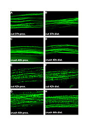
is cut or crushed, in which the part of the axon
Axon
An axon is a long, slender projection of a nerve cell, or neuron, that conducts electrical impulses away from the neuron's cell body or soma....
separated from the neuron
Neuron
A neuron is an electrically excitable cell that processes and transmits information by electrical and chemical signaling. Chemical signaling occurs via synapses, specialized connections with other cells. Neurons connect to each other to form networks. Neurons are the core components of the nervous...
's cell body degenerates distal to the injury. This is also known as anterograde degeneration, or orthograde degeneration. A related process known as 'Wallerian-like degeneration' occurs in many neurodegenerative diseases, especially those where axonal transport is impaired. Primary culture studies suggest that a failure to deliver sufficient quantities of the essential axonal protein NMNAT2
NMNAT2
Nicotinamide mononucleotide adenylyltransferase 2 is an enzyme that in humans is encoded by the NMNAT2 gene.-Further reading:...
is a key initiating event.
Wallerian degeneration occurs after axonal injury in both the peripheral nervous system
Peripheral nervous system
The peripheral nervous system consists of the nerves and ganglia outside of the brain and spinal cord. The main function of the PNS is to connect the central nervous system to the limbs and organs. Unlike the CNS, the PNS is not protected by the bone of spine and skull, or by the blood–brain...
(PNS) and central Nervous System
Central nervous system
The central nervous system is the part of the nervous system that integrates the information that it receives from, and coordinates the activity of, all parts of the bodies of bilaterian animals—that is, all multicellular animals except sponges and radially symmetric animals such as jellyfish...
(CNS). It occurs in the axon stump distal to a site of injury and usually begins within 24–36 hours of a lesion. Prior to degeneration, distal axon stumps tend to remain electrically excitable. After injury, the axonal skeleton disintegrates, and the axonal membrane breaks apart. The axonal degeneration is followed by degradation of the myelin sheath
Myelin
Myelin is a dielectric material that forms a layer, the myelin sheath, usually around only the axon of a neuron. It is essential for the proper functioning of the nervous system. Myelin is an outgrowth of a type of glial cell. The production of the myelin sheath is called myelination...
and infiltration by macrophage
Macrophage
Macrophages are cells produced by the differentiation of monocytes in tissues. Human macrophages are about in diameter. Monocytes and macrophages are phagocytes. Macrophages function in both non-specific defense as well as help initiate specific defense mechanisms of vertebrate animals...
s. The macrophages, accompanied by Schwann cells, serve to clear the debris from the degeneration.
The nerve fiber's neurolemma
Neurolemma
Neurolemma is the outermost nucleated cytoplasmic layer of Schwann cells that surrounds the axon of the neuron. It forms the outermost layer of the nerve fiber in the peripheral nervous system....
does not degenerate and remains as a hollow tube. Within 96 hours of the injury, the distal end of the portion of the nerve fiber proximal to the lesion sends out sprouts towards those tubes and these sprouts are attracted by growth factors produced by Schwann cells in the tubes. If a sprout reaches the tube, it grows into it and advances about 1 mm per day, eventually reaching and reinnervating the target tissue. If the sprouts cannot reach the tube, for instance because the gap is too wide or scar tissue has formed, surgery can help to guide the sprouts into the tubes. This regeneration is much slower in the spinal cord
Spinal cord
The spinal cord is a long, thin, tubular bundle of nervous tissue and support cells that extends from the brain . The brain and spinal cord together make up the central nervous system...
than in PNS. The crucial difference is that in the CNS, including in the spinal cord, myelin sheaths are produced by oligodendrocyte
Oligodendrocyte
Oligodendrocytes , or oligodendroglia , are a type of brain cell. They are a variety of neuroglia. Their main function is the insulation of axons in the central nervous system of some vertebrates...
s and not by Schwann cells.
History
Wallerian degeneration is named after Augustus Volney WallerAugustus Volney Waller
Augustus Volney Waller FRS was a British neurophysiologist. He was the first to describe the degeneration of severed nerve fibers, now known as Wallerian degeneration.-Life:...
. Waller experimented on frog
Frog
Frogs are amphibians in the order Anura , formerly referred to as Salientia . Most frogs are characterized by a short body, webbed digits , protruding eyes and the absence of a tail...
s in 1850, by severing their glossopharyngeal
Glossopharyngeal nerve
The glossopharyngeal nerve is the ninth of twelve pairs of cranial nerves . It exits the brainstem out from the sides of the upper medulla, just rostral to the vagus nerve...
and hypoglossal
Hypoglossal nerve
The hypoglossal nerve is the twelfth cranial nerve , leading to the tongue. The nerve arises from the hypoglossal nucleus and emerges from the medulla oblongata in the preolivary sulcus separating the olive and the pyramid. It then passes through the hypoglossal canal...
nerves. He then observed the distal nerves from the site of injury,
which were separated from their cell bodies in the brain stem.
Waller described the disintegration of myelin, which he referred to as "medulla", into separate particles of various sizes. The degenerated axons formed droplets that could be stained, thus allowing studies of the course of individual nerve fibres.
Axonal degeneration
Although most injury responses include a calciumCalcium
Calcium is the chemical element with the symbol Ca and atomic number 20. It has an atomic mass of 40.078 amu. Calcium is a soft gray alkaline earth metal, and is the fifth-most-abundant element by mass in the Earth's crust...
influx signaling to promote resealing of severed parts, axonal injuries initially lead to acute axonal degeneration (AAD), which is rapid separation of the proximal (the part nearer the cell body) and distal ends within 30 minutes of injury.
Degeneration follows with swelling of the axolemma
Axolemma
The axolemma is the cell membrane surrounding an axon. It is responsible for maintaining the membrane potential of the neuron, and it contains ion channels through which ions can flow. When this occurs, the voltage inside the axon changes, and depolarization or hyperpolarization of the membrane can...
, and eventually leads to bead like formation. The process takes about roughly 24 hours in the PNS, and longer in the CNS. The signaling pathways leading to axolemma degeneration are currently unknown. However, research has shown that this AAD process is calcium–independent.
Granular disintegration of the axonal cytoskeleton and inner organelle
Organelle
In cell biology, an organelle is a specialized subunit within a cell that has a specific function, and is usually separately enclosed within its own lipid bilayer....
s occurs after axolemma degradation. Early changes include accumulation of mitochondria in the paranodal regions
Nodes of Ranvier
Myelin sheath gaps or nodes of Ranvier are the gaps formed between the myelin sheaths generated by different cells. A myelin sheath is a many-layered coating, largely composed of a fatty substance called myelin, that wraps around the axon of a neuron and very efficiently insulates it...
at the site of injury. Endoplasmic reticulum
Endoplasmic reticulum
The endoplasmic reticulum is an organelle of cells in eukaryotic organisms that forms an interconnected network of tubules, vesicles, and cisternae...
degrades and mitochondria swell up and eventually disintegrate. The depolymerization of microtubule
Microtubule
Microtubules are a component of the cytoskeleton. These rope-like polymers of tubulin can grow as long as 25 micrometers and are highly dynamic. The outer diameter of microtubule is about 25 nm. Microtubules are important for maintaining cell structure, providing platforms for intracellular...
s occurs and is soon followed by degradation of the neurofilaments and other cytoskeleton components. The disintegration is dependent on Ubiquitin
Ubiquitin
Ubiquitin is a small regulatory protein that has been found in almost all tissues of eukaryotic organisms. Among other functions, it directs protein recycling.Ubiquitin can be attached to proteins and label them for destruction...
and Calpain
Calpain
A calpain is a protein belonging to the family of calcium-dependent, non-lysosomal cysteine proteases expressed ubiquitously in mammals and many other organisms. Calpains constitute the C2 family of protease clan CA in the MEROPS database...
proteases (caused by influx of calcium ion), suggesting that axonal degeneration is an active process and not a passive one as previously misunderstood.
Thus the axon undergoes complete fragmentation. The rate of degradation is dependent on the type of injury and is also slower in the CNS than in the PNS. Another factor that affects degradation rate is the diameter of the axon: larger axons require a longer time for the cytoskeleton to degrade and thus take a longer time to degenerate.
Myelin clearance
MyelinMyelin
Myelin is a dielectric material that forms a layer, the myelin sheath, usually around only the axon of a neuron. It is essential for the proper functioning of the nervous system. Myelin is an outgrowth of a type of glial cell. The production of the myelin sheath is called myelination...
is a phospholipid membrane that wraps around axons to provide them with insulation. It is produced by Schwann cells in the PNS, and by oligodendrocytes in the CNS. Myelin clearance is the next step in Wallerian degeneration following axonal degeneration. The cleaning up of myelin debris is different for PNS and CNS.
PNS is much faster and efficient at clearing myelin debris in comparison to CNS, and Schwann cells are the primary cause of this difference.
Another key aspect is the change in permeability of the blood-tissue barrier in the two systems.
In PNS, the permeability increases throughout the distal stump, but the barrier disruption in CNS is limited to
just the site of injury.
Clearance in PNS
The response of Schwann cells to axonal injury is rapid. The time period of responseis estimated to be prior to the onset of axonal degeneration. Neuregulins are believed
to be responsible for the rapid activation. They activate ErbB2 receptors in the Schwann
cell microvilli, which results in the activation of the mitogen-activated protein kinase
Mitogen-activated protein kinase
Mitogen-activated protein kinases are serine/threonine-specific protein kinases that respond to extracellular stimuli and regulate various cellular activities, such as gene expression, mitosis, differentiation, proliferation, and cell survival/apoptosis.-Activation:MAP kinases are activated...
(MAPK).
Although MAPK activity is observed, the injury sensing mechanism of Schwann cells is
yet to be fully understood. The sensing is followed by decreased synthesis of myelin lipids
and eventually stops within 48 hrs. The myelin sheaths separate from the axons at
the Schmidt-Lanterman incisures
Schmidt-Lanterman incisures
In the peripheral nervous system axons can be either myelinated or unmyelinated. Myelination means that the axon is insulated by surrounding layers of fatty membrane produced by Schwann cells...
first and then rapidly deteriorate and shorten to form
bead-like structures. Schwann cells continue to clear up the myelin debris by degrading their
own myelin, phagocytose extracellular myelin and attract macrophages to myelin debris for further phagocytosis. However, the macrophages are not attracted to the region for the first few days; hence the Schwann cells take the major role in myelin cleaning until then.
Schwann cells have been observed to recruit macrophages by release of cytokine
Cytokine
Cytokines are small cell-signaling protein molecules that are secreted by the glial cells of the nervous system and by numerous cells of the immune system and are a category of signaling molecules used extensively in intercellular communication...
s and
chemokine
Chemokine
Chemokines are a family of small cytokines, or proteins secreted by cells. Their name is derived from their ability to induce directed chemotaxis in nearby responsive cells; they are chemotactic cytokines...
s after sensing of axonal injury. The recruitment of macrophages helps
improve the clearing rate of myelin debris. The resident macrophages present in the
nerves release further chemokines and cytokines to attract further macrophages.
The degenerating nerve also produce macrophage chemotactic molecules.
Another source of macrophage recruitment factors is serum. Delayed macrophage recruitment
was observed in B-cell deficient mice lacking serum antibodies.
These signaling molecules together cause an influx of macrophages, which peaks during the third week
after injury. While Schwann cells mediate the initial stage of myelin debris clean up,
macrophages come in to finish the job. Macrophages are facilitated by opsonins, which label debris for removal.
The 3 major groups found in serum include complement
Complement system
The complement system helps or “complements” the ability of antibodies and phagocytic cells to clear pathogens from an organism. It is part of the immune system called the innate immune system that is not adaptable and does not change over the course of an individual's lifetime...
, pentraxins
Pentraxins
Pentraxins, also known as pentaxins, are an evolutionary conserved family of proteins characterised by containing a pentraxin protein domain. Proteins of the pentraxin family are involved in acute immunological responses...
, and antibodies. However, only complement has shown to
help in myelin debris phagocytosis.
Murinson et al. (2005) observed that non-myelinated or myelinated Schwann cells in contact with an injured
axon enter cell cycle thus leading to proliferation. Observed time duration for
Schwann cell divisions where approximately 3 days after injury.
Possible sources of proliferation signal are attributed to the ErbB2 receptors and the ErbB3 receptors.
This proliferation could further enhance the myelin cleaning rates and plays an essential role in
regeneration of axons observed in PNS. Schwann cells emit growth factors which attract new axonal sprouts growing from the proximal stump after complete degeneration of the injured distal stump.
This leads to possible reinnervation of the target cell or organ. However, the reinnervation is
not necessarily perfect as possible misleading occurs during reinnervation of the proximal axons to target cells.
Clearance in CNS
In comparison to Schwann cells, oligodendrocytes require axon signals to survive.In their developmental stages, oligodendrocytes that failed to make contact to axon and
receive any axon signals underwent apoptosis
Apoptosis
Apoptosis is the process of programmed cell death that may occur in multicellular organisms. Biochemical events lead to characteristic cell changes and death. These changes include blebbing, cell shrinkage, nuclear fragmentation, chromatin condensation, and chromosomal DNA fragmentation...
.
Experiments in Wallerian degeneration have shown that upon injury oligodendrocytes either undergo programmed cell death or enter a state of rest. Therefore, unlike Schwann cells, oligodendrocytes fail to clean up the myelin sheaths and their debris. In experiments conducted on rats
,
myelin sheaths were found for up to 22 months. Therefore, CNS rates of myelin sheath clearance are very slow and
could possibly be the cause for hindrance in the regeneration capabilities of the CNS axons as no growth factors
are available to attract the proximal axons. Another feature that results eventually is Glial scar
Glial scar
Glial scar formation is a reactive cellular process involving astrogliosis that occurs after injury to the Central Nervous System. As with scarring in other organs and tissues, the glial scar is the body's mechanism to protect and begin the healing process in the nervous system...
formation.
This further hinders chances for regeneration and reinnervation.
Oligodendrocytes fail to recruit macrophages for debris removal.
Macrophage entry in general into CNS site of injury is very slow. In contrast to PNS,
Microglia
Microglia
Microglia are a type of glial cell that are the resident macrophages of the brain and spinal cord, and thus act as the first and main form of active immune defense in the central nervous system . Microglia constitute 20% of the total glial cell population within the brain...
play a vital role in CNS wallerian degeneration. However, their recruitment is
slower in comparison to macrophage recruitment in PNS by approximately 3 days. Further, microglia might be activated but hypertrophy
Hypertrophy
Hypertrophy is the increase in the volume of an organ or tissue due to the enlargement of its component cells. It should be distinguished from hyperplasia, in which the cells remain approximately the same size but increase in number...
, and fail to transform
into fully phagocytic cells. Those microglia that do transform, clear out the debris effectively. Differentiating phagocytic microglia
can be accomplished by testing for expression of Major histocompatibility complex
Major histocompatibility complex
Major histocompatibility complex is a cell surface molecule encoded by a large gene family in all vertebrates. MHC molecules mediate interactions of leukocytes, also called white blood cells , which are immune cells, with other leukocytes or body cells...
(MHC) class I and II during wallerian degeneration.
The rate of clearance is very slow among microglia in comparison to macrophages. Possible source for variations in clearance rates could include lack of
opsonin activity around microglia, and the lack of increased permeability in the blood-brain barrier
Blood-brain barrier
The blood–brain barrier is a separation of circulating blood and the brain extracellular fluid in the central nervous system . It occurs along all capillaries and consists of tight junctions around the capillaries that do not exist in normal circulation. Endothelial cells restrict the diffusion...
.
The decreased permeability could further hinder macrophage infiltration to the site of injury.
These findings have suggested that the delay in Wallerian degeneration in CNS in comparison to PNS is
caused not due to a delay in axonal degeneration, but rather is due to the difference in clearance rates of myelin in CNS and PNS.
Regeneration
Regeneration follows degeneration. Regeneration is rapid in PNS, allowing for rates of up to 1 millimeter a day of regrowth. Grafts may also be needed to allow for appropriate reinnervation.It is supported by Schwann cells through growth factors release. CNS regeneration is much slower, and is almost absent in most species.
The primary cause for this could be the delay in clearing up myelin debris. Myelin debris, present in CNS or PNS, contains several
inhibitory factors. The elongated presence of myelin debris in CNS could possibly hinder the regeneration.
An experiment conducted on Newt
Newt
A newt is an aquatic amphibian of the family Salamandridae, although not all aquatic salamanders are considered newts. Newts are classified in the subfamily Pleurodelinae of the family Salamandridae, and are found in North America, Europe and Asia...
s, animals which have fast CNS axon regeneration capabilities, found that
Wallerian degeneration of an optic nerve injury took up to 10 to 14 days on average, further suggesting that slow clearance
inhibits regeneration.
Schwann cells and endoneural fibroblasts in PNS
In healthy nerves, Nerve growth factorNerve growth factor
Nerve growth factor is a small secreted protein that is important for the growth, maintenance, and survival of certain target neurons . It also functions as a signaling molecule. It is perhaps the prototypical growth factor, in that it is one of the first to be described...
(NGF) is produced in very small amounts. However, upon injury, NGF mRNA expression increases
by five to sevenfold within a period of 14 days. Nerve fibroblasts and Schwann cells play an important role in increased expression of
NGF mRNA.
Macrophages also stimulate Schwann cells and fibroblasts to produce NGF via macrophage-derived interleukin-1.
Other neurotrophic molecules produced by Schwann cells and fibroblasts together include Brain-derived neurotrophic factor
Brain-derived neurotrophic factor
Brain-derived neurotrophic factor, also known as BDNF, is a protein that, in humans, is encoded by the BDNF gene. BDNF is a member of the "neurotrophin" family of growth factors, which are related to the canonical "Nerve Growth Factor", NGF...
, Glial cell line-derived neurotrophic factor
Glial cell line-derived neurotrophic factor
Glial cell-derived neurotrophic factor, also known as GDNF is a protein that, in humans, is encoded by the GDNF gene.GDNF is a small protein that potently promotes the survival of many types of neurons.-Function:...
,
Ciliary neurotrophic factor, Leukemia inhibitory factor
Leukemia inhibitory factor
Leukemia inhibitory factor, or LIF, an interleukin 6 class cytokine, is a protein in cells that affects cell growth and development.-Function:LIF derives its name from its ability to induce the terminal differentiation of myeloid leukemic cells...
, Insulin-like growth factor
Insulin-like growth factor
The insulin-like growth factors are proteins with high sequence similarity to insulin. IGFs are part of a complex system that cells use to communicate with their physiologic environment...
, and Fibroblast growth factor
Fibroblast growth factor
Fibroblast growth factors, or FGFs, are a family of growth factors involved in angiogenesis, wound healing, and embryonic development. The FGFs are heparin-binding proteins and interactions with cell-surface associated heparan sulfate proteoglycans have been shown to be essential for FGF signal...
. These factors
together create a favorable environment for axonal growth and regeneration. Apart from growth factors, Schwann cells
also provide structural guidance to further enhance regeneration. During their proliferation phase, Schwann cells begin to form a line of cells called
Bands of Bungner within the basal laminar tube. Axons have been observed to regenerate
in close association to these cells.
Schwann cells upregulate the production of cell surface adhesion molecule ninjurin further
promoting growth.
These lines of cell guide the axon regeneration in proper direction.
The possible source of error that could result from this is possible mismatching of the
target cells as discussed earlier.
Due to lack of such favorable promoting factors in CNS, regeneration is stunted in CNS.
Delayed Wallerian degeneration
MiceHouse mouse
The house mouse is a small rodent, a mouse, one of the most numerous species of the genus Mus.As a wild animal the house mouse mainly lives associated with humans, causing damage to crops and stored food....
belonging to the strain C57BL/Wlds have delayed Wallerian degeneration,
and thus allow to study the roles of various cell types and the underlying cellular and molecular processes. Current understanding of the
process has been possible via experimentation on the Wlds strain of mice. The mutation occurred first in mice in
Harlan-Olac, a laboratory producing animals the United Kingdom. The Wlds mutation
is an autosomal dominant
Dominance relationship
Dominance in genetics is a relationship between two variant forms of a single gene, in which one allele masks the effect of the other in influencing some trait. In the simplest case, if a gene exists in two allelic forms , three combinations of alleles are possible: AA, AB, and BB...
mutation occurring in the mouse chromosome 4. The gene mutation is an 85-kb tandem triplication, occurring naturally.
The mutated region contains two associated genes: nicotinamide mononucleotide adenlyl transferase 1 (Nmnat-1) and ubiquitination factor e4b (Ube4b).
A linker region encoding 18 amino acid
Amino acid
Amino acids are molecules containing an amine group, a carboxylic acid group and a side-chain that varies between different amino acids. The key elements of an amino acid are carbon, hydrogen, oxygen, and nitrogen...
s is also part of the mutation.
The protein created, localizes within the nucleus and is undetectable in axons.
Effects of Mutation
The mutation causes no harm to the mouse. The only known effect is that the Wallerian degenerationis delayed by up to three weeks on average after injury of a nerve. Initially it was suspected that the
Wlds mutation slows down the macrophage infiltration, but recent studies suggest that the mutation protects axons rather than
slowing down the macrophages. The process by which the axonal protection is achieved is
poorly understood. However, studies suggest that the Wlds mutation leads to overexpression of the Nmnat-1 protein, which
leads to increased NAD
Nicotinamide adenine dinucleotide
Nicotinamide adenine dinucleotide, abbreviated NAD, is a coenzyme found in all living cells. The compound is a dinucleotide, since it consists of two nucleotides joined through their phosphate groups. One nucleotide contains an adenine base and the other nicotinamide.In metabolism, NAD is involved...
synthesis. This in turn activates
SIRT1-dependent process within the nucleus causing changes in gene transcription.
NAD+ by itself provides added axonal protection by increasing the axon's energy resources. More recent work, however, raises doubt that either NMNAT or NAD can substitute for the full length WldS gene. These authors demonstrated by both in vitro and in vivo methods that the protective effect of overexpression of NMNAT1 or the addition of NAD did not protect axons from degeneration. Thus, the underlying biological mechanism accounting for the WldS phenotype remains unknown.
The provided axonal protection delays the onset of Wallerian degeneration. Schwann cell activation
would be delayed, and they wouldn't detect axonal degradation signals from ErbB2 receptors. In experiments on Wlds mutated mice,
macrophage infiltration was considerably delayed by up to six to eight days.
However, once the axonal degradation has begun,
degeneration takes its normal course and respective of the nervous system, degradation follows at the above described rates.
Possible effects that could result due to this late onset would be weaker regenerative abilities in the mice.
See also
- AxonotmesisAxonotmesisAxonotmesis is a disruption of nerve cell axon, with Wallerian degeneration occurring below and slightly proximal to the site of injury. If axons, and their myelin sheath are damaged, but schwann cells, the endoneurium, perineurium and epineurium remain intact is called axonotmesis. Axonotmesis is...
- Connective tissue in the peripheral nervous systemConnective tissue in the peripheral nervous systemA peripheral nerve contains two types of tissue: nerve fibers, and connective tissue. Dendrites and axons with schwann cells and myelin sheath are surrounded by connective tissue. A nerve fiber in the peripheral nervous system consists of an axon or long dendrite, myelin sheath and their schwann...
- Diffuse axonal injuryDiffuse axonal injuryDiffuse axonal injury is one of the most common and devastating types of traumatic brain injury, meaning that damage occurs over a more widespread area than in focal brain injury. DAI, which refers to extensive lesions in white matter tracts, is one of the major causes of unconsciousness and...
- Digestion chambersDigestion chambersDigestion chambers are a histologic finding in nerves that are undergoing Wallerian degeneration.-Appearance:Digestion chambers consist of small globular fragments, which represent degenerating myelin sheaths.-External links:...
- Nerve injuryNerve injuryNerve injury is injury to nervous tissue. There is no single classification system that can describe all the many variations of nerve injury. Most systems attempt to correlate the degree of injury with symptoms, pathology and prognosis...
- Neuroregeneration
- Peripheral nerve injuryPeripheral nerve injuryPeripheral nerve damage is categorized in the Seddon classification based on the extent of damage to both the nerve and the surrounding connective tissue since the nervous system is characterized by dependence of neurons on their supporting glia. Unlike in the central nervous system, regeneration...
- Primary and secondary brain injuryPrimary and secondary brain injuryPrimary and secondary brain injury are ways to classify the injury processes that occur in brain injury. In traumatic brain injury , primary injury occurs during the initial insult, and results from displacement of the physical structures of the brain. On the other hand, secondary injury occurs...
- Seddon's classification