
Electroencephalography
Encyclopedia

Scalp
The scalp is the anatomical area bordered by the face anteriorly and the neck to the sides and posteriorly.-Layers:It is usually described as having five layers, which can conveniently be remembered as a mnemonic:...
. EEG measures voltage fluctuations resulting from ionic current flows within the neurons of the brain
Brain
The brain is the center of the nervous system in all vertebrate and most invertebrate animals—only a few primitive invertebrates such as sponges, jellyfish, sea squirts and starfishes do not have one. It is located in the head, usually close to primary sensory apparatus such as vision, hearing,...
. In clinical contexts, EEG refers to the recording of the brain's spontaneous electrical activity over a short period of time, usually 20–40 minutes, as recorded from multiple electrodes placed on the scalp
Scalp
The scalp is the anatomical area bordered by the face anteriorly and the neck to the sides and posteriorly.-Layers:It is usually described as having five layers, which can conveniently be remembered as a mnemonic:...
. In neurology
Neurology
Neurology is a medical specialty dealing with disorders of the nervous system. Specifically, it deals with the diagnosis and treatment of all categories of disease involving the central, peripheral, and autonomic nervous systems, including their coverings, blood vessels, and all effector tissue,...
, the main diagnostic application of EEG is in the case of epilepsy
Epilepsy
Epilepsy is a common chronic neurological disorder characterized by seizures. These seizures are transient signs and/or symptoms of abnormal, excessive or hypersynchronous neuronal activity in the brain.About 50 million people worldwide have epilepsy, and nearly two out of every three new cases...
, as epileptic activity can create clear abnormalities on a standard EEG study. A secondary clinical use of EEG is in the diagnosis of coma
Coma
In medicine, a coma is a state of unconsciousness, lasting more than 6 hours in which a person cannot be awakened, fails to respond normally to painful stimuli, light or sound, lacks a normal sleep-wake cycle and does not initiate voluntary actions. A person in a state of coma is described as...
, encephalopathies, and brain death
Brain death
Brain death is the irreversible end of all brain activity due to total necrosis of the cerebral neurons following loss of brain oxygenation. It should not be confused with a persistent vegetative state...
. EEG used to be a first-line method for the diagnosis of tumor
Tumor
A tumor or tumour is commonly used as a synonym for a neoplasm that appears enlarged in size. Tumor is not synonymous with cancer...
s, stroke
Stroke
A stroke, previously known medically as a cerebrovascular accident , is the rapidly developing loss of brain function due to disturbance in the blood supply to the brain. This can be due to ischemia caused by blockage , or a hemorrhage...
and other focal brain disorders, but this use has decreased with the advent of anatomical imaging techniques with high (<1 mm) spatial resolution such as MRI and CT
Computed tomography
X-ray computed tomography or Computer tomography , is a medical imaging method employing tomography created by computer processing...
. Despite limited spatial resolution, EEG continues to be a valuable tool for research and diagnosis, especially when millisecond-range temporal resolution (not possible with CT or MRI) is required.
Derivatives of the EEG technique include evoked potential
Evoked potential
An evoked potential is an electrical potential recorded from the nervous system of a human or other animal following presentation of a stimulus, as distinct from spontaneous potentials as detected by electroencephalography or electromyography .Evoked potential amplitudes tend to be low, ranging...
s (EP), which involves averaging the EEG activity time-locked to the presentation of a stimulus of some sort (visual, somatosensory, or auditory). Event-related potentials (ERPs) refer to averaged EEG responses that are time-locked to more complex processing of stimuli; this technique is used in cognitive science
Cognitive science
Cognitive science is the interdisciplinary scientific study of mind and its processes. It examines what cognition is, what it does and how it works. It includes research on how information is processed , represented, and transformed in behaviour, nervous system or machine...
, cognitive psychology
Cognitive psychology
Cognitive psychology is a subdiscipline of psychology exploring internal mental processes.It is the study of how people perceive, remember, think, speak, and solve problems.Cognitive psychology differs from previous psychological approaches in two key ways....
, and psychophysiological
Psychophysiology
Psychophysiology is the branch of psychology that is concerned with the physiological bases of psychological processes. While psychophysiology was a general broad field of research in the 1960s and 1970s, it has now become quite specialized, and has branched into subspecializations...
research.
Source of EEG activity
The brain's electrical charge is maintained by billions of neuronNeuron
A neuron is an electrically excitable cell that processes and transmits information by electrical and chemical signaling. Chemical signaling occurs via synapses, specialized connections with other cells. Neurons connect to each other to form networks. Neurons are the core components of the nervous...
s. Neurons are electrically charged (or "polarized") by membrane transport proteins that pump ions across their membranes. Neurons are constantly exchanging ions with the extracellular milieu, for example to maintain resting potential and to propagate action potentials. Ions of like charge repel each other, and when many ions are pushed out of many neurons at the same time, they can push their neighbours, who push their neighbours, and so on, in a wave. This process is known as volume conduction. When the wave of ions reaches the electrodes on the scalp, they can push or pull electrons on the metal on the electrodes. Since metal conducts the push and pull of electrons easily, the difference in push or voltage between any two electrodes can be measured by a voltmeter
Voltmeter
A voltmeter is an instrument used for measuring electrical potential difference between two points in an electric circuit. Analog voltmeters move a pointer across a scale in proportion to the voltage of the circuit; digital voltmeters give a numerical display of voltage by use of an analog to...
. Recording these voltages over time gives us the EEG.
The electric potential
Electric potential
In classical electromagnetism, the electric potential at a point within a defined space is equal to the electric potential energy at that location divided by the charge there...
s generated by single neurons are far too small to be picked by EEG or MEG. EEG activity therefore always reflects the summation of the synchronous activity of thousands or millions of neurons that have similar spatial orientation. If the cells do not have similar spatial orientation, their ions do not line up and create waves to be detected. Pyramidal neurons of the cortex are thought to produce the most EEG signal because they are well-aligned and fire together. Because voltage fields fall off with the square of distance, activity from deep sources is more difficult to detect than currents near the skull.
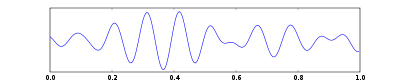
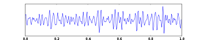
Scalp EEG activity shows oscillations
Neural oscillations
Neural oscillation is rhythmic or repetitive neural activity in the central nervous system. Neural tissue can generate oscillatory activity in many ways, driven either by mechanisms localized within individual neurons or by interactions between neurons...
at a variety of frequencies. Several of these oscillations have characteristic frequency ranges, spatial distributions and are associated with different states of brain functioning (e.g., waking and the various sleep stages
Sleep
Sleep is a naturally recurring state characterized by reduced or absent consciousness, relatively suspended sensory activity, and inactivity of nearly all voluntary muscles. It is distinguished from quiet wakefulness by a decreased ability to react to stimuli, and is more easily reversible than...
). These oscillations represent synchronized activity over a network of neurons. The neuronal networks underlying some of these oscillations are understood (e.g., the thalamocortical resonance underlying sleep spindles), while many others are not (e.g., the system that generates the posterior basic rhythm). Research that measures both EEG and neuron spiking finds the relationship between the two is complex with the power of surface EEG in only two bands (gamma
Gamma wave
A gamma wave is a pattern of neural oscillation in humans with a frequency between 25 to 100 Hz, though 40 Hz is prototypical.According to a popular theory, gamma waves may be implicated in creating the unity of conscious perception...
and delta
Delta wave
A delta wave is a high amplitude brain wave with a frequency of oscillation between 0–4 hertz. Delta waves, like other brain waves, are recorded with an electroencephalogram and are usually associated with the deepest stages of sleep , also known as slow-wave sleep , and aid in characterizing the...
) relating to neuron spike activity.
Clinical use
A routine clinical EEG recording typically lasts 20–30 minutes (plus preparation time) and usually involves recording from scalp electrodes. Routine EEG is typically used in the following clinical circumstances:- to distinguish epilepticEpilepsyEpilepsy is a common chronic neurological disorder characterized by seizures. These seizures are transient signs and/or symptoms of abnormal, excessive or hypersynchronous neuronal activity in the brain.About 50 million people worldwide have epilepsy, and nearly two out of every three new cases...
seizureSeizureAn epileptic seizure, occasionally referred to as a fit, is defined as a transient symptom of "abnormal excessive or synchronous neuronal activity in the brain". The outward effect can be as dramatic as a wild thrashing movement or as mild as a brief loss of awareness...
s from other types of spells, such as psychogenic non-epileptic seizuresPsychogenic non-epileptic seizuresPsychogenic non-epileptic seizures , also known as Non-Epileptic Attack Disorders, are events superficially resembling an epileptic seizure, but without the characteristic electrical discharges associated with epilepsy. Instead, PNES are psychological in origin, and may be thought of as similar to...
, syncope (fainting), sub-cortical movement disorderMovement disorderMovement disorders include:* Akathisia * Akinesia * Associated Movements * Athetosis...
s and migraineMigraineMigraine is a chronic neurological disorder characterized by moderate to severe headaches, and nausea...
variants. - to differentiate "organic" encephalopathyEncephalopathyEncephalopathy means disorder or disease of the brain. In modern usage, encephalopathy does not refer to a single disease, but rather to a syndrome of global brain dysfunction; this syndrome can be caused by many different illnesses.-Terminology:...
or deliriumDeliriumDelirium or acute confusional state is a common and severe neuropsychiatric syndrome with core features of acute onset and fluctuating course, attentional deficits and generalized severe disorganization of behavior...
from primary psychiatric syndromes such as catatoniaCatatoniaCatatonia is a state of neurogenic motor immobility, and behavioral abnormality manifested by stupor. It was first described in 1874: Die Katatonie oder das Spannungsirresein .... - to serve as an adjunct test of brain deathBrain deathBrain death is the irreversible end of all brain activity due to total necrosis of the cerebral neurons following loss of brain oxygenation. It should not be confused with a persistent vegetative state...
- to prognosticate, in certain instances, in patients with coma
- to determine whether to wean anti-epileptic medications
At times, a routine EEG is not sufficient, particularly when it is necessary to record a patient while he/she is having a seizure. In this case, the patient may be admitted to the hospital for days or even weeks, while EEG is constantly being recorded (along with time-synchronized video and audio recording). A recording of an actual seizure (i.e., an ictal
Ictal
Ictal refers to a physiologic state or event such as a seizure, stroke or headache. The word originates from the Latin ictus, meaning a blow or a stroke...
recording, rather than an inter-ictal recording of a possibly epileptic patient at some period between seizures) can give significantly better information about whether or not a spell is an epileptic seizure and the focus in the brain from which the seizure activity emanates.
Epilepsy monitoring is typically done:
- to distinguish epilepticEpilepsyEpilepsy is a common chronic neurological disorder characterized by seizures. These seizures are transient signs and/or symptoms of abnormal, excessive or hypersynchronous neuronal activity in the brain.About 50 million people worldwide have epilepsy, and nearly two out of every three new cases...
seizureSeizureAn epileptic seizure, occasionally referred to as a fit, is defined as a transient symptom of "abnormal excessive or synchronous neuronal activity in the brain". The outward effect can be as dramatic as a wild thrashing movement or as mild as a brief loss of awareness...
s from other types of spells, such as psychogenic non-epileptic seizuresPsychogenic non-epileptic seizuresPsychogenic non-epileptic seizures , also known as Non-Epileptic Attack Disorders, are events superficially resembling an epileptic seizure, but without the characteristic electrical discharges associated with epilepsy. Instead, PNES are psychological in origin, and may be thought of as similar to...
, syncope (fainting)Syncope (medicine)Syncope , the medical term for fainting, is precisely defined as a transient loss of consciousness and postural tone characterized by rapid onset, short duration, and spontaneous recovery due to global cerebral hypoperfusion that most often results from hypotension.Many forms of syncope are...
, sub-cortical movement disorderMovement disorderMovement disorders include:* Akathisia * Akinesia * Associated Movements * Athetosis...
s and migraineMigraineMigraine is a chronic neurological disorder characterized by moderate to severe headaches, and nausea...
variants. - to characterize seizuresSeizure typesThe numerous epileptic seizure types are most commonly defined and grouped according to the scheme proposed by the International League Against Epilepsy in 1981...
for the purposes of treatment - to localize the region of brain from which a seizure originates for work-up of possible seizure surgery
Additionally, EEG may be used to monitor certain procedures:
- to monitor the depth of anesthesiaAnesthesiaAnesthesia, or anaesthesia , traditionally meant the condition of having sensation blocked or temporarily taken away...
- as an indirect indicator of cerebral perfusion in carotid endarterectomyCarotid endarterectomyCarotid endarterectomy is a surgical procedure used to prevent stroke, by correcting stenosis in the common carotid artery...
- to monitor amobarbital effect during the Wada testWada testThe Wada test, named after Canadian neurologist and epileptologist Juhn Atsushi Wada, also known as the "intracarotid sodium amobarbital procedure" , is used to establish cerebral language and memory representation of each hemisphere.-Method:...
EEG can also be used in intensive care unit
Intensive Care Unit
thumb|220px|ICU roomAn intensive-care unit , critical-care unit , intensive-therapy unit/intensive-treatment unit is a specialized department in a hospital that provides intensive-care medicine...
s for brain function monitoring:
- to monitor for non-convulsive seizures/non-convulsive status epilepticus
- to monitor the effect of sedative/anesthesia in patients in medically induced coma (for treatment of refractory seizures or increased intracranial pressureIntracranial pressureIntracranial pressure is the pressure inside the skull and thus in the brain tissue and cerebrospinal fluid . The body has various mechanisms by which it keeps the ICP stable, with CSF pressures varying by about 1 mmHg in normal adults through shifts in production and absorption of CSF...
) - to monitor for secondary brain damage in conditions such as subarachnoid hemorrhageSubarachnoid hemorrhageA subarachnoid hemorrhage , or subarachnoid haemorrhage in British English, is bleeding into the subarachnoid space—the area between the arachnoid membrane and the pia mater surrounding the brain...
(currently a research method)
If a patient with epilepsy is being considered for resective surgery
Epilepsy surgery
Approximately 60% of all patients with epilepsy suffer from focal epilepsy syndromes. In 15 to 20% of these patients, the condition is not adequately controlled with anticonvulsive drugs. Such patients are potential candidates for surgical epilepsy treatment...
, it is often necessary to localize the focus (source) of the epileptic brain activity with a resolution greater than what is provided by scalp EEG. This is because the cerebrospinal fluid
Cerebrospinal fluid
Cerebrospinal fluid , Liquor cerebrospinalis, is a clear, colorless, bodily fluid, that occupies the subarachnoid space and the ventricular system around and inside the brain and spinal cord...
, skull and scalp smear the electrical potentials recorded by scalp EEG. In these cases, neurosurgeons typically implant strips and grids of electrodes (or penetrating depth electrodes) under the dura mater
Dura mater
The dura mater , or dura, is the outermost of the three layers of the meninges surrounding the brain and spinal cord. It is derived from Mesoderm. The other two meningeal layers are the pia mater and the arachnoid mater. The dura surrounds the brain and the spinal cord and is responsible for...
, through either a craniotomy
Craniotomy
A craniotomy is a surgical operation in which a bone flap is temporarily removed from the skull to access the brain. Craniotomies are often a critical operation performed on patients recording, brain imaging, and for neurological manipulations such as electrical stimulation and chemical...
or a burr hole. The recording of these signals is referred to as electrocorticography
Electrocorticography
Electrocorticography is the practice of using electrodes placed directly on the exposed surface of the brain to record electrical activity from the cerebral cortex. ECoG may be performed either in the operating room during surgery or outside of surgery...
(ECoG), subdural EEG (sdEEG) or intracranial EEG (icEEG)--all terms for the same thing. The signal recorded from ECoG is on a different scale of activity than the brain activity recorded from scalp EEG. Low voltage, high frequency components that cannot be seen easily (or at all) in scalp EEG can be seen clearly in ECoG. Further, smaller electrodes (which cover a smaller parcel of brain surface) allow even lower voltage, faster components of brain activity to be seen. Some clinical sites record from penetrating microelectrodes.
Research use
Event-related potential
An event-related potential is any measured brain response that is directly the result of a thought or perception. More formally, it is any stereotyped electrophysiological response to an internal or external stimulus....
are used extensively in neuroscience
Neuroscience
Neuroscience is the scientific study of the nervous system. Traditionally, neuroscience has been seen as a branch of biology. However, it is currently an interdisciplinary science that collaborates with other fields such as chemistry, computer science, engineering, linguistics, mathematics,...
, cognitive science
Cognitive science
Cognitive science is the interdisciplinary scientific study of mind and its processes. It examines what cognition is, what it does and how it works. It includes research on how information is processed , represented, and transformed in behaviour, nervous system or machine...
, cognitive psychology
Cognitive psychology
Cognitive psychology is a subdiscipline of psychology exploring internal mental processes.It is the study of how people perceive, remember, think, speak, and solve problems.Cognitive psychology differs from previous psychological approaches in two key ways....
, and psychophysiological
Psychophysiology
Psychophysiology is the branch of psychology that is concerned with the physiological bases of psychological processes. While psychophysiology was a general broad field of research in the 1960s and 1970s, it has now become quite specialized, and has branched into subspecializations...
research. Many EEG techniques used in research are not standardized sufficiently for clinical use.
A different method to study brain function is functional magnetic resonance imaging (fMRI)
Functional magnetic resonance imaging
Functional magnetic resonance imaging or functional MRI is a type of specialized MRI scan used to measure the hemodynamic response related to neural activity in the brain or spinal cord of humans or other animals. It is one of the most recently developed forms of neuroimaging...
. Some advantages of EEG over fMRI include:
- Hardware costs are significantly lower
- EEG sensors can be used in more places than a bulky, immobile fMRI machine can
- EEG has higher temporal resolution - milliseconds, rather than seconds
- EEG is relatively tolerant of subject movement (in fMRI the subject must remain completely still)
- EEG is silent, which allows for better study of the responses to auditory stimuli
- EEG does not aggravate claustrophobiaClaustrophobiaClaustrophobia is the fear of having no escape and being closed in small spaces or rooms...
- EEG does not involve exposure to high-intensity (>1 Tesla) magnetic fields (as in MRI)
In addition, EEG does not involve exposure to radioligands (unlike positron emission tomography
Positron emission tomography
Positron emission tomography is nuclear medicine imaging technique that produces a three-dimensional image or picture of functional processes in the body. The system detects pairs of gamma rays emitted indirectly by a positron-emitting radionuclide , which is introduced into the body on a...
)
Disadvantages of EEG relative to fMRI include:
- Significantly lower spatial resolution
- ERP studies require relatively simple paradigms, compared with block-design fMRI studies
Simultaneous EEG recordings and fMRI scans have been obtained successfully, though successful simultaneous recording requires that several technical difficulties be overcome, such as the presence of ballistocardiographic artifact, MRI pulse artifact and the induction of electrical currents in EEG wires that move within the strong magnetic fields of the MRI.
Another method of high temporal resolution is magnetoencephalography
Magnetoencephalography
Magnetoencephalography is a technique for mapping brain activity by recording magnetic fields produced by electrical currents occurring naturally in the brain, using arrays of SQUIDs...
, which benefits from the lack of interference by the skull, but it has low spatial resolution and requires expensive and bulky equipment consisting of liquid helium
Liquid helium
Helium exists in liquid form only at extremely low temperatures. The boiling point and critical point depend on the isotope of the helium; see the table below for values. The density of liquid helium-4 at its boiling point and 1 atmosphere is approximately 0.125 g/mL Helium-4 was first liquefied...
-cooled detectors that can be used only in magnetically shielded rooms.
EEG also has some characteristics that compare favorably with behavioral testing:
- EEG can detect covert processing (i.e., processing that does not require a response)
- EEG can be used in subjects who are incapable of making a motor response
- Some ERP components can be detected even when the subject is not attending to the stimuli
- Unlike other means of studying reaction time, ERPs can elucidate stages of processing (rather than just the final end result)
Method
.jpg)
Electrode
An electrode is an electrical conductor used to make contact with a nonmetallic part of a circuit...
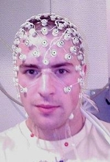
s on the scalp with a conductive gel or paste, usually after preparing the scalp area by light abrasion to reduce impedance
Electrical impedance
Electrical impedance, or simply impedance, is the measure of the opposition that an electrical circuit presents to the passage of a current when a voltage is applied. In quantitative terms, it is the complex ratio of the voltage to the current in an alternating current circuit...
due to dead skin cells. Many systems typically use electrodes, each of which is attached to an individual wire. Some systems use caps or nets into which electrodes are embedded; this is particularly common when high-density arrays of electrodes are needed.
Electrode locations and names are specified by the International 10–20 system
10-20 system (EEG)
right|200pxThe 10-20 system or International 10-20 system is an internationally recognized method to describe and apply the location of scalp electrodes in the context of an EEG test or experiment. This method was developed to ensure standardized reproducibility so that a subject's studies could be...
for most clinical and research applications (except when high-density arrays are used). This system ensures that the naming of electrodes is consistent across laboratories. In most clinical applications, 19 recording electrodes (plus ground and system reference) are used. A smaller number of electrodes are typically used when recording EEG from neonates
Infant
A newborn or baby is the very young offspring of a human or other mammal. A newborn is an infant who is within hours, days, or up to a few weeks from birth. In medical contexts, newborn or neonate refers to an infant in the first 28 days after birth...
. Additional electrodes can be added to the standard set-up when a clinical or research application demands increased spatial resolution for a particular area of the brain. High-density arrays (typically via cap or net) can contain up to 256 electrodes more-or-less evenly spaced around the scalp.
Each electrode is connected to one input of a differential amplifier
Differential amplifier
A differential amplifier is a type of electronic amplifier that amplifies the difference between two voltages but does not amplify the particular voltages.- Theory :Many electronic devices use differential amplifiers internally....
(one amplifier per pair of electrodes); a common system reference electrode is connected to the other input of each differential amplifier. These amplifiers amplify the voltage between the active electrode and the reference (typically 1,000–100,000 times, or 60–100 dB
Decibel
The decibel is a logarithmic unit that indicates the ratio of a physical quantity relative to a specified or implied reference level. A ratio in decibels is ten times the logarithm to base 10 of the ratio of two power quantities...
of voltage gain). In analog EEG, the signal is then filtered (next paragraph), and the EEG signal is output as the deflection of pens as paper passes underneath. Most EEG systems these days, however, are digital, and the amplified signal is digitized via an analog-to-digital converter
Analog-to-digital converter
An analog-to-digital converter is a device that converts a continuous quantity to a discrete time digital representation. An ADC may also provide an isolated measurement...
, after being passed through an anti-aliasing filter
Anti-aliasing filter
An anti-aliasing filter is a filter used before a signal sampler, to restrict the bandwidth of a signal to approximately satisfy the sampling theorem....
. Analog-to-digital sampling typically occurs at 256–512 Hz in clinical scalp EEG; sampling rates of up to 20 kHz are used in some research applications.
During the recording, a series of activation procedures may be used. These procedures may induce normal or abnormal EEG activity that might not otherwise be seen. These procedures include hyperventilation, photic stimulation (with a strobe light), eye closure, mental activity, sleep and sleep deprivation. During (inpatient) epilepsy monitoring, a patient's typical seizure medications may be withdrawn.
The digital EEG signal is stored electronically and can be filtered for display. Typical settings for the high-pass filter
High-pass filter
A high-pass filter is a device that passes high frequencies and attenuates frequencies lower than its cutoff frequency. A high-pass filter is usually modeled as a linear time-invariant system...
and a low-pass filter
Low-pass filter
A low-pass filter is an electronic filter that passes low-frequency signals but attenuates signals with frequencies higher than the cutoff frequency. The actual amount of attenuation for each frequency varies from filter to filter. It is sometimes called a high-cut filter, or treble cut filter...
are 0.5-1 Hz
Hertz
The hertz is the SI unit of frequency defined as the number of cycles per second of a periodic phenomenon. One of its most common uses is the description of the sine wave, particularly those used in radio and audio applications....
and 35–70 Hz, respectively. The high-pass filter typically filters out slow artifact, such as electrogalvanic
Galvanic skin response
Skin conductance, also known as galvanic skin response , electrodermal response , psychogalvanic reflex , skin conductance response or skin conductance level , is a method of measuring the electrical conductance of the skin, which varies with its moisture level...
signals and movement artifact, whereas the low-pass filter filters out high-frequency artifacts, such as electromyographic
Electromyography
Electromyography is a technique for evaluating and recording the electrical activity produced by skeletal muscles. EMG is performed using an instrument called an electromyograph, to produce a record called an electromyogram. An electromyograph detects the electrical potential generated by muscle...
signals. An additional notch filter
Band-stop filter
In signal processing, a band-stop filter or band-rejection filter is a filter that passes most frequencies unaltered, but attenuates those in a specific range to very low levels. It is the opposite of a band-pass filter...
is typically used to remove artifact caused by electrical power lines (60 Hz in the United States and 50 Hz in many other countries).
As part of an evaluation for epilepsy surgery, it may be necessary to insert electrodes near the surface of the brain, under the surface of the dura mater
Dura mater
The dura mater , or dura, is the outermost of the three layers of the meninges surrounding the brain and spinal cord. It is derived from Mesoderm. The other two meningeal layers are the pia mater and the arachnoid mater. The dura surrounds the brain and the spinal cord and is responsible for...
. This is accomplished via burr hole or craniotomy
Craniotomy
A craniotomy is a surgical operation in which a bone flap is temporarily removed from the skull to access the brain. Craniotomies are often a critical operation performed on patients recording, brain imaging, and for neurological manipulations such as electrical stimulation and chemical...
. This is referred to variously as "electrocorticography (ECoG)"
Electrocorticography
Electrocorticography is the practice of using electrodes placed directly on the exposed surface of the brain to record electrical activity from the cerebral cortex. ECoG may be performed either in the operating room during surgery or outside of surgery...
, "intracranial EEG (I-EEG)" or "subdural EEG (SD-EEG)". Depth electrodes may also be placed into brain structures, such as the amygdala
Amygdala
The ' are almond-shaped groups of nuclei located deep within the medial temporal lobes of the brain in complex vertebrates, including humans. Shown in research to perform a primary role in the processing and memory of emotional reactions, the amygdalae are considered part of the limbic system.-...
or hippocampus
Hippocampus
The hippocampus is a major component of the brains of humans and other vertebrates. It belongs to the limbic system and plays important roles in the consolidation of information from short-term memory to long-term memory and spatial navigation. Humans and other mammals have two hippocampi, one in...
, structures, which are common epileptic foci and may not be "seen" clearly by scalp EEG. The electrocorticographic signal is processed in the same manner as digital scalp EEG (above), with a couple of caveats. ECoG is typically recorded at higher sampling rates than scalp EEG because of the requirements of Nyquist theorem
Nyquist–Shannon sampling theorem
The Nyquist–Shannon sampling theorem, after Harry Nyquist and Claude Shannon, is a fundamental result in the field of information theory, in particular telecommunications and signal processing. Sampling is the process of converting a signal into a numeric sequence...
—the subdural signal is composed of a higher predominance of higher frequency components. Also, many of the artifacts that affect scalp EEG do not impact ECoG, and therefore display filtering is often not needed.
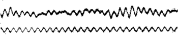
A typical adult human EEG signal is about 10µV to 100 µV in amplitude when measured from the scalp and is about 10–20 mV when measured from subdural electrodes.
Since an EEG voltage signal represents a difference between the voltages at two electrodes, the display of the EEG for the reading encephalographer may be set up in one of several ways. The representation of the EEG channels is referred to as a montage.
Bipolar montage : Each channel (i.e., waveform) represents the difference between two adjacent electrodes. The entire montage consists of a series of these channels. For example, the channel "Fp1-F3" represents the difference in voltage between the Fp1 electrode and the F3 electrode. The next channel in the montage, "F3-C3," represents the voltage difference between F3 and C3, and so on through the entire array of electrodes.
Referential montage: Each channel represents the difference between a certain electrode and a designated reference electrode. There is no standard position for this reference; it is, however, at a different position than the "recording" electrodes. Midline positions are often used because they do not amplify the signal in one hemisphere vs. the other. Another popular reference is "linked ears," which is a physical or mathematical average of electrodes attached to both earlobes or mastoids
Mastoid process
The mastoid process is a conical prominence projecting from the undersurface of the mastoid portion of the temporal bone. It is located just behind the external acoustic meatus, and lateral to the styloid process...
.
Average reference montage : The outputs of all of the amplifiers are summed and averaged, and this averaged signal is used as the common reference for each channel.
Laplacian montage : Each channel represents the difference between an electrode and a weighted average of the surrounding electrodes.
When analog (paper) EEGs are used, the technologist switches between montages during the recording in order to highlight or better characterize certain features of the EEG. With digital EEG, all signals are typically digitized and stored in a particular (usually referential) montage; since any montage can be constructed mathematically from any other, the EEG can be viewed by the electroencephalographer in any display montage that is desired.
The EEG is read by a clinical neurophysiologist
Clinical neurophysiology
Clinical neurophysiology is a medical specialty that studies the central and peripheral nervous systems through the recording of bioelectrical activity, whether spontaneous or stimulated....
or neurologist
Neurologist
A neurologist is a physician who specializes in neurology, and is trained to investigate, or diagnose and treat neurological disorders.Neurology is the medical specialty related to the human nervous system. The nervous system encompasses the brain, spinal cord, and peripheral nerves. A specialist...
(depending on local custom and law regardingmedical specialities
Specialty (medicine)
A specialty in medicine is a branch of medical science. After completing medical school, physicians or surgeons usually further their medical education in a specific specialty of medicine by completing a multiple year residency to become a medical specialist.-History of medical specialization:To...
), optimally one who has specific training in the interpretation of EEGs for clinical purposes. This is done by visual inspection of the waveforms, called graphoelements. The use of computer signal processing of the EEG—so-called quantitative EEG—is somewhat controversial when used for clinical purposes (although there are many research uses).
Limitations
EEG has several limitations. Most important is its poor spatial resolution. EEG is most sensitive to a particular set of post-synaptic potentials: those generated in superficial layers of the cortex, on the crests of gyriGyrus
A gyrus is a ridge on the cerebral cortex. It is generally surrounded by one or more sulci .-Notable gyri:* Superior frontal gyrus, lat. gyrus frontalis superior* Middle frontal gyrus, lat. gyrus frontalis medius...
directly abutting the skull and radial to the skull. Dendrites, which are deeper in the cortex, inside sulci
Sulcus (neuroanatomy)
In neuroanatomy, a sulcus is a depression or fissure in the surface of the brain.It surrounds the gyri, creating the characteristic appearance of the brain in humans and other large mammals....
, in midline or deep structures (such as the cingulate gyrus or hippocampus
Hippocampus
The hippocampus is a major component of the brains of humans and other vertebrates. It belongs to the limbic system and plays important roles in the consolidation of information from short-term memory to long-term memory and spatial navigation. Humans and other mammals have two hippocampi, one in...
), or producing currents that are tangential to the skull, have far less contribution to the EEG signal.
The meninges
Meninges
The meninges is the system of membranes which envelopes the central nervous system. The meninges consist of three layers: the dura mater, the arachnoid mater, and the pia mater. The primary function of the meninges and of the cerebrospinal fluid is to protect the central nervous system.-Dura...
, cerebrospinal fluid
Cerebrospinal fluid
Cerebrospinal fluid , Liquor cerebrospinalis, is a clear, colorless, bodily fluid, that occupies the subarachnoid space and the ventricular system around and inside the brain and spinal cord...
and skull "smear" the EEG signal, obscuring its intracranial source.
It is mathematically impossible to reconstruct a unique intracranial current source for a given EEG signal, as some currents produce potentials that cancel each other out. This is referred to as the inverse problem
Inverse problem
An inverse problem is a general framework that is used to convert observed measurements into information about a physical object or system that we are interested in...
. However, much work has been done to produce remarkably good estimates of, at least, a localized electric dipole that represents the recorded currents.
EEG vs fMRI and PET
EEG has several strong points as a tool for exploring brain activity. EEG's can detect changes over milliseconds, which is excellent considering an action potentialAction potential
In physiology, an action potential is a short-lasting event in which the electrical membrane potential of a cell rapidly rises and falls, following a consistent trajectory. Action potentials occur in several types of animal cells, called excitable cells, which include neurons, muscle cells, and...
takes approximately 0.5-130 milliseconds to propagate across a single neuron, depending on the type of neuron. Other methods of looking at brain activity, such as PET and fMRI have time resolution between seconds and minutes. EEG measures the brain's electrical activity directly, while other methods record changes in blood flow (e.g., SPECT
Single photon emission computed tomography
Single-photon emission computed tomography is a nuclear medicine tomographic imaging technique using gamma rays. It is very similar to conventional nuclear medicine planar imaging using a gamma camera. However, it is able to provide true 3D information...
, fMRI
Functional magnetic resonance imaging
Functional magnetic resonance imaging or functional MRI is a type of specialized MRI scan used to measure the hemodynamic response related to neural activity in the brain or spinal cord of humans or other animals. It is one of the most recently developed forms of neuroimaging...
) or metabolic activity (e.g., PET
Positron emission tomography
Positron emission tomography is nuclear medicine imaging technique that produces a three-dimensional image or picture of functional processes in the body. The system detects pairs of gamma rays emitted indirectly by a positron-emitting radionuclide , which is introduced into the body on a...
), which are indirect markers of brain electrical activity. EEG can be used simultaneously with fMRI
Functional magnetic resonance imaging
Functional magnetic resonance imaging or functional MRI is a type of specialized MRI scan used to measure the hemodynamic response related to neural activity in the brain or spinal cord of humans or other animals. It is one of the most recently developed forms of neuroimaging...
so that high-temporal-resolution data can be recorded at the same time as high-spatial-resolution data, however, since the data derived from each occurs over a different time course, the data sets do not necessarily represent exactly the same brain activity. There are technical difficulties associated with combining these two modalities, including the need to remove the MRI gradient artifact present during MRI acquisition and the ballistocardiographic artifact (resulting from the pulsatile motion of blood and tissue) from the EEG. Furthermore, currents can be induced in moving EEG electrode wires due to the magnetic field of the MRI.
EEG vs MEG
EEG reflects correlated synaptic activity caused by post-synaptic potentials of cortical neurons. The ionic currents involved in the generation of fast action potentials may not contribute greatly to the averaged field potentialsLocal field potential
A local field potential is a particular class of electrophysiological signals, which is dominated by the electrical current flowing from all nearby dendritic synaptic activity within a volume of tissue. A voltage is produced by the summed synaptic current flowing across the resistance of the local...
representing the EEG . More specifically, the scalp electrical potentials that produce EEG are generally thought to be caused by the extracellular ionic currents caused by dendritic
Dendrite
Dendrites are the branched projections of a neuron that act to conduct the electrochemical stimulation received from other neural cells to the cell body, or soma, of the neuron from which the dendrites project...
electrical activity, whereas the fields producing magnetoencephalographic
Magnetoencephalography
Magnetoencephalography is a technique for mapping brain activity by recording magnetic fields produced by electrical currents occurring naturally in the brain, using arrays of SQUIDs...
signals are associated with intracellular ionic currents .
EEG can be recorded at the same time as MEG
Magnetoencephalography
Magnetoencephalography is a technique for mapping brain activity by recording magnetic fields produced by electrical currents occurring naturally in the brain, using arrays of SQUIDs...
so that data from these complementary high-time-resolution techniques can be combined.
Normal activity
EEGLAB
EEGLAB is a MATLAB toolbox distributed under the free GNU GPL license for processing data from electroencephalography , magnetoencephalography , and other electrophysiological signals. Along with all the basic processing tools, EEGLAB implements independent component analysis , time/frequency...
.
Most of the cerebral signal observed in the scalp EEG falls in the range of 1–20 Hz (activity below or above this range is likely to be artifactual, under standard clinical recording techniques).
Comparison table
| Type | Frequency (Hz) | Location | Normally | Pathologically |
|---|---|---|---|---|
| Delta Delta wave A delta wave is a high amplitude brain wave with a frequency of oscillation between 0–4 hertz. Delta waves, like other brain waves, are recorded with an electroencephalogram and are usually associated with the deepest stages of sleep , also known as slow-wave sleep , and aid in characterizing the... |
up to 4 | frontally in adults, posteriorly in children; high amplitude waves |
|
|
| Theta | 4 – 8 | Found in locations not related to task at hand |
|
|
| Alpha Alpha Waves Alpha Waves is an early 3D game that combines labyrinthine exploration with platform gameplay. By most definitions of the genre it could be considered to be the first 3D platform game, released in 1990, 6 years before the genre's seminal classic Super Mario 64... |
8 – 13 | posterior regions of head, both sides, higher in amplitude on dominant side. Central sites (c3-c4) at rest |
|
|
| Beta Beta wave Beta wave, or beta rhythm, is the term used to designate the frequency range of human brain activity between 12 and 30 Hz . Beta waves are split into three sections: High Beta Waves ; Beta Waves ; and Low Beta Waves... |
>13 – 30 | both sides, symmetrical distribution, most evident frontally; low amplitude waves |
|
|
| Gamma Gamma wave A gamma wave is a pattern of neural oscillation in humans with a frequency between 25 to 100 Hz, though 40 Hz is prototypical.According to a popular theory, gamma waves may be implicated in creating the unity of conscious perception... |
30 – 100+ | Somatosensory cortex |
|
|
| Mu Mu wave Mu waves, also known as the comb or wicket rhythm, are electromagnetic oscillations in the frequency range of 8–13 Hz and appear in bursts of at 9 – 11 Hz. Mu wave patterns arise from synchronous and coherent electrical activity of large groups of neurons in the human brain... |
8 – 13 | Sensorimotor cortex |
|
Autism Autism is a disorder of neural development characterized by impaired social interaction and communication, and by restricted and repetitive behavior. These signs all begin before a child is three years old. Autism affects information processing in the brain by altering how nerve cells and their... . |
It should be noted that while these are the universally recognized ranges, they are not concrete definitions of the range of brain-waves. While researchers tend to follow these guidelines, many scholars use their own specific boundaries depending on the range they choose to focus on. Additionally, some researchers define the bands using decimal values rather than rounding to whole numbers (for example, one researcher may define the lower Beta band cut-off as 12.1, while another may use the value 13), while still others sometimes divide the bands into sub-bands. Generally, this is only done for the sake of analysis.
Wave patterns
- DeltaDelta waveA delta wave is a high amplitude brain wave with a frequency of oscillation between 0–4 hertz. Delta waves, like other brain waves, are recorded with an electroencephalogram and are usually associated with the deepest stages of sleep , also known as slow-wave sleep , and aid in characterizing the...
is the frequency range up to 4 Hz. It tends to be the highest in amplitude and the slowest waves. It is seen normally in adults in slow wave sleepNREMNon-rapid eye movement, or NREM is, collectively, sleep stages 1 – 3, previously known as stages 1 – 4. Rapid eye movement sleep is not included. There are distinct electroencephalographic and other characteristics seen in each stage. Unlike REM sleep, there is usually little or no eye movement...
. It is also seen normally in babies. It may occur focally with subcortical lesions and in general distribution with diffuse lesions, metabolic encephalopathy hydrocephalus or deep midline lesions. It is usually most prominent frontally in adults (e.g. FIRDA - Frontal Intermittent Rhythmic Delta) and posteriorly in children (e.g. OIRDA - Occipital Intermittent Rhythmic Delta).

- Theta is the frequency range from 4 Hz to 7 Hz. Theta is seen normally in young children. It may be seen in drowsiness or arousal in older children and adults; it can also be seen in meditationMeditationMeditation is any form of a family of practices in which practitioners train their minds or self-induce a mode of consciousness to realize some benefit....
. Excess theta for age represents abnormal activity. It can be seen as a focal disturbance in focal subcortical lesions; it can be seen in generalized distribution in diffuse disorder or metabolic encephalopathy or deep midline disorders or some instances of hydrocephalus. On the contrary this range has been associated with reports of relaxed, meditative, and creative states.
- AlphaAlpha WavesAlpha Waves is an early 3D game that combines labyrinthine exploration with platform gameplay. By most definitions of the genre it could be considered to be the first 3D platform game, released in 1990, 6 years before the genre's seminal classic Super Mario 64...
is the frequency range from 8 HzHertzThe hertz is the SI unit of frequency defined as the number of cycles per second of a periodic phenomenon. One of its most common uses is the description of the sine wave, particularly those used in radio and audio applications....
to 12 Hz. Hans BergerHans BergerHans Berger was born in Neuses near Coburg, Bavaria, Germany. He is best known as the first to record human electroencephalograms in 1924, for which he invented the electroencephalogram , and the discoverer of the alpha wave rhythm known as "Berger's wave".- Biography :After attending...
named the first rhythmic EEG activity he saw as the "alpha wave". This was the "posterior basic rhythm" (also called the "posterior dominant rhythm" or the "posterior alpha rhythm"), seen in the posterior regions of the head on both sides, higher in amplitude on the dominant side. It emerges with closing of the eyes and with relaxation, and attenuates with eye opening or mental exertion. The posterior basic rhythm is actually slower than 8 Hz in young children (therefore technically in the theta range).In addition to the posterior basic rhythm, there are other normal alpha rhythms such as the mu rhythm Mu rhythmMu rhythm is a kind of brain wave rhythm measured using Electroencephalography that has a maximal amplitude of somatosensory cortices at rest. It is also called arciform rhythm because of the shape of the waveforms.-Description:...
(alpha activity in the contralateral sensorySensory cortexThe sensory cortex can refer informally to the primary somatosensory cortex, or it can be used as an umbrella term for the primary and secondary cortices of the different senses : the visual cortex on the occipital lobes, the auditory cortex on the temporal lobes, the primary olfactory cortex on...
and motorMotor cortexMotor cortex is a term that describes regions of the cerebral cortex involved in the planning, control, and execution of voluntary motor functions.-Anatomy of the motor cortex :The motor cortex can be divided into four main parts:...
cortical areas that emerges when the hands and arms are idle; and the "third rhythm" (alpha activity in the temporal or frontal lobes). Alpha can be abnormal; for example, an EEG that has diffuse alpha occurring in coma and is not responsive to external stimuli is referred to as "alpha coma".

- BetaBeta waveBeta wave, or beta rhythm, is the term used to designate the frequency range of human brain activity between 12 and 30 Hz . Beta waves are split into three sections: High Beta Waves ; Beta Waves ; and Low Beta Waves...
is the frequency range from 12 Hz to about 30 Hz. It is seen usually on both sides in symmetrical distribution and is most evident frontally. Beta activity is closely linked to motor behavior and is generally attenuated during active movements. Low amplitude beta with multiple and varying frequencies is often associated with active, busy or anxious thinking and active concentration. Rhythmic beta with a dominant set of frequencies is associated with various pathologies and drug effects, especially benzodiazepines. It may be absent or reduced in areas of cortical damage. It is the dominant rhythm in patients who are alert or anxious or who have their eyes open.
- GammaGamma waveA gamma wave is a pattern of neural oscillation in humans with a frequency between 25 to 100 Hz, though 40 Hz is prototypical.According to a popular theory, gamma waves may be implicated in creating the unity of conscious perception...
is the frequency range approximately 30–100 Hz. Gamma rhythms are thought to represent binding of different populations of neurons together into a network for the purpose of carrying out a certain cognitive or motor function.
- MuMu waveMu waves, also known as the comb or wicket rhythm, are electromagnetic oscillations in the frequency range of 8–13 Hz and appear in bursts of at 9 – 11 Hz. Mu wave patterns arise from synchronous and coherent electrical activity of large groups of neurons in the human brain...
ranges 8–13 Hz., and partly overlaps with other frequencies. It reflects the synchronous firing of motor neurons in rest state. Mu suppression is thought to reflect motor mirror neuron systems, because when an action is observed, the pattern extinguishes, possibly because of the normal neuronal system and the mirror neuron system "go out of sync", and interfere with each other.
"Ultra-slow" or "near-DC" (Direct current
Direct current
Direct current is the unidirectional flow of electric charge. Direct current is produced by such sources as batteries, thermocouples, solar cells, and commutator-type electric machines of the dynamo type. Direct current may flow in a conductor such as a wire, but can also flow through...
) activity is recorded using DC amplifiers in some research contexts. It is not typically recorded in a clinical context because the signal at these frequencies is susceptible to a number of artifacts.
Some features of the EEG are transient rather than rhythmic. Spikes and sharp waves may represent seizure activity or interictal activity in individuals with epilepsy or a predisposition toward epilepsy. Other transient features are normal: vertex waves and sleep spindles are seen in normal sleep.
Note that there are types of activity that are statistically uncommon, but not associated with dysfunction or disease. These are often referred to as "normal variants." The mu rhythm is an example of a normal variant.
The normal Electroencephalography (EEG) varies by age. The neonatal EEG is quite different from the adult EEG. The EEG in childhood generally has slower frequency oscillations than the adult EEG.
The normal EEG also varies depending on state. The EEG is used along with other measurements (EOG
Electrooculography
Electrooculography is a technique for measuring the resting potential of the retina. The resulting signal is called the electrooculogram. The main applications are in ophthalmological diagnosis and in recording eye movements...
, EMG
Electromyography
Electromyography is a technique for evaluating and recording the electrical activity produced by skeletal muscles. EMG is performed using an instrument called an electromyograph, to produce a record called an electromyogram. An electromyograph detects the electrical potential generated by muscle...
) to define sleep stages
Sleep
Sleep is a naturally recurring state characterized by reduced or absent consciousness, relatively suspended sensory activity, and inactivity of nearly all voluntary muscles. It is distinguished from quiet wakefulness by a decreased ability to react to stimuli, and is more easily reversible than...
in polysomnography
Polysomnography
Polysomnography , also known as a sleep study, is a multi-parametric test used in the study of sleep and as a diagnostic tool in sleep medicine. The test result is called a polysomnogram, also abbreviated PSG...
. Stage I sleep (equivalent to drowsiness in some systems) appears on the EEG as drop-out of the posterior basic rhythm. There can be an increase in theta frequencies. Santamaria and Chiappa cataloged a number of the variety of patterns associated with drowsiness. Stage II sleep is characterized by sleep spindles—transient runs of rhythmic activity in the 12–14 Hz range (sometimes referred to as the "sigma" band) that have a frontal-central maximum. Most of the activity in Stage II is in the 3–6 Hz range. Stage III and IV sleep are defined by the presence of delta frequencies and are often referred to collectively as "slow-wave sleep." Stages I-IV comprise non-REM (or "NREM") sleep. The EEG in REM (rapid eye movement) sleep appears somewhat similar to the awake EEG.
EEG under general anesthesia depends on the type of anesthetic employed. With halogenated anesthetics, such as halothane
Halothane
Halothane is an inhalational general anesthetic. Its IUPAC name is 2-bromo-2-chloro-1,1,1-trifluoroethane. It is the only inhalational anesthetic agent containing a bromine atom; there are several other halogenated anesthesia agents which lack the bromine atom and do contain the fluorine and...
or intravenous agents, such as propofol
Propofol
Propofol is a short-acting, intravenously administered hypnotic agent. Its uses include the induction and maintenance of general anesthesia, sedation for mechanically ventilated adults, and procedural sedation. Propofol is also commonly used in veterinary medicine...
, a rapid (alpha or low beta), nonreactive EEG pattern is seen over most of the scalp, especially anteriorly; in some older terminology this was known as a WAR (widespread anterior rapid) pattern, contrasted with a WAIS (widespread slow) pattern associated with high doses of opiate
Opiate
In medicine, the term opiate describes any of the narcotic opioid alkaloids found as natural products in the opium poppy plant.-Overview:Opiates are so named because they are constituents or derivatives of constituents found in opium, which is processed from the latex sap of the opium poppy,...
s. Anesthetic effects on EEG signals are beginning to be understood at the level of drug actions on different kinds of synapses and the circuits that allow synchronized neuronal activity (see: http://www.stanford.edu/group/maciverlab/).
Biological artifacts
Electrical signals detected along the scalp by an EEG, but that originate from non-cerebral origin are called artifacts. EEG data is almost always contaminated by such artifacts. The amplitude of artifacts can be quite large relative to the size of amplitude of the cortical signals of interest. This is one of the reasons why it takes considerable experience to correctly interpret EEGs clinically. Some of the most common types of biological artifacts include:- Eye-induced artifacts (includes eye blinks, eye movements and extra-ocular muscle activity)
- ECGElectrocardiogramElectrocardiography is a transthoracic interpretation of the electrical activity of the heart over a period of time, as detected by electrodes attached to the outer surface of the skin and recorded by a device external to the body...
(cardiac) artifacts - EMGElectromyographyElectromyography is a technique for evaluating and recording the electrical activity produced by skeletal muscles. EMG is performed using an instrument called an electromyograph, to produce a record called an electromyogram. An electromyograph detects the electrical potential generated by muscle...
(muscle activation)-induced artifacts - Glossokinetic artifacts
The most prominent eye-induced artifacts are caused by the potential difference between the cornea
Cornea
The cornea is the transparent front part of the eye that covers the iris, pupil, and anterior chamber. Together with the lens, the cornea refracts light, with the cornea accounting for approximately two-thirds of the eye's total optical power. In humans, the refractive power of the cornea is...
and retina
Retina
The vertebrate retina is a light-sensitive tissue lining the inner surface of the eye. The optics of the eye create an image of the visual world on the retina, which serves much the same function as the film in a camera. Light striking the retina initiates a cascade of chemical and electrical...
, which is quite large compared to cerebral potentials. When the eyes and eyelids are completely still, this corneo-retinal dipole does not affect EEG. However, blinks occur several times per minute, the eyes movements occur several times per second. Eyelid movements, occurring mostly during blinking or vertical eye movements, elicit a large potential seen mostly in the difference between the Electrooculography
Electrooculography
Electrooculography is a technique for measuring the resting potential of the retina. The resulting signal is called the electrooculogram. The main applications are in ophthalmological diagnosis and in recording eye movements...
(EOG) channels above and below the eyes. An established explanation of this potential regards the eyelids as sliding electrodes that short-circuit the positively charged cornea to the extra-ocular skin.
Rotation of the eyeballs, and consequently of the corneo-retinal dipole, increases the potential in electrodes towards which the eyes are rotated, and decrease the potentials in the opposing electrodes.
Eye movements called saccades also generate transient electromyographic
Electromyography
Electromyography is a technique for evaluating and recording the electrical activity produced by skeletal muscles. EMG is performed using an instrument called an electromyograph, to produce a record called an electromyogram. An electromyograph detects the electrical potential generated by muscle...
potentials, known as saccadic spike potentials (SPs). The spectrum of these SPs overlaps the gamma-band (see Gamma wave
Gamma wave
A gamma wave is a pattern of neural oscillation in humans with a frequency between 25 to 100 Hz, though 40 Hz is prototypical.According to a popular theory, gamma waves may be implicated in creating the unity of conscious perception...
), and seriously confounds analysis of induced gamma-band responses, requiring tailored artifact correction approaches. Purposeful or reflexive eye blinking also generates electromyographic
Electromyography
Electromyography is a technique for evaluating and recording the electrical activity produced by skeletal muscles. EMG is performed using an instrument called an electromyograph, to produce a record called an electromyogram. An electromyograph detects the electrical potential generated by muscle...
potentials, but more importantly there is reflexive movement of the eyeball during blinking that gives a characteristic artifactual appearance of the EEG (see Bell's phenomenon
Bell's phenomenon
Bell's phenomenon is a medical sign that allows observers to notice an upward and outward movement of the eye, when an attempt is made to close the eyes. The upward movement of the eye is present in the majority of the population, and is a defensive mechanism...
).
Eyelid fluttering artifacts of a characteristic type were previously called Kappa rhythm (or Kappa waves). It is usually seen in the prefrontal leads, that is, just over the eyes. Sometimes they are seen with mental activity. They are usually in the Theta (4–7 Hz) or Alpha (8–13 Hz) range. They were named because they were believed to originate from the brain. Later study revealed they were generated by rapid fluttering of the eyelids, sometimes so minute that it was difficult to see. They are in fact noise in the EEG reading, and should not technically be called a rhythm or wave. Therefore, current usage in electroencephalography refers to the phenomenon as an eyelid fluttering artifact, rather than a Kappa rhythm (or wave).
Some of these artifacts can be useful in various applications. The EOG signals, for instance, can be used to detect and track eye-movements
Eye tracking
Eye tracking is the process of measuring either the point of gaze or the motion of an eye relative to the head. An eye tracker is a device for measuring eye positions and eye movement. Eye trackers are used in research on the visual system, in psychology, in cognitive linguistics and in product...
, which are very important in polysomnography
Polysomnography
Polysomnography , also known as a sleep study, is a multi-parametric test used in the study of sleep and as a diagnostic tool in sleep medicine. The test result is called a polysomnogram, also abbreviated PSG...
, and is also in conventional EEG for assessing possible changes in alertness, drowsiness or sleep.
EKG artifacts are quite common and can be mistaken for spike activity. Because of this, modern EEG acquisition commonly includes a one-channel EKG from the extremities. This also allows the EEG to identify cardiac arrhythmias that are an important differential diagnosis
Differential diagnosis
A differential diagnosis is a systematic diagnostic method used to identify the presence of an entity where multiple alternatives are possible , and may also refer to any of the included candidate alternatives A differential diagnosis (sometimes abbreviated DDx, ddx, DD, D/Dx, or ΔΔ) is a...
to syncope or other episodic/attack disorders.
Glossokinetic artifacts are caused by the potential difference between the base and the tip of the tongue. Minor tongue movements can contaminate the EEG, especially in parkinsonian
Parkinson's disease
Parkinson's disease is a degenerative disorder of the central nervous system...
and tremor
Tremor
A tremor is an involuntary, somewhat rhythmic, muscle contraction and relaxation involving to-and-fro movements of one or more body parts. It is the most common of all involuntary movements and can affect the hands, arms, eyes, face, head, vocal folds, trunk, and legs. Most tremors occur in the...
disorders.
Environmental artifacts
In addition to artifacts generated by the body, many artifacts originate from outside the body. Movement by the patient, or even just settling of the electrodes, may cause electrode pops, spikes originating from a momentary change in the impedanceElectrical impedance
Electrical impedance, or simply impedance, is the measure of the opposition that an electrical circuit presents to the passage of a current when a voltage is applied. In quantitative terms, it is the complex ratio of the voltage to the current in an alternating current circuit...
of a given electrode. Poor grounding
Ground (electricity)
In electrical engineering, ground or earth may be the reference point in an electrical circuit from which other voltages are measured, or a common return path for electric current, or a direct physical connection to the Earth....
of the EEG electrodes can cause significant 50 or 60 Hz artifact, depending on the local power system's frequency
Utility frequency
The utility frequency, line frequency or mains frequency is the frequency at which alternating current is transmitted from a power plant to the end-user. In most parts of the world this is 50 Hz, although in the Americas it is typically 60 Hz...
. A third source of possible interference can be the presence of an IV drip
Intravenous therapy
Intravenous therapy or IV therapy is the infusion of liquid substances directly into a vein. The word intravenous simply means "within a vein". Therapies administered intravenously are often called specialty pharmaceuticals...
; such devices can cause rhythmic, fast, low-voltage bursts, which may be confused for spikes.
Artifact correction
Recently, independent component analysisIndependent component analysis
Independent component analysis is a computational method for separating a multivariate signal into additive subcomponents supposing the mutual statistical independence of the non-Gaussian source signals...
techniques have been used to correct or remove EEG contaminates. These techniques attempt to "unmix" the EEG signals into some number of underlying components. There are many source separation algorithms, often assuming various behaviors or natures of EEG. Regardless, the principle behind any particular method usually allow "remixing" only those components that would result in "clean" EEG by nullifying (zeroing) the weight of unwanted components. Fully automated artifact rejection methods, which use ICA, have also been developed.
Abnormal activity
Abnormal activity can broadly be separated into epileptiform and non-epileptiform activity. It can also be separated into focal or diffuse.Focal epileptiform discharges represent fast, synchronous potentials in a large number of neurons in a somewhat discrete area of the brain. These can occur as interictal activity, between seizures, and represent an area of cortical irritability that may be predisposed to producing epileptic seizures. Interictal discharges are not wholly reliable for determining whether a patient has epilepsy nor where his/her seizure might originate. (See focal epilepsy
Focal Epilepsy
Focal epilepsy, also called partial epilepsy, is a seizure disorder in which seizures are preceded by an isolated disturbance of a cerebral function. Such disturbances may include a twitching of a part of the body, such as a limb; a deceptive or illusory sensation or feeling, or some other mental...
.)
Generalized epileptiform discharges often have an anterior maximum, but these are seen synchronously throughout the entire brain. They are strongly suggestive of a generalized epilepsy.
Focal non-epileptiform abnormal activity may occur over areas of the brain where there is focal damage of the cortex or white matter
White matter
White matter is one of the two components of the central nervous system and consists mostly of myelinated axons. White matter tissue of the freshly cut brain appears pinkish white to the naked eye because myelin is composed largely of lipid tissue veined with capillaries. Its white color is due to...
. It often consists of an increase in slow frequency rhythms and/or a loss of normal higher frequency rhythms. It may also appear as focal or unilateral decrease in amplitude of the EEG signal.
Diffuse non-epileptiform abnormal activity may manifest as diffuse abnormally slow rhythms or bilateral slowing of normal rhythms, such as the PBR.
Intracortical Encephalogram electrodes and sub-dural electrodes can be used in tandem to discriminate and discretize artifact from epileptiform and other severe neurological events.
More advanced measures of abnormal EEG signals have also recently received attention as possible biomarkers for different disorders such as Alzheimer's disease
Alzheimer's disease
Alzheimer's disease also known in medical literature as Alzheimer disease is the most common form of dementia. There is no cure for the disease, which worsens as it progresses, and eventually leads to death...
.
History
A timeline of the history of EEG is given by Swartz. Richard CatonRichard Caton
Richard Caton , of Liverpool, England, was a scientist who was crucial in discovering the electrical nature of the brain and laid the groundwork for Hans Berger to discover Alpha wave activity in the human brain....
(1842–1926), a physician practicing in Liverpool
Liverpool
Liverpool is a city and metropolitan borough of Merseyside, England, along the eastern side of the Mersey Estuary. It was founded as a borough in 1207 and was granted city status in 1880...
, presented his findings about electrical phenomena of the exposed cerebral hemispheres of rabbits and monkeys in the British Medical Journal in 1875. In 1890, Polish physiologist Adolf Beck
Adolf Beck (physiologist)
Adolf Beck was a Polish Jew, physician of and professor of physiology at the University of Lemberg....
published an investigation of spontaneous electrical activity of the brain of rabbits and dogs that included rhythmic oscillations altered by light.
In 1912, Russian physiologist, Vladimir Vladimirovich Pravdich-Neminsky
Vladimir Pravdich-Neminsky
Vladimir Pravdich-Neminsky was a Ukrainian physiologist who published the first EEG and the evoked potential of the mammalian brain. He was a representative of the Kazan and Kiev Physiological Schools.-Works:* Pravdich-Neminsky VV. Ein Versuch der Registrierung der elektrischen Gehirnerscheinungen...
published the first animal EEG and the evoked potential
Evoked potential
An evoked potential is an electrical potential recorded from the nervous system of a human or other animal following presentation of a stimulus, as distinct from spontaneous potentials as detected by electroencephalography or electromyography .Evoked potential amplitudes tend to be low, ranging...
of the mammal
Mammal
Mammals are members of a class of air-breathing vertebrate animals characterised by the possession of endothermy, hair, three middle ear bones, and mammary glands functional in mothers with young...
ian (dog). In 1914, Napoleon Cybulski
Napoleon Cybulski
Napoleon Cybulski was a Polish physiologist and one of pioneers of endocrinology and electroencephalography. The discoverer of adrenaline, he was the first to isolate and identify the substance in 1895.-References:...
and Jelenska-Macieszyna photographed EEG-recordings of experimentally induced seizures.
German physiologist and psychiatrist Hans Berger
Hans Berger
Hans Berger was born in Neuses near Coburg, Bavaria, Germany. He is best known as the first to record human electroencephalograms in 1924, for which he invented the electroencephalogram , and the discoverer of the alpha wave rhythm known as "Berger's wave".- Biography :After attending...
(1873–1941) recorded the first human EEG in 1924. Expanding on work previously conducted on animals by Richard Caton and others, Berger also invented the electroencephalogram (giving the device its name), an invention described "as one of the most surprising, remarkable, and momentous developments in the history of clinical neurology". His discoveries were first confirmed by British scientists Edgar Douglas Adrian and B. H. C. Matthews in 1934 and developed by them.
In 1934, Fisher and Lowenback first demonstrated epileptiform spikes. In 1935 Gibbs, Davis and Lennox described interictal spike waves and the 3 cycles/s pattern of clinical absence seizure
Absence seizure
Absence seizures are one of several kinds of seizures. These seizures are sometimes referred to as petit mal seizures ....
s, which began the field of clinical electroencephalography. Subsequently, in 1936 Gibbs and Jasper reported the interictal spike as the focal signature of epilepsy. The same year, the first EEG laboratory opened at Massachusetts General Hospital.
Franklin Offner (1911–1999), professor of biophysics at Northwestern University
Northwestern University
Northwestern University is a private research university in Evanston and Chicago, Illinois, USA. Northwestern has eleven undergraduate, graduate, and professional schools offering 124 undergraduate degrees and 145 graduate and professional degrees....
developed a prototype of the EEG that incorporated a piezoelectric inkwriter called a Crystograph (the whole device was typically known as the Offner Dynograph).
In 1947, The American EEG Society was founded and the first International EEG congress was held. In 1953 Aserinsky and Kleitman describe REM sleep.
In the 1950s, William Grey Walter
William Grey Walter
W. Grey Walter was a neurophysiologist and robotician.-Overview:Walter was born in Kansas City, Missouri, in 1910. His ancestry was German/British on his father's side, and American/British on his mother's side. He was brought to England in 1915, and educated at Westminster School and afterwards...
developed an adjunct to EEG called EEG topography, which allowed for the mapping of electrical activity across the surface of the brain. This enjoyed a brief period of popularity in the 1980s and seemed especially promising for psychiatry. It was never accepted by neurologists and remains primarily a research tool.
Various uses
The EEG has been used for many purposes besides the conventional uses of clinical diagnosis and conventional cognitive neuroscience. Long-term EEG recordings in epilepsy patients are used for seizure predictionSeizure prediction
Seizure prediction refers to the forecasting of epileptic seizures based on physiological parameters, particularly of the electroencephalogram of epilepsy patients.-Background:...
. Neurofeedback
Neurofeedback
Neurofeedback , also called neurotherapy, neurobiofeedback or EEG biofeedback is a type of biofeedback that uses realtime displays of electroencephalography or functional magnetic resonance imaging to illustrate brain activity, often with a goal of controlling central nervous system activity...
remains an important extension, and in its most advanced form is also attempted as the basis of brain computer interfaces
Brain-computer interface
A brain–computer interface , sometimes called a direct neural interface or a brain–machine interface , is a direct communication pathway between the brain and an external device...
. The EEG is also used quite extensively in the field of neuromarketing
Neuromarketing
Neuromarketing is a new field of marketing that studies consumers' sensorimotor, cognitive, and affective response to marketing stimuli. Researchers use technologies such as functional magnetic resonance imaging to measure changes in activity in parts of the brain, electroencephalography and...
. There are many commercial products substantially based on the EEG.
Honda is attempting to develop a system to enable an operator to control its Asimo
ASIMO
is a humanoid robot created by Honda. Introduced in 2000, ASIMO, which is an acronym for "Advanced Step in Innovative MObility", was created to be a helper to people. With aspirations of helping people who lack full mobility, ASIMO is used to encourage young people to study science and mathematics...
robot using EEG, a technology it eventually hopes to incorporate into its automobiles.
EEGs have been used as evidence in trials in the India
India
India , officially the Republic of India , is a country in South Asia. It is the seventh-largest country by geographical area, the second-most populous country with over 1.2 billion people, and the most populous democracy in the world...
n state of Maharastra.
EEG and Telepathy
The United States Army Research Office budgeted $4 million in 2009 to researchers at the University of California, Irvine to develop EEG processing techniques to identify correlates of imagined speechImagined speech
Imagined speech is thinking in the form of sound – “hearing” one’s own voice silently to oneself, without the intentional movement of any extremities such as the lips, tongue, or hands...
and intended direction to enable soldiers on the battlefield to communicate via computer-mediated telepathy
Telepathy
Telepathy , is the induction of mental states from one mind to another. The term was coined in 1882 by the classical scholar Fredric W. H. Myers, a founder of the Society for Psychical Research, and has remained more popular than the more-correct expression thought-transference...
.
Games
Recently a few companies have scaled back medical grade EEG technology (and in one case, NeuroSky, rebuilt the technology from the ground up) to create inexpensive devices based on EEG.Two of these companies, NeuroSky and OCZ, have even built commercial EEG devices retailing for under $100.
- In 2007 NeuroSkyNeuroSkyNeuroSky, Inc. is a manufacturer of Brain-Computer Interface technologies for consumer product applications. It was founded in 2004 and is a Silicon Valley based company. The company claims that their mission is to make BCI technology available to any industry...
released the first affordable consumer based EEG along with the game NeuroBoy. This was also the first large scale EEG device to use dry sensor technology. - In 2008 OCZ TechnologyOCZ TechnologyOCZ Technology is a manufacturer of computer hardware based in San Jose, California, USA. Since entering the memory market in 2002, OCZ has targeted its products primarily at the computer hardware enthusiast market, first producing performance DDR RAM, Video Cards, USB drives, and various Cooling...
developed device for use in video games relying primarily on electromyographyElectromyographyElectromyography is a technique for evaluating and recording the electrical activity produced by skeletal muscles. EMG is performed using an instrument called an electromyograph, to produce a record called an electromyogram. An electromyograph detects the electrical potential generated by muscle...
.
- In 2008 the Final FantasyFinal Fantasyis a media franchise created by Hironobu Sakaguchi, and is developed and owned by Square Enix . The franchise centers on a series of fantasy and science-fantasy role-playing video games , but includes motion pictures, anime, printed media, and other merchandise...
developer Square EnixSquare Enixis a Japanese video game and publishing company best known for its console role-playing game franchises, which include the Final Fantasy series, the Dragon Quest series, and the action-RPG Kingdom Hearts series...
announced that it was partnering with NeuroSky to create a game, Judecca. - In 2009 MattelMattelMattel, Inc. is the world's largest toy company based on revenue. The products it produces include Fisher Price, Barbie dolls, Hot Wheels and Matchbox toys, Masters of the Universe, American Girl dolls, board games, and, in the early 1980s, video game consoles. The company's name is derived from...
partnered with NeuroSky to release the MindflexMindflexMindflex is a toy by Mattel which uses brain waves to steer a ball through an obstacle course. The brain waves are captured with the enclosed EEG headset, which allows the user to speed up or slow down a fan, thus lifting or lowering the blue styrofoam ball. The game was released in the fall of 2009...
, a game that used an EEG to steer a ball through an obstacle course. By far the best selling consumer based EEG to date. - In 2009 Uncle Milton Industries partnered with NeuroSky to release the StarWars Force TrainerForce TrainerThe Force Trainer is a Star Wars-themed toy being produced by Uncle Milton Industries that is said to allow its user to create the illusion of performing Force-powered telekinesis....
, a game designed to create the illusion of possessing The ForceForce (Star Wars)The Force is a binding, metaphysical and ubiquitous power in the fictional universe of the Star Wars galaxy created by George Lucas. Mentioned in the first film in the series, it is integral to all subsequent incarnations of Star Wars, including the expanded universe of comic books, novels, and...
. - In 2009 Emotiv released the EPOC, a 14 channel EEG device. The EPOC is the first commercial BCI to not use dry sensor technology, requiring users to apply a saline solution to their head.
- In 2010 NeuroSky added blink an electromyography function to the MindSet.
- NeuroSky released the MindWave. An EEG device designed for educational purposes and games. The MindWave won the Guinness Book of World Records award for “Heaviest machine moved using a brain control interface”.
See also
External links
- Scholarpedia EEG
- FASTER A fully automated, unsupervised method for processing of high density EEG data. FASTER has been peer-reviewed, it is free and the software is open source. The FASTER software is available here.
- Video demonstration of placement of electrodes
- OpenEEG The OpenEEG project makes hardware plans and software for do-it-yourself EEG devices in an Open SourceOpen sourceThe term open source describes practices in production and development that promote access to the end product's source materials. Some consider open source a philosophy, others consider it a pragmatic methodology...
manner. The hardware is aimed toward amateurs who would like to experiment with EEG. - http://www.caet.org Canadian association of EEG techs (CAET)