

Visual cortex
Encyclopedia
Brain
The brain is the center of the nervous system in all vertebrate and most invertebrate animals—only a few primitive invertebrates such as sponges, jellyfish, sea squirts and starfishes do not have one. It is located in the head, usually close to primary sensory apparatus such as vision, hearing,...
is the part of the cerebral cortex
Cerebral cortex
The cerebral cortex is a sheet of neural tissue that is outermost to the cerebrum of the mammalian brain. It plays a key role in memory, attention, perceptual awareness, thought, language, and consciousness. It is constituted of up to six horizontal layers, each of which has a different...
responsible for processing visual information. It is located in the occipital lobe
Occipital lobe
The occipital lobe is the visual processing center of the mammalian brain containing most of the anatomical region of the visual cortex. The primary visual cortex is Brodmann area 17, commonly called V1...
, in the back of the brain.
The term visual cortex refers to the primary visual cortex (also known as striate cortex or V1) and extrastriate visual cortical areas
Extrastriate cortex
The extrastriate cortex is the region of the occipital cortex of the mammalian brain located next to the primary visual cortex, which is also named striate cortex because of its appeareance in the microscope. The extrastriate cortex encompasses multiple functional areas, including V3, V4, V5/MT...
such as V2, V3, V4, and V5. The primary visual cortex is anatomically equivalent to Brodmann area
Brodmann area
A Brodmann area is a region of the cerebral cortex defined based on its cytoarchitectonics, or structure and organization of cells.-History:...
17, or BA17. The extrastriate cortical areas consist of Brodmann area 18
Brodmann area 18
-Human:Brodmann area 18, or BA18, is part of the occipital cortex in the human brain. It accounts for the bulk of the volume of the occipital lobe....
and Brodmann area 19
Brodmann area 19
-Human:Brodmann area 19, or BA19, is part of the occipital lobe cortex in the human brain. Along with area 18, it comprises the extrastriate cortex...
.
There is a visual cortex in each hemisphere of the brain
Cerebral hemisphere
A cerebral hemisphere is one of the two regions of the eutherian brain that are delineated by the median plane, . The brain can thus be described as being divided into left and right cerebral hemispheres. Each of these hemispheres has an outer layer of grey matter called the cerebral cortex that is...
. The left hemisphere visual cortex receives signals from the right visual field
Visual field
The term visual field is sometimes used as a synonym to field of view, though they do not designate the same thing. The visual field is the "spatial array of visual sensations available to observation in introspectionist psychological experiments", while 'field of view' "refers to the physical...
and the right visual cortex from the left visual field.
The body of this article describes the primate
Primate
A primate is a mammal of the order Primates , which contains prosimians and simians. Primates arose from ancestors that lived in the trees of tropical forests; many primate characteristics represent adaptations to life in this challenging three-dimensional environment...
(especially, human) visual cortex.
Introduction
The primary visual cortex, V1, is the koniocortex (sensory type) located in and around the calcarine fissureCalcarine fissure
The calcarine fissure is an anatomical landmark located at the caudal end of the medial surface of the brain. Its name comes from the Latin "calcar" meaning "spur."-Anatomy:...
in the occipital lobe
Occipital lobe
The occipital lobe is the visual processing center of the mammalian brain containing most of the anatomical region of the visual cortex. The primary visual cortex is Brodmann area 17, commonly called V1...
. Each hemisphere's V1 receives information directly from its ipsilateral lateral geniculate nucleus
Lateral geniculate nucleus
The lateral geniculate nucleus is the primary relay center for visual information received from the retina of the eye. The LGN is found inside the thalamus of the brain....
.
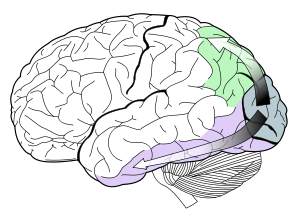
Each V1 transmits information to two primary pathways, called the dorsal stream and the ventral stream:
- The dorsal stream begins with V1, goes through Visual area V2, then to the dorsomedial areaDorsomedial areaThe Dorsomedial area, also known as DM or V6, is a subdivision of the visual cortex of primates first described by John Allman and Jon Kaas in 1975...
and Visual area MT (also known as V5) and to the posterior parietal cortexPosterior parietal cortexThe posterior parietal cortex plays an important role in producing planned movements. Before an effective movement can be initiated, the nervous system must know the original positions of the body parts that are to be moved, and the positions of any external objects with which the body is going to...
. The dorsal stream, sometimes called the "Where Pathway" or "How Pathway", is associated with motion, representation of object locations, and control of the eyes and arms, especially when visual information is used to guide saccadeSaccadeA saccade is a fast movement of an eye, head or other part of an animal's body or device. It can also be a fast shift in frequency of an emitted signal or other quick change. Saccades are quick, simultaneous movements of both eyes in the same direction...
s or reaching. - The ventral stream begins with V1, goes through visual area V2, then through visual area V4, and to the inferior temporal cortex. The ventral stream, sometimes called the "What Pathway", is associated with form recognition and object representation. It is also associated with storage of long-term memoryLong-term memoryLong-term memory is memory in which associations among items are stored, as part of the theory of a dual-store memory model. According to the theory, long term memory differs structurally and functionally from working memory or short-term memory, which ostensibly stores items for only around 20–30...
.
The dichotomy of the dorsal/ventral pathways
Two Streams hypothesis
The two-streams hypothesis is a widely accepted, but still controversial, account of visual processing. As visual information exits the occipital lobe, it follows two main channels, or "streams". The ventral stream travels to the temporal lobe and is involved with object identification...
(also called the "where/what" or "action/perception" streams) was first defined by Ungerleider and Mishkin and is still contentious among vision scientists and psychologists. It is probably an over-simplification of the true state of affairs in the visual cortex. It is based on the findings that visual illusions such as the Ebbinghaus illusion
Ebbinghaus illusion
The Ebbinghaus illusion or Titchener circles is an optical illusion of relative size perception. In the best-known version of the illusion, two circles of identical size are placed near to each other and one is surrounded by large circles while the other is surrounded by small circles; the first...
may distort judgements of a perceptual nature, but when the subject responds with an action, such as grasping, no distortion occurs. However, recent work suggests that both the action and perception systems are equally fooled by such illusions.
Neurons in the visual cortex fire action potential
Action potential
In physiology, an action potential is a short-lasting event in which the electrical membrane potential of a cell rapidly rises and falls, following a consistent trajectory. Action potentials occur in several types of animal cells, called excitable cells, which include neurons, muscle cells, and...
s when visual stimuli appear within their receptive field
Receptive field
The receptive field of a sensory neuron is a region of space in which the presence of a stimulus will alter the firing of that neuron. Receptive fields have been identified for neurons of the auditory system, the somatosensory system, and the visual system....
. By definition, the receptive field is the region within the entire visual field which elicits an action potential
Action potential
In physiology, an action potential is a short-lasting event in which the electrical membrane potential of a cell rapidly rises and falls, following a consistent trajectory. Action potentials occur in several types of animal cells, called excitable cells, which include neurons, muscle cells, and...
. But for any given neuron, it may respond best to a subset of stimuli within its receptive field. This property is called neuronal tuning
Neuronal tuning
Neuronal tuning refers to the property of brain cells to selectively represent a particular kind of sensory, motor, or cognitive information. For example, an auditory system neuron best responding to the sound of particular frequency is said to be tuned to that frequency. In the visual system,...
. In the earlier visual areas, neurons have simpler tuning. For example, a neuron in V1 may fire to any vertical stimulus in its receptive field. In the higher visual areas, neurons have complex tuning. For example, in the inferior temporal cortex (IT), a neuron may only fire when a certain face appears in its receptive field.
The visual cortex receives its blood supply primarily from the calcarine branch of the posterior cerebral artery
Posterior cerebral artery
-External links: - Posterior Cerebral Artery Stroke* at strokecenter.org* at State University of New York Upstate Medical University* at psyweb.com* at neuropat.dote.hu...
.
Current research
Research on the primary visual cortex can involve recording action potentialAction potential
In physiology, an action potential is a short-lasting event in which the electrical membrane potential of a cell rapidly rises and falls, following a consistent trajectory. Action potentials occur in several types of animal cells, called excitable cells, which include neurons, muscle cells, and...
s from electrodes within the brain of cat
Cat
The cat , also known as the domestic cat or housecat to distinguish it from other felids and felines, is a small, usually furry, domesticated, carnivorous mammal that is valued by humans for its companionship and for its ability to hunt vermin and household pests...
s, ferret
Ferret
The ferret is a domesticated mammal of the type Mustela putorius furo. Ferrets are sexually dimorphic predators with males being substantially larger than females. They typically have brown, black, white, or mixed fur...
s, rat
Rat
Rats are various medium-sized, long-tailed rodents of the superfamily Muroidea. "True rats" are members of the genus Rattus, the most important of which to humans are the black rat, Rattus rattus, and the brown rat, Rattus norvegicus...
s, mice
Mouse
A mouse is a small mammal belonging to the order of rodents. The best known mouse species is the common house mouse . It is also a popular pet. In some places, certain kinds of field mice are also common. This rodent is eaten by large birds such as hawks and eagles...
, or monkeys, or through recording intrinsic optical signals from animals or EEG
EEG
EEG commonly refers to electroencephalography, a measurement of the electrical activity of the brain.EEG may also refer to:* Emperor Entertainment Group, a Hong Kong-based entertainment company...
, MEG, or fMRI signals from human and monkey V1.
One recent discovery concerning the human V1 is that signals measured by fMRI show very large attentional modulation. This result strongly contrasts with macaque physiology research showing very small changes (or no changes) in firing associated with attentional modulation. Research with the macaque monkey is usually performed by measuring spiking activity from single neurons. The neural basis of the fMRI signal on the other hand is mostly related to post synaptic potentiation (PSP) . This difference therefore does not necessarily indicate a difference between macaque and human physiology.
Other current work on V1 seeks to fully characterize its tuning properties, and to use it as a model area for the canonical cortical circuit.
Lesions to primary visual cortex usually lead to a scotoma
Scotoma
A scotoma is an area of partial alteration in one's field of vision consisting of a partially diminished or entirely degenerated visual acuity which is surrounded by a field of normal - or relatively well-preserved - vision.Every normal mammalian eye has a scotoma in its field of vision, usually...
, or hole in the visual field. Interestingly, patients with scotomas are often able to make use of visual information presented to their scotomas, despite being unable to consciously perceive it. This phenomenon, called blindsight
Blindsight
Blindsight is a phenomenon in which people who are perceptually blind in a certain area of their visual field demonstrate some response to visual stimuli...
, is widely studied by scientists interested in the neural correlates of consciousness
Consciousness
Consciousness is a term that refers to the relationship between the mind and the world with which it interacts. It has been defined as: subjectivity, awareness, the ability to experience or to feel, wakefulness, having a sense of selfhood, and the executive control system of the mind...
.
Primary visual cortex (V1)
Brain
The brain is the center of the nervous system in all vertebrate and most invertebrate animals—only a few primitive invertebrates such as sponges, jellyfish, sea squirts and starfishes do not have one. It is located in the head, usually close to primary sensory apparatus such as vision, hearing,...
. In all mammals studied, it is located in the posterior pole of the occipital cortex (the occipital cortex is responsible for processing visual
Visual perception
Visual perception is the ability to interpret information and surroundings from the effects of visible light reaching the eye. The resulting perception is also known as eyesight, sight, or vision...
stimuli). It is the simplest, earliest cortical visual area. It is highly specialized for processing information about static and moving objects and is excellent in pattern recognition
Pattern recognition
In machine learning, pattern recognition is the assignment of some sort of output value to a given input value , according to some specific algorithm. An example of pattern recognition is classification, which attempts to assign each input value to one of a given set of classes...
.
The functionally defined primary visual cortex is approximately equivalent to the anatomically defined striate cortex. The name "striate cortex" is derived from the stria of Gennari, a distinctive stripe visible to the naked eye that represents myelin
Myelin
Myelin is a dielectric material that forms a layer, the myelin sheath, usually around only the axon of a neuron. It is essential for the proper functioning of the nervous system. Myelin is an outgrowth of a type of glial cell. The production of the myelin sheath is called myelination...
ated axons from the lateral geniculate body terminating in layer 4 of the gray matter
Gray Matter
"Gray Matter" is a short story by Stephen King, first published in the October 1973 issue of Cavalier magazine, and later collected in King's 1978 collection Night Shift. It is set in the same area as King's novel Dreamcatcher.-Setting:...
.
The primary visual cortex is divided into six functionally distinct layers, labeled 1 through 6. Layer 4, which receives most visual input from the lateral geniculate nucleus
Lateral geniculate nucleus
The lateral geniculate nucleus is the primary relay center for visual information received from the retina of the eye. The LGN is found inside the thalamus of the brain....
(LGN), is further divided into 4 layers, labelled 4A, 4B, 4Cα, and 4Cβ. Sublamina 4Cα receives most magnocellular
Magnocellular part
Magnocellular cells, also called M-cells, are neurons located within the magnocellular layer of the lateral geniculate nucleus of the thalamus. The cells are part of the visual system...
input from the LGN, while layer 4Cβ receives input from parvocellular pathways.
The average number of neurons in the adult human primary visual cortex, in each hemisphere, has been estimated at around 140 million (Leuba & Kraftsik, Anatomy and Embryology, 1994).
Function
V1 has a very well-defined map of the spatial information in vision. For example, in humans the upper bank of the calcarine sulcus responds strongly to the lower half of visual fieldVisual field
The term visual field is sometimes used as a synonym to field of view, though they do not designate the same thing. The visual field is the "spatial array of visual sensations available to observation in introspectionist psychological experiments", while 'field of view' "refers to the physical...
(below the center), and the lower bank of the calcarine to the upper half of visual field. Conceptually, this retinotopic
Retinotopy
Retinotopy describes the spatial organization of the neuronal responses to visual stimuli. In many locations within the brain, adjacent neurons have receptive fields that include slightly different, but overlapping portions of the visual field. The position of the center of these receptive fields...
mapping is a transformation of the visual image from retina
Retina
The vertebrate retina is a light-sensitive tissue lining the inner surface of the eye. The optics of the eye create an image of the visual world on the retina, which serves much the same function as the film in a camera. Light striking the retina initiates a cascade of chemical and electrical...
to V1. The correspondence between a given location in V1 and in the subjective visual field is very precise: even the blind spots
Blind spot (vision)
A blind spot, also known as a scotoma, is an obscuration of the visual field. A particular blind spot known as the blindspot, or physiological blind spot, or punctum caecum in medical literature, is the place in the visual field that corresponds to the lack of light-detecting photoreceptor cells on...
are mapped into V1. Evolutionarily, this correspondence is very basic and found in most animals that possess a V1. In human and animals with a fovea
Fovea
The fovea centralis, also generally known as the fovea , is a part of the eye, located in the center of the macula region of the retina....
in the retina, a large portion of V1 is mapped to the small, central portion of visual field, a phenomenon known as cortical magnification
Cortical magnification
Cortical magnification describes how many neurons in an area of the visual cortex are 'responsible' for processing a stimulus of a given size, as a function of visual field location. In the center of the visual field, corresponding to the fovea of the retina, a very large number of neurons process...
. Perhaps for the purpose of accurate spatial encoding, neurons in V1 have the smallest receptive field size of any visual cortex microscopic regions.
The tuning properties of V1 neurons (what the neurons respond to) differ greatly over time. Early in time (40 ms and further) individual V1 neurons have strong tuning to a small set of stimuli. That is, the neuronal responses can discriminate small changes in visual orientations, spatial frequencies and color
Color
Color or colour is the visual perceptual property corresponding in humans to the categories called red, green, blue and others. Color derives from the spectrum of light interacting in the eye with the spectral sensitivities of the light receptors...
s. Furthermore, individual V1 neurons in human and animals with binocular vision
Binocular vision
Binocular vision is vision in which both eyes are used together. The word binocular comes from two Latin roots, bini for double, and oculus for eye. Having two eyes confers at least four advantages over having one. First, it gives a creature a spare eye in case one is damaged. Second, it gives a...
have ocular dominance, namely tuning to one of the two eyes. In V1, and primary sensory cortex in general, neurons with similar tuning properties tend to cluster together as cortical columns. David Hubel and Torsten Wiesel
Torsten Wiesel
Torsten Nils Wiesel was a Swedish co-recipient with David H. Hubel of the 1981 Nobel Prize in Physiology or Medicine, for their discoveries concerning information processing in the visual system; the prize was shared with Roger W...
proposed the classic ice-cube organization model of cortical columns for two tuning properties: ocular dominance and orientation. However, this model cannot accommodate the color, spatial frequency and many other features to which neurons are tuned . The exact organization of all these cortical columns within V1 remains a hot topic of current research.
Current consensus seems to be that early responses of V1 neurons consists of tiled sets of selective spatiotemporal filters. In the spatial domain, the functioning of V1 can be thought of as similar to many spatially local, complex Fourier transform
Fourier transform
In mathematics, Fourier analysis is a subject area which grew from the study of Fourier series. The subject began with the study of the way general functions may be represented by sums of simpler trigonometric functions...
s, or more accurately, Gabor transform
Gabor transform
The Gabor transform, named after Dennis Gabor, is a special case of the short-time Fourier transform. It is used to determine the sinusoidal frequency and phase content of local sections of a signal as it changes over time...
s. Theoretically, these filters together can carry out neuronal processing of spatial frequency, orientation, motion, direction, speed (thus temporal frequency), and many other spatiotemporal features. Experiments of neurons substantiate these theories, but also raise new questions.
Later in time (after 100 ms) neurons in V1 are also sensitive to the more global organisation of the scene (Lamme & Roelfsema, 2000). These response properties probably stem from recurrent processing (the influence of higher-tier cortical areas on lower-tier cortical areas) and lateral connections from pyramidal neurons (Hupe et al. 1998). While feedforward connections are mainly driving, feedback connections are mostly modulatory in their effects (Angelucci et al., 2003; Hupe et al., 2001). Evidence shows that feedback originating in higher level areas such as V4, IT or MT, with bigger and more complex receptive fields, can modify and shape V1 responses, accounting for contextual or extra-classical receptive field effects (Guo et al., 2007; Harrison et al., 2007; Huang et al., 2007; Sillito et al., 2006).
The visual information relayed to V1 is not coded in terms of spatial (or optical) imagery, but rather as the local contrast. As an example, for an image comprising half side black and half side white, the divide line between black and white has strongest local contrast and is encoded, while few neurons code the brightness information (black or white per se). As information is further relayed to subsequent visual areas, it is coded as increasingly non-local frequency/phase signals. Importantly, at these early stages of cortical visual processing, spatial location of visual information is well preserved amid the local contrast encoding.
V2
Visual area V2, also called prestriate cortex, is the second major area in the visual cortex, and the first region within the visual association area. It receives strong feedforward connections from V1 (direct and via the pulvinar) and sends strong connections to V3, V4, and V5. It also sends strong feedbackFeedback
Feedback describes the situation when output from an event or phenomenon in the past will influence an occurrence or occurrences of the same Feedback describes the situation when output from (or information about the result of) an event or phenomenon in the past will influence an occurrence or...
connections to V1.
Anatomically, V2 is split into four quadrants, a dorsal
Dorsum (biology)
In anatomy, the dorsum is the upper side of animals that typically run, fly, or swim in a horizontal position, and the back side of animals that walk upright. In vertebrates the dorsum contains the backbone. The term dorsal refers to anatomical structures that are either situated toward or grow...
and ventral representation in the left and the right hemispheres
Cerebral hemisphere
A cerebral hemisphere is one of the two regions of the eutherian brain that are delineated by the median plane, . The brain can thus be described as being divided into left and right cerebral hemispheres. Each of these hemispheres has an outer layer of grey matter called the cerebral cortex that is...
. Together these four regions provide a complete map of the visual world. Functionally, V2 has many properties in common with V1. Cells are tuned to simple properties such as orientation, spatial frequency, and color. The responses of many V2 neurons are also modulated by more complex properties, such as the orientation of illusory contours and whether the stimulus is part of the figure or the ground (Qiu and von der Heydt, 2005).
Recent research has shown that V2 cells show a small amount of attentional modulation (more than V1, less than V4), are tuned for moderately complex patterns, and may be driven by multiple orientations at different subregions within a single receptive field.
It is argued that the entire ventral visual-to-hippocampal stream is important for visual memory. This theory, unlike the dominant one, predicts that object-recognition memory (ORM) alterations could result from the manipulation in V2, an area that is highly interconnected within the ventral stream of visual cortices. In the monkey brain, this area receives strong feedforward connections from the primary visual cortex (V1) and sends strong projections to other secondary visual cortices (V3, V4, and V5) . Most of the neurons of this area are tuned to simple visual characteristics such as orientation, spatial frequency, size, color, and shape . V2 cells also respond to various complex shape characteristics, such as the orientation of illusory contours and whether the stimulus is part of the figure or the ground . Anatomical studies implicate layer 3 of area V2 in visual-information processing. In contrast to layer 3, layer 6 of the visual cortex is composed of many types of neurons, and their response to visual stimuli is more complex.
In a recent study, the Layer 6 cells of the V2 cortex were found to play a very important role in the storage of Object Recognition Memory as well as the conversion of short-term object memories into long-term memories.
Third visual complex, including area V3
The term third visual complex refers to the region of cortex located immediately in front of V2, which includes the region named visual area V3 in humans. The "complex" nomenclature is justified by the fact that some controversy still exists regarding the exact extent of area V3, with some researchers proposing that the cortex located in front of V2 may include two or three functional subdivisions. For example, David Van Essen and others (1986) have proposed the existence of a "dorsal V3" in the upper part of the cerebral hemisphere, which is distinct from the "ventral V3" (or ventral posterior area, VP) located in the lower part of the brain. Dorsal and ventral V3 have distinct connections with other parts of the brain, appear different in sections stained with a variety of methods, and contain neurons that respond to different combinations of visual stimulus (for example, colour-selective neurons are more common in the ventral V3). Additional subdivisions, including V3A and V3B have also been reported in humans. These subdivisions are located near dorsal V3, but do not adjoin V2.Dorsal V3 is normally considered to be part of the dorsal stream, receiving inputs from V2 and from the primary visual area and projecting to the posterior parietal cortex. It may be anatomically located in Brodmann area 19
Brodmann area 19
-Human:Brodmann area 19, or BA19, is part of the occipital lobe cortex in the human brain. Along with area 18, it comprises the extrastriate cortex...
. Recent work with fMRI has suggested that area V3/V3A may play a role in the processing of global motion Other studies prefer to consider dorsal V3 as part of a larger area, named the dorsomedial area
Dorsomedial area
The Dorsomedial area, also known as DM or V6, is a subdivision of the visual cortex of primates first described by John Allman and Jon Kaas in 1975...
(DM), which contains a representation of the entire visual field. Neurons in area DM respond to coherent motion of large patterns covering extensive portions of the visual field (Lui and collaborators, 2006).
Ventral V3 (VP), has much weaker connections from the primary visual area, and stronger connections with the inferior temporal cortex. While earlier studies proposed that VP only contained a representation of the upper part of the visual field (above the point of fixation), more recent work indicates that this area is more extensive than previously appreciated, and like other visual areas it may contain a complete visual representation. The revised, more extensive VP is referred to as the ventrolateral posterior area (VLP) by Rosa and Tweedale.
V4
Visual area V4 is one of the visual areas in the extrastriate visual cortex. It is located anterior to V2 and posterior to posterior inferotemporal area (PIT)Inferior temporal gyrus
The inferior temporal gyrus is placed below the middle temporal sulcus, and is connected behind with the inferior occipital gyrus; it also extends around the infero-lateral border on to the inferior surface of the temporal lobe, where it is limited by the inferior sulcus...
. It comprises at least four regions (left and right V4d, left and right V4v), and some groups report that it contains rostral and caudal subdivisions as well. It is unknown what the human homologue of V4 is, and this issue is currently the subject of much scrutiny.
V4 is the third cortical area in the ventral stream, receiving strong feedforward input from V2 and sending strong connections to the PIT
Inferior temporal gyrus
The inferior temporal gyrus is placed below the middle temporal sulcus, and is connected behind with the inferior occipital gyrus; it also extends around the infero-lateral border on to the inferior surface of the temporal lobe, where it is limited by the inferior sulcus...
. It also receives direct inputs from V1, especially for central space. In addition, it has weaker connections to V5 and dorsal prelunate gyrus
Angular gyrus
The angular gyrus is a region of the brain in the parietal lobe, that lies near the superior edge of the temporal lobe, and immediately posterior to the supramarginal gyrus; it is involved in a number of processes related to language, mathematics and cognition...
(DP).
V4 is the first area in the ventral stream to show strong attentional modulation. Most studies indicate that selective attention can change firing rates in V4 by about 20%. A seminal paper by Moran and Desimone characterizing these effects was the first paper to find attention effects anywhere in the visual cortex http://www.sciencemag.org/cgi/content/abstract/229/4715/782).
Like V1, V4 is tuned for orientation, spatial frequency, and color. Unlike V1, V4 is tuned for object features of intermediate complexity, like simple geometric shapes, although no one has developed a full parametric description of the tuning space for V4. Visual area V4 is not tuned for complex objects such as faces, as areas in the inferotemporal cortex
Inferior temporal gyrus
The inferior temporal gyrus is placed below the middle temporal sulcus, and is connected behind with the inferior occipital gyrus; it also extends around the infero-lateral border on to the inferior surface of the temporal lobe, where it is limited by the inferior sulcus...
are.
The firing properties of V4 were first described by Semir Zeki
Semir Zeki
Semir Zeki is a professor of neuroesthetics at University College London. His main interest is the organization of the primate visual brain. He published his first scientific paper in 1967...
in the late 1970s, who also named the area. Before that, V4 was known by its anatomical description, the prelunate gyrus. Originally, Zeki argued that the purpose of V4 was to process color information. Work in the early 1980s proved that V4 was as directly involved in form recognition as earlier cortical areas. This research supported the Two Streams hypothesis
Two Streams hypothesis
The two-streams hypothesis is a widely accepted, but still controversial, account of visual processing. As visual information exits the occipital lobe, it follows two main channels, or "streams". The ventral stream travels to the temporal lobe and is involved with object identification...
, first presented by Ungerleider and Mishkin in 1982.
Recent work has shown that V4 exhibits long-term plasticity, encodes stimulus salience, is gated by signals coming from the frontal eye fields
Frontal eye fields
The frontal eye fields is a region located in the premotor cortex, which is part of the frontal cortex of the primate brain.-Function:...
and shows changes in the spatial profile of its receptive fields with attention.
V5/MT
Visual area V5, also known as visual area MT (middle temporal), is a region of extrastriate visual cortex that is thought to play a major role in the perception of motionMotion perception
Motion perception is the process of inferring the speed and direction of elements in a scene based on visual, vestibular and proprioceptive inputs...
, the integration of local motion signals into global percepts and the guidance of some eye movements.
Connections
MT is connected to a wide array of cortical and subcortical brain areas. Its inputs include the visual cortical areas V1, V2, and dorsal V3 (dorsomedial areaDorsomedial area
The Dorsomedial area, also known as DM or V6, is a subdivision of the visual cortex of primates first described by John Allman and Jon Kaas in 1975...
), the koniocellular
Koniocellular
A koniocellular cell is a neuron whose cell body is located in the koniocellular layer of the lateral geniculate nucleus in primates, including humans. The koniocellular cells receive their input from bistratified retinal ganglion cells exiting the optic tract, and send their information via...
regions of the LGN, and the inferior pulvinar
Pulvinar
The pulvinar nuclei are a collection of nuclei located in the pulvinar thalamus. The pulvinar part is the most posterior region of the thalamus....
. The pattern of projections to MT changes somewhat between the representations of the foveal and peripheral visual fields, with the latter receiving inputs from areas located in the midline cortex and retrosplenial region
Retrosplenial region
The retrosplenial region is a brain area and part of the cingulate cortex. It is defined by Brodmann area 26, Brodmann area 29 and Brodmann area 30.-Function:...
A standard view is that V1 provides the "most important" input to MT. Nonetheless, several studies have demonstrated that neurons in MT are capable of responding to visual information, often in a direction-selective manner, even after V1 has been destroyed or inactivated. Moreover, research by Semir Zeki
Semir Zeki
Semir Zeki is a professor of neuroesthetics at University College London. His main interest is the organization of the primate visual brain. He published his first scientific paper in 1967...
and collaborators has suggested that certain types of visual information may reach MT before it even reaches V1.
MT sends its major outputs to areas located in the cortex immediately surrounding it, including areas FST, MST and V4t (middle temporal crescent). Other projections of MT target the eye movement-related areas of the frontal and parietal lobes (frontal eye field and lateral intraparietal area).
Function
The first studies of the electrophysiological properties of neurons in MT showed that a large portion of the cells were tunedNeuronal tuning
Neuronal tuning refers to the property of brain cells to selectively represent a particular kind of sensory, motor, or cognitive information. For example, an auditory system neuron best responding to the sound of particular frequency is said to be tuned to that frequency. In the visual system,...
to the speed and direction of moving visual stimuli These results suggested that MT played a significant role in the processing of visual motion.
Lesion
Lesion
A lesion is any abnormality in the tissue of an organism , usually caused by disease or trauma. Lesion is derived from the Latin word laesio which means injury.- Types :...
studies have also supported the role of MT in motion perception and eye movements. Neuropsychological
Neuropsychology
Neuropsychology studies the structure and function of the brain related to specific psychological processes and behaviors. The term neuropsychology has been applied to lesion studies in humans and animals. It has also been applied to efforts to record electrical activity from individual cells in...
studies of a patient who could not see motion, seeing the world in a series of static "frames" instead, suggested that MT in the primate is homologous to V5 in the human.
However, since neurons in V1 are also tuned to the direction and speed of motion, these early results left open the question of precisely what MT could do that V1 could not. Much work has been carried out on this region as it appears to integrate local visual motion signals into the global motion of complex objects.
For example, lesion to the V5 lead to deficits in perceiving motion and processing of complex stimuli. It contains many neurons selective for the motion of complex visual features (line ends, corners). Microstimulation of a neuron located in the V5 affects the perception of motion. For example, if one finds a neuron with preference for upward motion, and then we use an electrode to stimulate it, the monkey becomes more likely to report 'upward' motion.
There is still much controversy over the exact form of the computations carried out in area MT and some research suggests that feature motion is in fact already available at lower levels of the visual system such as V1.
Functional organization
MT was shown to be organized in direction columns. DeAngelis argued that MT neurons were also organized based on their tuning for binocular disparity.See also
- Brodmann areaBrodmann areaA Brodmann area is a region of the cerebral cortex defined based on its cytoarchitectonics, or structure and organization of cells.-History:...
- Cortical areaCortical areaA cortical area is a part of the cerebral cortex.-Functionally Defined:Often, a cortical area is functionally defined, i.e. its neurons share certain distinguishing functional properties...
- Cortical blindnessCortical blindnessCortical blindness is the total or partial loss of vision in a normal-appearing eye caused by damage to the visual area in the brain's occipital cortex. This damage is most often caused by loss of blood flow to the occipital cortex from either unilateral or bilateral posterior cerebral artery...
- Feature integration theoryFeature integration theoryThe feature integration theory, developed by Anne Treisman and Garry Gelade since the early 1980s, posits that different kinds of attention are responsible for binding different features into consciously experienced wholes...
- List of regions in the human brain
- RetinotopyRetinotopyRetinotopy describes the spatial organization of the neuronal responses to visual stimuli. In many locations within the brain, adjacent neurons have receptive fields that include slightly different, but overlapping portions of the visual field. The position of the center of these receptive fields...
External links
- The Primary Visual Cortex by Matthew Schmolesky at University of UtahUniversity of UtahThe University of Utah, also known as the U or the U of U, is a public, coeducational research university in Salt Lake City, Utah, United States. The university was established in 1850 as the University of Deseret by the General Assembly of the provisional State of Deseret, making it Utah's oldest...
- Architecture of the Visual Cortex, by David Hubel at Harvard UniversityHarvard UniversityHarvard University is a private Ivy League university located in Cambridge, Massachusetts, United States, established in 1636 by the Massachusetts legislature. Harvard is the oldest institution of higher learning in the United States and the first corporation chartered in the country...
- striate area 17 - Brodmann area 17 in guenon - Simulator for computational modeling of visual cortex maps at topographica.org