
List of regions in the human brain
Encyclopedia
Anatomical
regions of the brain are listed vertically, following hierarchies that are standard in neuroanatomy
. Functional
, connective, and developmental
regions are listed horizontally in parentheses where appropriate.
Myelencephalon
Metencephalon
Midbrain
Epithalamus
Thalamus
Hypothalamus
Pituitary gland
Telencephalon (cerebrum)
White matter
Rhinencephalon
Cerebral cortex
Neural pathway
Motor systems
Vascular systems
Anatomy
Anatomy is a branch of biology and medicine that is the consideration of the structure of living things. It is a general term that includes human anatomy, animal anatomy , and plant anatomy...
regions of the brain are listed vertically, following hierarchies that are standard in neuroanatomy
Neuroanatomy
Neuroanatomy is the study of the anatomy and organization of the nervous system. In contrast to animals with radial symmetry, whose nervous system consists of a distributed network of cells, animals with bilateral symmetry have segregated, defined nervous systems, and thus we can begin to speak of...
. Functional
Physiology
Physiology is the science of the function of living systems. This includes how organisms, organ systems, organs, cells, and bio-molecules carry out the chemical or physical functions that exist in a living system. The highest honor awarded in physiology is the Nobel Prize in Physiology or...
, connective, and developmental
Embryology
Embryology is a science which is about the development of an embryo from the fertilization of the ovum to the fetus stage...
regions are listed horizontally in parentheses where appropriate.
MyelencephalonMyelencephalonThe myelencephalon is categorized as a secondary vesicle in the development of the central nervous system. The prefix "myelen" is derived from Greek for medulla...
- medulla oblongataMedulla oblongataThe medulla oblongata is the lower half of the brainstem. In discussions of neurology and similar contexts where no ambiguity will result, it is often referred to as simply the medulla...
- medullary pyramids
- Medullary cranial nerve nuclei
- Inferior salivatory nucleusInferior salivatory nucleusIn the brain, the inferior salivatory nucleus is a cluster of neurons controlling the parasympathetic input to the parotid gland. It is one of the components of the glossopharyngeal nerve .-Location:...
- Nucleus ambiguusNucleus ambiguusThe nucleus ambiguus is a region of histologically disparate cells located just dorsal to the inferior olivary nucleus in the lateral portion of the upper medulla...
- Dorsal nucleus of vagus nerve
- Hypoglossal nucleusHypoglossal nucleusThe hypoglossal nucleus is a cranial nerve nucleus, and it extends the length of the medulla, and being a motor nucleus, is close to the midline...
- Solitary nucleusSolitary nucleusThe solitary tract and nucleus are structures in the brainstem that carry and receive visceral sensation and taste from the facial , glossopharyngeal and vagus cranial nerves.-Anatomy:...
- Inferior salivatory nucleus
MetencephalonMetencephalonThe metencephalon is a developmental categorization of portions of the central nervous system. The metencephalon is composed of the pons and the cerebellum; contains a portion of the fourth ventricle; and the trigeminal nerve , abducens nerve , facial nerve , and a portion of the vestibulocochlear...
- ponsPonsThe pons is a structure located on the brain stem, named after the Latin word for "bridge" or the 16th-century Italian anatomist and surgeon Costanzo Varolio . It is superior to the medulla oblongata, inferior to the midbrain, and ventral to the cerebellum. In humans and other bipeds this means it...
- Respiratory centres
- pneumotactic centre
- apneustic centre
- Pontine cranial nerve nuclei
- chief or pontine nucleus of the trigeminal nerveTrigeminal nerveThe trigeminal nerve contains both sensory and motor fibres. It is responsible for sensation in the face and certain motor functions such as biting, chewing, and swallowing. Sensory information from the face and body is processed by parallel pathways in the central nervous system...
sensory nucleus (V) - motor nucleus for the trigeminal nerve (V)
- abducens nucleusAbducens nucleusThe abducens nucleus is the originating nucleus from which the abducens nerve emerges - a cranial nerve nucleus. This nucleus is located beneath the fourth ventricle in the caudal portion of the pons, medial to the sulcus limitans....
(VI) - facial nerve nucleus (VII)
- vestibulocochlearVestibulocochlear nerveThe vestibulocochlear nerve is the eighth of twelve cranial nerves, and is responsible for transmitting sound and equilibrium information from the inner ear to the brain...
nuclei (vestibular nucleiVestibular nucleiThe vestibular nuclei are the cranial nuclei for the vestibular nerve.In Terminologia Anatomica they are grouped in both the pons and medulla.-Subnuclei:There are 4 subnuclei; they are situated at the floor of the fourth ventricle....
and cochlear nucleiCochlear nucleiThe cochlear nuclei are two heterogeneous collections of neurons in the mammalian brainstem that receive input from the cochlear nerve, which carry sound information from the cochleae...
) (VIII) - Superior salivatory nucleus
- chief or pontine nucleus of the trigeminal nerve
- Respiratory centres
- paramedian pontine reticular formationParamedian pontine reticular formationThe paramedian pontine reticular formation, or PPRF, is part of the pontine reticular formation, a brain region without clearly defined borders in the center of the pons. It is involved in the coordination of eye movements, particularly horizontal gaze and saccades.- Input, Output, and Function...
- cerebellumCerebellumThe cerebellum is a region of the brain that plays an important role in motor control. It may also be involved in some cognitive functions such as attention and language, and in regulating fear and pleasure responses, but its movement-related functions are the most solidly established...
- cerebellar vermisCerebellar vermisThe cerebellar vermis is located in the medial, cortico-nuclear zone of the cerebellum, residing in the posterior fossa of the cranium. The primary fissure in the vermis curves ventrolaterally to the superior surface of the cerebellum, dividing it into anterior and posterior lobes....
- cerebellar hemispheres
- anterior lobe
- posterior lobe
- flocculonodular lobeFlocculonodular lobeThe flocculonodular lobe is a lobe of the cerebellum consisting of the nodule and the flocculus.It is closely associated with the vestibulocerebellum. This lobe is involved in the maintenance of equilibrium.-External links:...
- cerebellar nuclei
- fastigial nucleusFastigial nucleusThe fastigial nucleus or nucleus fastigii refers specifically to the concentration of gray matter nearest to the middle line at the anterior end of the superior vermis, and immediately over the roof of the fourth ventricle, from which it is separated by a thin layer of white matter...
- globose nucleusGlobose nucleusThe globose nucleus is one of the deep cerebellar nuclei. It is located medial to the emboliform nucleus and lateral to the fastigial nucleus. This nucleus contains primarily large and small multipolar neurons....
- emboliform nucleusEmboliform nucleusThe emboliform nucleus lies immediately to the medial side of the nucleus dentatus, and partly covering its hilus. It is one among the four pairs of cerebellar nuclei, which are from lateral to medial: the dentate, interposed , and fastigial nuclei...
- dentate nucleusDentate nucleusThe dentate nucleus is located within the deep white matter of each cerebellar hemisphere, and it is the largest single structure linking the cerebellum to the rest of the brain. It is the largest and most lateral, or farthest from the midline, of the four pairs of deep cerebellar nuclei, the...
- fastigial nucleus
- cerebellar vermis
MidbrainMesencephalonThe midbrain or mesencephalon is a portion of the central nervous system associated with vision, hearing, motor control, sleep/wake, arousal , and temperature regulation....
(mesencephalon)
- tectum
- inferior colliculiInferior colliculusThe inferior colliculus is the principal midbrain nucleus of the auditory pathway and receives input from several more peripheral brainstem nuclei in the auditory pathway, as well as inputs from the auditory cortex...
- superior colliculiSuperior colliculusThe optic tectum or simply tectum is a paired structure that forms a major component of the vertebrate midbrain. In mammals this structure is more commonly called the superior colliculus , but, even in mammals, the adjective tectal is commonly used. The tectum is a layered structure, with a...
- inferior colliculi
- mesencephalic duct (cerebral aqueduct, Aqueduct of Sylvius)
- cerebral peduncleCerebral peduncleMainly, the three common areas that give rise to the cerebral peduncles are the cortex, the spinal cord and the cerebellum. The cerebral peduncle, by most classifications, is everything in the mesencephalon except the tectum. The region includes the midbrain tegmentum, crus cerebri and pretectum...
- interpeduncular nucleusInterpeduncular nucleusThe Interpeduncular nucleus is a unpaired, ovoid cell group at the base of the midbrain tegmentum. It is located in the mesencephalon below the interpeduncular fossa...
- interpeduncular nucleus
- midbrain tegmentumMidbrain tegmentumThe midbrain tegmentum is the part of the midbrain extending from the substantia nigra to the cerebral aqueduct in a horizontal section of the midbrain. It forms the floor of the midbrain which surrounds the cerebral aqueduct.-External links:*...
- ventral tegmental area
- Red NucleusRed nucleusThe red nucleus is a structure in the rostral midbrain involved in motor coordination. It comprises a caudal magnocellular and a rostral parvocellular part.-Function:...
- crus cerebri
- pretectumPretectumThe pretectum, also known as the pretectal area, is a region of neurons found between the thalamus and midbrain. It receives binocular sensory input from retinal ganglion cells of the eyes, and is the region responsible for maintaining the pupillary light reflex.-Outputs:The pretectum, after...
EpithalamusEpithalamusThe epithalamus is a dorsal posterior segment of the diencephalon which includes the habenula, the stria medullaris and the pineal body...
- pineal bodyPineal glandThe pineal gland is a small endocrine gland in the vertebrate brain. It produces the serotonin derivative melatonin, a hormone that affects the modulation of wake/sleep patterns and seasonal functions...
- habenular nucleiHabenular nucleiThe habenular nuclei comprise a small group of nuclei that are part of the epithalamus of the diencephalon, situated at the posterior end of the thalamus, on its upper surface.The habenular nuclei are typically divided into:* lateral habenular nucleus...
- stria medullares
- tenia thalami
ThalamusThalamusThe thalamus is a midline paired symmetrical structure within the brains of vertebrates, including humans. It is situated between the cerebral cortex and midbrain, both in terms of location and neurological connections...
- anterior nuclear group
- anteroventral nucleus
- anterodorsal nucleus
- anteromedial nucleus
- medial nuclear group
- medial dorsal nucleusMedial dorsal nucleusThe medial dorsal nucleus is a large nucleus in the thalamus.It is believed to play a role in memory.-Anatomy:...
- midline nuclear groupMidline nuclear groupThe midline nuclear group a region of the thalamus consisting of the following nuclei:* paraventricular nucleus of thalamus - not to be confused with paraventricular nucleus of hypothalamus* paratenial nucleus * reuniens nucleus * rhomboidal nucleus The midline nuclear group (or midline thalamic...
- paratenial nucleus
- reuniens nucleus
- rhomboidal nucleus
- intralaminar nuclear group
- centromedial nucleus
- parafascicular nucleus
- paracentral nucleus
- central lateral nucleus
- central medial nucleus
- medial dorsal nucleus
- lateral nuclear groupLateral nuclear groupThe lateral nuclear group is a collection of nuclei on the lateral side of the thalamus. According to MeSH, it consists of the following:* lateral dorsal nucleus* lateral posterior nucleus* pulvinar...
- lateral dorsal nucleus
- lateral posterior nucleus
- pulvinarPulvinarThe pulvinar nuclei are a collection of nuclei located in the pulvinar thalamus. The pulvinar part is the most posterior region of the thalamus....
- ventral nuclear groupVentral nuclear groupThe ventral nuclear group is a collection of nuclei on the ventral side of the thalamus. According to MeSH, it consists of the following:* ventral anterior nucleus* ventral lateral nucleus* ventral posterior nucleus...
- ventral anterior nucleusVentral anterior nucleus-Inputs and outputs:The ventral anterior nucleus receives neuronal inputs from the basal ganglia which includes the substantia nigra and the globus pallidus. It also has inputs from the cerebellum. It sends out neuronal input to the supplementary motor cortex and to primary motor...
- ventral lateral nucleusVentral lateral nucleus-Inputs and outputs:It receives neuronal inputs from the basal ganglia which includes the substantia nigra and the globus pallidus . It also has inputs from the cerebellum ....
- ventral posterior nucleusVentral posterior nucleusThe ventral posterior nucleus is the somato-sensory relay nucleus in thalamus of the brain.-Input and output:The ventral posterior nucleus receives neuronal input from the medial lemniscus, spinal lemniscus, spinothalamic tracts, and trigeminothalamic tract...
- Ventral posterior lateral nucleus
- Ventral posterior medial nucleus
- ventral anterior nucleus
- metathalamusMetathalamusThe metathalamus is a composite structure of the thalamus, consisting of the medial geniculate nucleus and the lateral geniculate nucleus.-External links:*...
- medial geniculate body
- lateral geniculate body
- thalamic reticular nucleus
HypothalamusHypothalamusThe Hypothalamus is a portion of the brain that contains a number of small nuclei with a variety of functions...
(limbic systemLimbic systemThe limbic system is a set of brain structures including the hippocampus, amygdala, anterior thalamic nuclei, septum, limbic cortex and fornix, which seemingly support a variety of functions including emotion, behavior, long term memory, and olfaction. The term "limbic" comes from the Latin...
) (HPA axis)
- Anterior
- Medial area
- Parts of preoptic areaPreoptic areaThe preoptic area is a region of the hypothalamus. According to the MeSH classification, it is considered part of the anterior hypothalamus. There are four nuclei in this region, according to Terminologia Anatomica .-Functions:The preoptic area is responsible for thermoregulation and receives...
- Medial preoptic nucleus
- Suprachiasmatic nucleusSuprachiasmatic nucleusThe suprachiasmatic nucleus or nuclei, abbreviated SCN, is a tiny region on the brain's midline, situated directly above the optic chiasm. It is responsible for controlling circadian rhythms...
- Paraventricular nucleusParaventricular nucleusThe paraventricular nucleus is a neuronal nucleus in the hypothalamus. It contains multiple subpopulations of neurons that are activated by a variety of stressful and/or physiological changes. Many PVN neurons project directly to the posterior pituitary where they release oxytocin or vasopressin...
- Supraoptic nucleusSupraoptic nucleusThe supraoptic nucleus is a nucleus of magnocellular neurosecretory cells in the hypothalamus of the mammalian brain. The nucleus is situated at the base of the brain, adjacent to the optic chiasm...
(mainly) - Anterior hypothalamic nucleusAnterior hypothalamic nucleusThe anterior hypothalamic nucleus is a nucleus of the hypothalamus.Its function is thermoregulation of the body.The anterior hypothalamus plays a role in regulating sleep....
- Parts of preoptic area
- Lateral area
- Parts of preoptic areaPreoptic areaThe preoptic area is a region of the hypothalamus. According to the MeSH classification, it is considered part of the anterior hypothalamus. There are four nuclei in this region, according to Terminologia Anatomica .-Functions:The preoptic area is responsible for thermoregulation and receives...
- Lateral preoptic nucleus
- Anterior part of Lateral nucleusLateral hypothalamusThe lateral hypothalamus or lateral hypothalamic area is a part of the hypothalamus.It is concerned with hunger. Damage to this area can cause reduced food intake...
- Part of supraoptic nucleus
- Parts of preoptic area
- Other nuclei of preoptic areaPreoptic areaThe preoptic area is a region of the hypothalamus. According to the MeSH classification, it is considered part of the anterior hypothalamus. There are four nuclei in this region, according to Terminologia Anatomica .-Functions:The preoptic area is responsible for thermoregulation and receives...
- median preoptic nucleus
- periventricular preoptic nucleus
- Medial area
- Tuberal
- Medial area
- Dorsomedial hypothalamic nucleusDorsomedial hypothalamic nucleusThe Dorsomedial hypothalamic nucleus is a nucleus of the hypothalamus. It is involved in feeding, drinking, and body weight regulation....
- Ventromedial nucleusVentromedial nucleusThe ventromedial nucleus is a nucleus of the hypothalamus.-Division:It has four subdivisions:* anterior * dorsomedial * ventrolateral * central ....
- Arcuate nucleusArcuate nucleusThe arcuate nucleus is an aggregation of neurons in the mediobasal hypothalamus, adjacent to the third ventricle and the median eminence...
- Dorsomedial hypothalamic nucleus
- Lateral area
- Tuberal part of Lateral nucleusLateral hypothalamusThe lateral hypothalamus or lateral hypothalamic area is a part of the hypothalamus.It is concerned with hunger. Damage to this area can cause reduced food intake...
- Lateral tuberal nuclei
- Tuberal part of Lateral nucleus
- Medial area
- Posterior
- Medial area
- Mammillary nuclei (part of mammillary bodiesMammillary bodyThe mammillary bodies are a pair of small round bodies, located on the undersurface of the brain, that, as part of the diencephalon form part of the limbic system. They are located at the ends of the anterior arches of the fornix...
) - Posterior nucleusPosterior nucleus (hypothalamus)The posterior nucleus of the hypothalamus is one of the many nuclei that make up the hypothalamic region of the brain.Its function is thermoregulation of the body.Damage or destruction of this nucleus causes hypothermia...
- Mammillary nuclei (part of mammillary bodies
- Lateral area
- Posterior part of Lateral nucleusLateral hypothalamusThe lateral hypothalamus or lateral hypothalamic area is a part of the hypothalamus.It is concerned with hunger. Damage to this area can cause reduced food intake...
- Posterior part of Lateral nucleus
- Medial area
- optic chiasmOptic chiasmThe optic chiasm or optic chiasma is the part of the brain where the optic nerves partially cross...
- subfornical organSubfornical organThe subfornical organ, situated on the ventral surface of the fornix, at the interventricular foramina , is one of the circumventricular organs of the brain.-Relations with other circumventricular organs:...
- periventricular nucleusPeriventricular nucleusThe Periventricular nucleus is a thin sheet of small neurons located in the wall of the third ventricle, a composite structure of the hypothalamus. Functions in analgesia....
- infundibulumInfundibulumAn infundibulum is a funnel-shaped cavity or organ.* Lungs: The alveolar sacs of the lungs from which the air chambers open are called infundibula...
- tuber cinereumTuber cinereumThe tuber cinereum is a hollow eminence of gray matter situated between the mammillary bodies and the optic chiasm. The tuber cinereum is part of the hypothalamus.-Structure:...
- tuberal nucleus
- tuberomamillary nucleus
- tuberal region
- mammillary bodies
- mammillary nucleus
Pituitary glandPituitary glandIn vertebrate anatomy the pituitary gland, or hypophysis, is an endocrine gland about the size of a pea and weighing 0.5 g , in humans. It is a protrusion off the bottom of the hypothalamus at the base of the brain, and rests in a small, bony cavity covered by a dural fold...
(HPA axis)
- neurohypophysisPosterior pituitaryThe posterior pituitary comprises the posterior lobe of the pituitary gland and is part of the endocrine system. Despite its name, the posterior pituitary gland is not a gland, per se; rather, it is largely a collection of axonal projections from the hypothalamus that terminate behind the anterior...
- intermediate pituitary
- adenohypophysisAnterior pituitaryA major organ of the endocrine system, the anterior pituitary, also called the adenohypophysis, is the glandular, anterior lobe of the pituitary gland...
Telencephalon (cerebrum)TelencephalonThe cerebrum or telencephalon, together with the diencephalon, constitutes the forebrain. The cerebrum is the most anterior region of the vertebrate central nervous system. Telencephalon refers to the embryonic structure, from which the mature cerebrum develops...
Cerebral hemisphereCerebral hemisphereA cerebral hemisphere is one of the two regions of the eutherian brain that are delineated by the median plane, . The brain can thus be described as being divided into left and right cerebral hemispheres. Each of these hemispheres has an outer layer of grey matter called the cerebral cortex that is...
s
 |
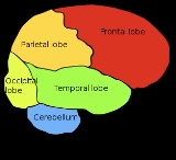
red:frontal lobe Frontal lobe The frontal lobe is an area in the brain of humans and other mammals, located at the front of each cerebral hemisphere and positioned anterior to the parietal lobe and superior and anterior to the temporal lobes... orange:parietal lobe Parietal lobe The parietal lobe is a part of the Brain positioned above the occipital lobe and behind the frontal lobe.The parietal lobe integrates sensory information from different modalities, particularly determining spatial sense and navigation. For example, it comprises somatosensory cortex and the... yellow:occipital lobe Occipital lobe The occipital lobe is the visual processing center of the mammalian brain containing most of the anatomical region of the visual cortex. The primary visual cortex is Brodmann area 17, commonly called V1... green: temporal lobe Temporal lobe The temporal lobe is a region of the cerebral cortex that is located beneath the Sylvian fissure on both cerebral hemispheres of the mammalian brain.... blue:cerebellum Cerebellum The cerebellum is a region of the brain that plays an important role in motor control. It may also be involved in some cognitive functions such as attention and language, and in regulating fear and pleasure responses, but its movement-related functions are the most solidly established... black:brainstem Brain stem In vertebrate anatomy the brainstem is the posterior part of the brain, adjoining and structurally continuous with the spinal cord. The brain stem provides the main motor and sensory innervation to the face and neck via the cranial nerves... |
White matterWhite matterWhite matter is one of the two components of the central nervous system and consists mostly of myelinated axons. White matter tissue of the freshly cut brain appears pinkish white to the naked eye because myelin is composed largely of lipid tissue veined with capillaries. Its white color is due to...
- Corona radiataCorona radiataIn neuroanatomy, the corona radiata is a white matter sheet that continues caudally as the internal capsule and rostrally as the centrum semiovale. This sheet of axons contains both descending and ascending axons that carry nearly all of the neural traffic from and to the cerebral cortex...
- Internal capsuleInternal capsuleThe internal capsule is an area of white matter in the brain that separates the caudate nucleus and the thalamus from the lenticular nucleus. The internal capsule contains both ascending and descending axons....
- External capsuleExternal capsuleThe external capsule is a series of white matter fiber tracts in the brain. These fibers run between the most lateral segment of the lentiform nucleus and the claustrum....
- Extreme capsuleExtreme capsuleThe extreme capsule is a long association fiber pathway of white matter in the brain that provides bidirectional communication between such areas as the claustrum and the insular cortex, and the inferior frontal gyrus and the middle-posterior portion of the superior temporal gyrus...
- Arcuate fasciculusArcuate fasciculusThe arcuate fasciculus is the neural pathway connecting the posterior part of the temporoparietal junction with the frontal cortex in the brain and is now considered as part of the superior longitudinal fasciculus..-Neuroanatomy:...
- Uncinate fasciculusUncinate fasciculusThe uncinate fasciculus is a white matter tract in the human brain that connects parts of the limbic system such as the hippocampus and amygdala in the temporal lobe with frontal ones such as the orbitofrontal cortex. Its function is unknown though it is affected in several psychiatric conditions...
Subcortical
- HippocampusHippocampusThe hippocampus is a major component of the brains of humans and other vertebrates. It belongs to the limbic system and plays important roles in the consolidation of information from short-term memory to long-term memory and spatial navigation. Humans and other mammals have two hippocampi, one in...
(Medial Temporal Lobe) - AmygdalaAmygdalaThe ' are almond-shaped groups of nuclei located deep within the medial temporal lobes of the brain in complex vertebrates, including humans. Shown in research to perform a primary role in the processing and memory of emotional reactions, the amygdalae are considered part of the limbic system.-...
(limbic systemLimbic systemThe limbic system is a set of brain structures including the hippocampus, amygdala, anterior thalamic nuclei, septum, limbic cortex and fornix, which seemingly support a variety of functions including emotion, behavior, long term memory, and olfaction. The term "limbic" comes from the Latin...
) (limbic lobeLimbic lobeThe limbic lobe is an arc-shaped region of cortex on the medial surface of each cerebral hemisphere of the mammalian brain, consisting of parts of the frontal, parietal and temporal lobes...
) (paleopalliumPaleopalliumIn anatomy of animals, the paleopallium is a region within the telencephalon in the brain.It is a primitive and evolutionarily ancient region of the brain, first appearing in amphibians....
)- Central nucleus (autonomic nervous systemAutonomic nervous systemThe autonomic nervous system is the part of the peripheral nervous system that acts as a control system functioning largely below the level of consciousness, and controls visceral functions. The ANS affects heart rate, digestion, respiration rate, salivation, perspiration, diameter of the pupils,...
) - Medial nucleus (accessory olfactory system)
- Cortical and basomedial nuclei (main olfactory system)
- LateralLateral nucleusLateral nucleus can refer to:* Lateral hypothalamus* Lateral vestibular nucleus...
and basolateral nuclei (frontotemporal cortical system)
- Central nucleus (autonomic nervous system
- base head (limbic systemLimbic systemThe limbic system is a set of brain structures including the hippocampus, amygdala, anterior thalamic nuclei, septum, limbic cortex and fornix, which seemingly support a variety of functions including emotion, behavior, long term memory, and olfaction. The term "limbic" comes from the Latin...
) (limbic lobeLimbic lobeThe limbic lobe is an arc-shaped region of cortex on the medial surface of each cerebral hemisphere of the mammalian brain, consisting of parts of the frontal, parietal and temporal lobes...
) (archipalliumArchipalliumIn anatomy of animals, the archipallium is the oldest region of the brain's pallium.The archipallium is often considered contiguous with the olfactory cortex, but the extent of the archipallium varies among species. In older species, such as fish, the archipallium makes up most of the cerebrum...
)- Dentate gyrusDentate gyrusThe dentate gyrus is part of the hippocampal formation. It is thought to contribute to new memories as well as other functional roles. It is notable as being one of a select few brain structures currently known to have high rates of neurogenesis in adult rats, .The dentate gyrus cells receive...
- Cornu ammonis (CA fields)
- Dentate gyrus
- ClaustrumClaustrumThe claustrum, which is suspected to be present in all mammals, is a fairly thin vertical curved sheet of subcortical gray matter...
- Basal gangliaBasal gangliaThe basal ganglia are a group of nuclei of varied origin in the brains of vertebrates that act as a cohesive functional unit. They are situated at the base of the forebrain and are strongly connected with the cerebral cortex, thalamus and other brain areas...
- StriatumStriatumThe striatum, also known as the neostriatum or striate nucleus, is a subcortical part of the forebrain. It is the major input station of the basal ganglia system. The striatum, in turn, gets input from the cerebral cortex...
(archipalliumArchipalliumIn anatomy of animals, the archipallium is the oldest region of the brain's pallium.The archipallium is often considered contiguous with the olfactory cortex, but the extent of the archipallium varies among species. In older species, such as fish, the archipallium makes up most of the cerebrum...
)- Dorsal striatumDorsal striatumThe dorsal striatum, corpus striatum or striated body is a compound structure consisting of the caudate nucleus, and the lentiform nucleus...
- PutamenPutamenThe putamen is a round structure located at the base of the forebrain . The putamen and caudate nucleus together form the dorsal striatum. It is also one of the structures that comprises the basal ganglia. Through various pathways, the putamen is connected to the substantia nigra and globus pallidus...
- Caudate nucleusCaudate nucleusThe caudate nucleus is a nucleus located within the basal ganglia of the brains of many animal species. The caudate nucleus is an important part of the brain's learning and memory system.-Anatomy:...
- Putamen
- Ventral striatumVentral striatumThe ventral striatum is generally considered that part of the striatum that is connectionally associated with limbic structures, such as the amygdala, hippocampus, midline thalamus, and certain regions of the prefrontal cortex...
- Nucleus accumbensNucleus accumbensThe nucleus accumbens , also known as the accumbens nucleus or as the nucleus accumbens septi , is a collection of neurons and forms the main part of the ventral striatum...
- Olfactory tubercleOlfactory tubercleThe olfactory tubercle is a structure involved in Olfaction.It is present in humans, but much smaller than it is in some other animals.It is a frequent subject of research.-External links:...
- Nucleus accumbens
- Dorsal striatum
- Globus pallidusGlobus pallidusThe globus pallidus also known as paleostriatum, is a sub-cortical structure of the brain. Topographically, it is part of the telencephalon, but retains close functional ties with the subthalamus - both of which are part of the extrapyramidal motor system...
(forms nucleus lentiformis with putamenPutamenThe putamen is a round structure located at the base of the forebrain . The putamen and caudate nucleus together form the dorsal striatum. It is also one of the structures that comprises the basal ganglia. Through various pathways, the putamen is connected to the substantia nigra and globus pallidus...
) - Subthalamic nucleusSubthalamic nucleusThe subthalamic nucleus is a small lens-shaped nucleus in the brain where it is, from a functional point of view, part of the basal ganglia system. Anatomically, it is the major part of subthalamus. As suggested by its name, the subthalamic nucleus is located ventral to the thalamus. It is also...
- Substantia nigraSubstantia nigraThe substantia nigra is a brain structure located in the mesencephalon that plays an important role in reward, addiction, and movement. Substantia nigra is Latin for "black substance", as parts of the substantia nigra appear darker than neighboring areas due to high levels of melanin in...
- Striatum
RhinencephalonRhinencephalonIn animal anatomy, the rhinencephalon is a part of the brain involved with olfaction.-Components:The term rhinencephalon has been used to describe different structures at different points in time....
(paleopalliumPaleopalliumIn anatomy of animals, the paleopallium is a region within the telencephalon in the brain.It is a primitive and evolutionarily ancient region of the brain, first appearing in amphibians....
)
- Olfactory bulbOlfactory bulbThe olfactory bulb is a structure of the vertebrate forebrain involved in olfaction, the perception of odors.-Anatomy:In most vertebrates, the olfactory bulb is the most rostral part of the brain. In humans, however, the olfactory bulb is on the inferior side of the brain...
- Piriform cortexPiriform cortexIn anatomy of animals, the piriform cortex, or pyriform cortex is a region in the brain.-Anatomy and function:The piriform cortex is part of the rhinencephalon situated in the telencephalon....
- Anterior olfactory nucleusAnterior olfactory nucleusThe anterior olfactory nucleus is a portion of the forebrain of vertebrates.It is involved in olfaction.-Location:...
- Olfactory tractOlfactory tractThe olfactory tract is a bundle of axons connecting the mitral and tufted cells of the olfactory bulb to several target regions in the brain, including piriform cortex, amygdala, and entorhinal cortex...
- Anterior commissureAnterior commissureThe anterior commissure is a bundle of nerve fibers , connecting the two cerebral hemispheres across the midline, and placed in front of the columns of the fornix...
Cerebral cortexCerebral cortexThe cerebral cortex is a sheet of neural tissue that is outermost to the cerebrum of the mammalian brain. It plays a key role in memory, attention, perceptual awareness, thought, language, and consciousness. It is constituted of up to six horizontal layers, each of which has a different...
(neopallium)
- Frontal lobeFrontal lobeThe frontal lobe is an area in the brain of humans and other mammals, located at the front of each cerebral hemisphere and positioned anterior to the parietal lobe and superior and anterior to the temporal lobes...
- Cortex
- Primary motor cortexPrimary motor cortexThe primary motor cortex is a brain region that in humans is located in the posterior portion of the frontal lobe. Itworks in association with pre-motor areas to plan and execute movements. M1 contains large neurons known as Betz cells, which send long axons down the spinal cord to synapse onto...
(Precentral gyrus, M1)
- Primary motor cortex
- Brodmann areaBrodmann areaA Brodmann area is a region of the cerebral cortex defined based on its cytoarchitectonics, or structure and organization of cells.-History:...
4Brodmann area 4Brodmann area 4 comprises the primary motor cortex of the human brain. It is located in the posterior portion of the frontal lobe.Brodmann area 4 is about the same as the precentral gyrus...
(Primary motor cortexPrimary motor cortexThe primary motor cortex is a brain region that in humans is located in the posterior portion of the frontal lobe. Itworks in association with pre-motor areas to plan and execute movements. M1 contains large neurons known as Betz cells, which send long axons down the spinal cord to synapse onto...
)- Prefrontal cortexPrefrontal cortexThe prefrontal cortex is the anterior part of the frontal lobes of the brain, lying in front of the motor and premotor areas.This brain region has been implicated in planning complex cognitive behaviors, personality expression, decision making and moderating correct social behavior...
- Supplementary motor cortex
- Premotor cortexPremotor cortexThe premotor cortex is an area of motor cortex lying within the frontal lobe of the brain. It extends 3 mm anterior to the primary motor cortex, near the Sylvian fissure, before narrowing to approximately 1 mm near the medial longitudinal fissure, which serves as the posterior border for...
- Prefrontal cortex
- GyrusGyrusA gyrus is a ridge on the cerebral cortex. It is generally surrounded by one or more sulci .-Notable gyri:* Superior frontal gyrus, lat. gyrus frontalis superior* Middle frontal gyrus, lat. gyrus frontalis medius...
- Superior frontal gyrusSuperior frontal gyrusThe superior frontal gyrus makes up about one-third of the frontal lobe of the human brain. It is bounded laterally by the superior frontal sulcus....
- Middle frontal gyrusMiddle frontal gyrusThe middle frontal gyrus makes up about one-third of the frontal lobe of the human brain....
- Inferior frontal gyrusInferior frontal gyrusThe inferior frontal gyrus is a gyrus of the frontal lobe . It is labelled gyrus frontalis inferior, its Latin name...
- Superior frontal gyrus
- Brodmann areaBrodmann areaA Brodmann area is a region of the cerebral cortex defined based on its cytoarchitectonics, or structure and organization of cells.-History:...
s: 6Brodmann area 6- Human :Brodmann area 6 is part of the frontal cortex in the human brain. Situated just anterior to the primary motor cortex , it is composed of the premotor cortex and, medially, the supplementary motor area, or SMA...
, 8Brodmann area 8Brodmann area 8 is one of Brodmann's cytologically defined regions of the brain. It is involved in planning complex movements.-Human:Brodmann area 8, or BA8, is part of the frontal cortex in the human brain...
, 9Brodmann area 9-Human:Brodmann area 9, or BA9, is part of the frontal cortex in the human brain. It contributes to the dorsolateral prefrontal cortex.-Guenon:The term Brodmann area 9 refers to a cytoarchitecturally defined portion of the frontal lobe of the guenon...
, 10Brodmann area 10Brodmann area 10, or BA10 is the frontopolar part of the frontal cortex in the human brain. BA10 was originally defined in terms of cytoarchitectonic traits in autopsy brains; modern functional imaging research cannot directly identify these boundaries and the terms anterior prefrontal, rostral...
, 11Brodmann area 11Brodmann area 11 is one of Brodmann's cytologically defined regions of the brain. It is involved in planning, reasoning, and decision making.-Human:Brodmann area 11, or BA11, is part of the frontal cortex in the human brain...
, 12Brodmann area 12Brodmann area 12 is a subdivision of the cerebral cortex of the guenon defined on the basis of cytoarchitecture. It occupies the most rostral portion of the frontal lobe. Brodmann-1909 did not regard it as homologous, either topographically or cytoarchitecturally, to rostral area 12 of the human...
, 24Brodmann area 24-Human:In the human this area is known as ventral anterior cingulate area 24, and it refers to a subdivision of the cytoarchitecturally defined cingulate cortex region of cerebral cortex . It occupies most of the anterior cingulate gyrus in an arc around the genu of corpus callosum. Its outer...
, 25Brodmann area 25Brodmann area 25 is an area in the cerebral cortex of the brain and delineated based on its cytoarchitectonic characteristics.It is also called the subgenual area, area subgenualis or subgenual cingulate....
, 32Brodmann area 32The Brodmann area 32, also known in the human brain as the dorsal anterior cingulate area 32, refers to a subdivision of the cytoarchitecturally defined cingulate region of cerebral cortex. In the human it forms an outer arc around the anterior cingulate gyrus...
, 33Brodmann area 33Brodmann area 33, also known as pregenual area 33, is a subdivision of the cytoarchitecturally defined cingulate region of cerebral cortex. It is a narrow band located in the anterior cingulate gyrus adjacent to the supracallosal gyrus in the depth of the callosal sulcus...
, 44Brodmann area 44Brodmann area 44, or BA44, is part of the frontal cortex in the human brain. Situated just anterior to premotor cortex and on the lateral surface, inferior to BA9....
, 45Brodmann area 45Brodmann area 45 , is part of the frontal cortex in the human brain. Situated on the lateral surface, inferior to BA9 and adjacent to BA46.This area is also known as pars triangular ...
, 46Brodmann area 46Brodmann area 46, or BA46, is part of the frontal cortex in the human brain. It is between BA10 and BA45.BA46 is known as middle frontal area 46. In the human brain it occupies approximately the middle third of the middle frontal gyrus and the most rostral portion of the inferior frontal gyrus...
, 47Brodmann area 47Brodmann area 47, or BA47, is part of the frontal cortex in the human brain. Curving from the lateral surface of the frontal lobe into the ventral frontal cortex. It is below areas BA10 and BA45, and beside BA11....
- Cortex
- Parietal lobeParietal lobeThe parietal lobe is a part of the Brain positioned above the occipital lobe and behind the frontal lobe.The parietal lobe integrates sensory information from different modalities, particularly determining spatial sense and navigation. For example, it comprises somatosensory cortex and the...
- Cortex
- Primary somatosensory cortex (S1)
- S2
- Posterior parietal cortexPosterior parietal cortexThe posterior parietal cortex plays an important role in producing planned movements. Before an effective movement can be initiated, the nervous system must know the original positions of the body parts that are to be moved, and the positions of any external objects with which the body is going to...
- Gyrus
- Postcentral gyrusPostcentral gyrusThe lateral postcentral gyrus is a prominent structure in the parietal lobe of the human brain and an important landmark. It is the location of primary somatosensory cortex, the main sensory receptive area for the sense of touch...
(Primary somesthetic area)
- Postcentral gyrus
- Other
- PrecuneusPrecuneusThe precuneus is a part of the superior parietal lobule hidden in the medial longitudinal fissure between the two cerebral hemispheres. It is sometimes described as the medial area of the superior parietal cortex. It is involved with episodic memory, visuospatial processing, reflections upon self,...
- Precuneus
- Brodmann areaBrodmann areaA Brodmann area is a region of the cerebral cortex defined based on its cytoarchitectonics, or structure and organization of cells.-History:...
s 1, 2, 3 (Primary somesthetic area)Primary sensory areasThe primary sensory areas are the main cerebral areas that receive sensory information from thalamic nerve projections.Though some areas of the human brain that receive primary sensory information remain poorly defined, each of the five sensory modalities has been recognized to relate to specific...
; 5Brodmann area 5Brodmann area 5 is one of Brodmann's cytologically defined regions of the brain. It is involved in somatosensory processing and association.-Human:Brodmann area 5 is part of the parietal cortex in the human brain...
, 7Brodmann area 7Brodmann area 7 is one of Brodmann's cytologically defined regions of the brain. It is involved in locating objects in space. It serves as a point of convergence between vision and proprioception to determine where objects are in relation to parts of the body....
, 23Brodmann area 23Brodmann area 23 is a region in the brain corresponding to some portion of the posterior cingulate cortex. It lies between Brodmann area 30 and Brodmann area 31 and is located on the medial wall of the cingulate gyrus between the callosal sulcus and the cingulate sulcus.-Human:This area is also...
, 26Brodmann area 26-Human:In the human this area is called ectosplenial area 26. It is a cytoarchitecturally defined portion of the retrosplenial region of the cerebral cortex. It is a narrow band located in the isthmus of cingulate gyrus adjacent to the fasciolar gyrus internally...
, 29Brodmann area 29Brodmann area 29, also known as granular retrolimbic area 29, is a cytoarchitecturally defined portion of the retrosplenial region of the cerebral cortex. In the human it is a narrow band located in the isthmus of cingulate gyrus. Cytoarchitecturally it is bounded internally by the ectosplenial...
, 31Brodmann area 31Brodmann area 31, also known as dorsal posterior cingulate area 31, is a subdivision of the cytoarchitecturally defined cingulate region of the cerebral cortex. In the human it occupies portions of the posterior cingulate gyrus and medial aspect of the parietal lobe. Approximate boundaries are the...
, 39Brodmann area 39Brodmann area 39, or BA39, is part of the parietal cortex in the human brain. BA39 encompasses the angular gyrus, lying near to the junction of temporal, occipital and parietal lobes....
, 40Brodmann area 40Brodmann area 40, or BA40, is part of the parietal cortex in the human brain. The inferior part of BA40 is in the area of the supramarginal gyrus, which lies at the posterior end of the lateral fissure, in the inferior lateral part of the parietal lobe....
- Cortex
- Occipital lobeOccipital lobeThe occipital lobe is the visual processing center of the mammalian brain containing most of the anatomical region of the visual cortex. The primary visual cortex is Brodmann area 17, commonly called V1...
- Cortex
- Primary visual cortex (V1)
- V2
- Gyrus
- Lateral occipital gyrus
- Other
- CuneusCuneusThe cuneus is a portion of the human brain in the occipital lobe.The cuneus receives visual information from the contralateral superior retina representing the inferior visual field. It is most known for its involvement in basic visual processing. Pyramidal cells in the cuneus project to...
- Cuneus
- Brodmann areaBrodmann areaA Brodmann area is a region of the cerebral cortex defined based on its cytoarchitectonics, or structure and organization of cells.-History:...
s 17 (V1, primary visual cortex); 18Brodmann area 18-Human:Brodmann area 18, or BA18, is part of the occipital cortex in the human brain. It accounts for the bulk of the volume of the occipital lobe....
, 19Brodmann area 19-Human:Brodmann area 19, or BA19, is part of the occipital lobe cortex in the human brain. Along with area 18, it comprises the extrastriate cortex...
- Cortex
- Temporal lobeTemporal lobeThe temporal lobe is a region of the cerebral cortex that is located beneath the Sylvian fissure on both cerebral hemispheres of the mammalian brain....
- Cortex
- Primary auditory cortexPrimary auditory cortexThe primary auditory cortex is the region of the brain that is responsible for the processing of auditory information. Corresponding roughly with Brodmann areas 41 and 42, it is located on the temporal lobe, and performs the basics of hearing—pitch and volume...
(A1) - A2
- Inferior temporal cortex
- Posterior inferior temporal cortex
- Primary auditory cortex
- Gyrus
- Superior temporal gyrusSuperior temporal gyrusThe superior temporal gyrus is one of three gyri in the temporal lobe of the human brain, which is located laterally to the head, situated somewhat above the external ear.The superior temporal gyrus is bounded by:* the lateral sulcus above;...
- Middle temporal gyrusMiddle temporal gyrusMiddle temporal gyrus is a gyrus in the brain on the Temporal lobe. It is located between the superior temporal gyrus and inferior temporal gyrus. Its exact function is unknown, but it has been connected with processes as different as contemplating distance, recognition of known faces, and...
- Inferior temporal gyrusInferior temporal gyrusThe inferior temporal gyrus is placed below the middle temporal sulcus, and is connected behind with the inferior occipital gyrus; it also extends around the infero-lateral border on to the inferior surface of the temporal lobe, where it is limited by the inferior sulcus...
- Fusiform gyrusFusiform gyrusThe fusiform gyrus is part of the temporal lobe in Brodmann Area 37. It is also known as the occipitotemporal gyrus. Other sources have the fusiform gyrus above the occipitotemporal gyrus and underneath the parahippocampal gyrus....
- Parahippocampal gyrusParahippocampal gyrusThe parahippocampal gyrus is a grey matter cortical region of the brain that surrounds the hippocampus. This region plays an important role in memory encoding and retrieval....
- Superior temporal gyrus
- Brodmann areaBrodmann areaA Brodmann area is a region of the cerebral cortex defined based on its cytoarchitectonics, or structure and organization of cells.-History:...
s: 9Brodmann area 9-Human:Brodmann area 9, or BA9, is part of the frontal cortex in the human brain. It contributes to the dorsolateral prefrontal cortex.-Guenon:The term Brodmann area 9 refers to a cytoarchitecturally defined portion of the frontal lobe of the guenon...
, 20Brodmann area 20-Human:Brodmann area 20, or BA20, is part of the temporal cortex in the human brain. The region encompasses most of the ventral temporal cortex, a region believed to play a part in high-level visual processing and recognition memory....
, 21Brodmann area 21Brodmann area 21, or BA21, is part of the temporal cortex in the human brain. The region encompasses most of the lateral temporal cortex, a region believed to play a part in auditory processing and language. Language function is left lateralized in most individuals. BA21 is superior to BA20 and...
, 22Brodmann area 22Brodmann area 22 is one of Brodmann's cytologically defined regions of the brain. It is involved in auditory processing.-Human:Brodmann area 22 is a region of the human brain . On the left side of the brain this area helps with generation and understanding of individual words...
, 27Brodmann area 27Area 27 of Brodmann-1909 is a cytoarchitecturally defined cortical area that is a rostral part of the parahippocampal gyrus of the guenon . It is commonly regarded as a synonym of presubiculum .-External links:...
, 34Brodmann area 34Brodmann area 34 is a part of the brain.It has been described as part of the entorhinal area.It has been described as part of the superior temporal gyrus....
, 35Brodmann area 35Brodmann area 35, together with Brodmann area 36, is most frequently referred to as perirhinal cortex.-Human:This area is known as perirhinal area 35. It is a subdivision of the cytoarchitecturally defined hippocampal region of the cerebral cortex. In the human it is located along the rhinal sulcus...
, 36Brodmann area 36Ectorhinal area 36 is a subdivision of the cytoarchitecturally defined temporal region of cerebral cortex. With its medial boundary corresponding approximately to the rhinal sulcus it is located primarily in the fusiform gyrus...
, 37Brodmann area 37Brodmann area 37, or BA37, is part of the temporal cortex in the human brain.This area is known as occipitotemporal area 37 . It is a subdivision of the cytoarchitecturally defined temporal region of cerebral cortex...
, 38Brodmann area 38Brodmann area 38, also BA38 or temporopolar area 38 , is part of the temporal cortex in the human brain. BA 38 is at the anterior end of the temporal lobe, known as the temporal pole....
, 41, 42
- Cortex
- Insular cortexInsular cortexIn each hemisphere of the mammalian brain the insular cortex is a portion of the cerebral cortex folded deep within the lateral sulcus between the temporal lobe and the frontal lobe. The cortical area overlying it towards the lateral surface of the brain is the operculum...
- Cingulate cortexCingulate cortexThe cingulate cortex is a part of the brain situated in the medial aspect of the cortex. It includes the cortex of the cingulate gyrus, which lies immediately above the corpus callosum, and the continuation of this in the cingulate sulcus...
- Subgenual area 25
- Anterior cingulate
- Posterior cingulatePosterior cingulateThe posterior cingulate cortex is the backmost part of the cingulate cortex, lying behind the anterior cingulate cortex.This is the upper part of the "limbic lobe". The cingulate cortex is made up of an area around the midline of the brain...
- Retrosplenial cortexRetrosplenial regionThe retrosplenial region is a brain area and part of the cingulate cortex. It is defined by Brodmann area 26, Brodmann area 29 and Brodmann area 30.-Function:...
- Indusium griseum
- Brodmann areaBrodmann areaA Brodmann area is a region of the cerebral cortex defined based on its cytoarchitectonics, or structure and organization of cells.-History:...
s 23Brodmann area 23Brodmann area 23 is a region in the brain corresponding to some portion of the posterior cingulate cortex. It lies between Brodmann area 30 and Brodmann area 31 and is located on the medial wall of the cingulate gyrus between the callosal sulcus and the cingulate sulcus.-Human:This area is also...
, 24Brodmann area 24-Human:In the human this area is known as ventral anterior cingulate area 24, and it refers to a subdivision of the cytoarchitecturally defined cingulate cortex region of cerebral cortex . It occupies most of the anterior cingulate gyrus in an arc around the genu of corpus callosum. Its outer...
; 26Brodmann area 26-Human:In the human this area is called ectosplenial area 26. It is a cytoarchitecturally defined portion of the retrosplenial region of the cerebral cortex. It is a narrow band located in the isthmus of cingulate gyrus adjacent to the fasciolar gyrus internally...
, 29Brodmann area 29Brodmann area 29, also known as granular retrolimbic area 29, is a cytoarchitecturally defined portion of the retrosplenial region of the cerebral cortex. In the human it is a narrow band located in the isthmus of cingulate gyrus. Cytoarchitecturally it is bounded internally by the ectosplenial...
, 30Brodmann area 30Brodmann area 30, also known as agranular retrolimbic area 30, is a subdivision of the cytoarchitecturally defined retrosplenial region of the cerebral cortex. In the human it is located in the isthmus of cingulate gyrus...
(retrosplenial areas); 31Brodmann area 31Brodmann area 31, also known as dorsal posterior cingulate area 31, is a subdivision of the cytoarchitecturally defined cingulate region of the cerebral cortex. In the human it occupies portions of the posterior cingulate gyrus and medial aspect of the parietal lobe. Approximate boundaries are the...
, 32Brodmann area 32The Brodmann area 32, also known in the human brain as the dorsal anterior cingulate area 32, refers to a subdivision of the cytoarchitecturally defined cingulate region of cerebral cortex. In the human it forms an outer arc around the anterior cingulate gyrus...
Neural pathwayNeural pathwayA neural pathway, neural tract, or neural face, connects one part of the nervous system with another and usually consists of bundles of elongated, myelin-insulated neurons, known collectively as white matter...
s
- arcuate fasciculusArcuate fasciculusThe arcuate fasciculus is the neural pathway connecting the posterior part of the temporoparietal junction with the frontal cortex in the brain and is now considered as part of the superior longitudinal fasciculus..-Neuroanatomy:...
- cerebral peduncleCerebral peduncleMainly, the three common areas that give rise to the cerebral peduncles are the cortex, the spinal cord and the cerebellum. The cerebral peduncle, by most classifications, is everything in the mesencephalon except the tectum. The region includes the midbrain tegmentum, crus cerebri and pretectum...
- corpus callosumCorpus callosumThe corpus callosum , also known as the colossal commissure, is a wide, flat bundle of neural fibers beneath the cortex in the eutherian brain at the longitudinal fissure. It connects the left and right cerebral hemispheres and facilitates interhemispheric communication...
- pyramidal or corticospinal tract
- Major dopamine pathways dopamine system
- mesocortical pathwayMesocortical pathwayThe mesocortical pathway is a neural pathway that connects the ventral tegmentum to the cerebral cortex, particularly the frontal lobes. It is one of the four major dopamine pathways in the brain...
- mesolimbic pathwayMesolimbic pathwayThe mesolimbic pathway is one of the dopaminergic pathways in the brain. The pathway begins in the ventral tegmental area of the midbrain and connects to the limbic system via the nucleus accumbens, the amygdala, and the hippocampus as well as to the medial prefrontal cortex...
- nigrostriatal pathwayNigrostriatal pathwayThe nigrostriatal pathway is a neural pathway that connects the substantia nigra with the striatum. It is one of the four major dopamine pathways in the brain, and is particularly involved in the production of movement, as part of a system called the basal ganglia motor loop.Loss of dopamine...
- tuberoinfundibular pathwayTuberoinfundibular pathwayThe tuberoinfundibular pathway refers to a population of dopamine neurons in the arcuate nucleus of the mediobasal hypothalamus that project to the median eminence . It is one of the four major dopamine pathways in the brain...
- mesocortical pathway
- SerotoninSerotoninSerotonin or 5-hydroxytryptamine is a monoamine neurotransmitter. Biochemically derived from tryptophan, serotonin is primarily found in the gastrointestinal tract, platelets, and in the central nervous system of animals including humans...
Pathways serotonin system- Raphe NucleiRaphe nucleiThe raphe nuclei are a moderate-size cluster of nuclei found in the brain stem. Their main function is to release serotonin to the rest of the brain...
- Raphe Nuclei
Motor systemsVolitionVolition may refer to:*Volition , the cognitive process by which an individual decides on and commits to a particular course of action...
- motor system
- extrapyramidal systemExtrapyramidal systemIn human anatomy, the extrapyramidal system is a neural network located in the brain that is part of the motor system involved in the coordination of movement. The system is called "extrapyramidal" to distinguish it from the tracts of the motor cortex that reach their targets by traveling through...
- pyramidal tract
- alpha system
- gamma system
- extrapyramidal system
Nerves
- Spinal cordSpinal cordThe spinal cord is a long, thin, tubular bundle of nervous tissue and support cells that extends from the brain . The brain and spinal cord together make up the central nervous system...
- brain stemBrain stemIn vertebrate anatomy the brainstem is the posterior part of the brain, adjoining and structurally continuous with the spinal cord. The brain stem provides the main motor and sensory innervation to the face and neck via the cranial nerves...
- cranial nervesCranial nervesCranial nerves are nerves that emerge directly from the brain, in contrast to spinal nerves, which emerge from segments of the spinal cord. In humans, there are traditionally twelve pairs of cranial nerves...
- Cranial nerve, (0)
- Olfactory nerve, (I)Olfactory nerveThe olfactory nerve, or cranial nerve I, is the first of twelve cranial nerves. It is instrumental in the sense of smell. Derived from the embryonic nasal placode, the olfactory nerve is capable of regeneration.-Anatomy:...
- Optic nerve (II)Optic nerveThe optic nerve, also called cranial nerve 2, transmits visual information from the retina to the brain. Derived from the embryonic retinal ganglion cell, a diverticulum located in the diencephalon, the optic nerve doesn't regenerate after transection.-Anatomy:The optic nerve is the second of...
- Oculomotor nerve (III)Oculomotor nerveThe oculomotor nerve is the 3rd of 12 paired cranial nerves. It enters the orbit via the superior orbital fissure and controls most of the eye's movements, including constriction of the pupil and maintaining an open eyelid by innervating the Levator palpebrae superiors muscle. The optic nerve is...
- Trochlear nerve(IV)Trochlear nerveThe trochlear nerve is a motor nerve that innervates a single muscle: the superior oblique muscle of the eye....
- Trigeminal nerve (V)Trigeminal nerveThe trigeminal nerve contains both sensory and motor fibres. It is responsible for sensation in the face and certain motor functions such as biting, chewing, and swallowing. Sensory information from the face and body is processed by parallel pathways in the central nervous system...
- Abducens nerve (VI)
- Facial nerve (VII)Facial nerveThe facial nerve is the seventh of twelve paired cranial nerves. It emerges from the brainstem between the pons and the medulla, and controls the muscles of facial expression, and functions in the conveyance of taste sensations from the anterior two-thirds of the tongue and oral cavity...
- Vestibulocochlear nerve (VIII)Vestibulocochlear nerveThe vestibulocochlear nerve is the eighth of twelve cranial nerves, and is responsible for transmitting sound and equilibrium information from the inner ear to the brain...
- Glossopharyngeal nerve (IX)Glossopharyngeal nerveThe glossopharyngeal nerve is the ninth of twelve pairs of cranial nerves . It exits the brainstem out from the sides of the upper medulla, just rostral to the vagus nerve...
- Vagus nerve (X)Vagus nerveThe vagus nerve , also called pneumogastric nerve or cranial nerve X, is the tenth of twelve paired cranial nerves...
- Accessory nerve (XI)Accessory nerveIn anatomy, the accessory nerve is a nerve that controls specific muscles of the shoulder and neck. As part of it was formerly believed to originate in the brain, it is considered a cranial nerve...
- Hypoglossal nerve (XII)Hypoglossal nerveThe hypoglossal nerve is the twelfth cranial nerve , leading to the tongue. The nerve arises from the hypoglossal nucleus and emerges from the medulla oblongata in the preolivary sulcus separating the olive and the pyramid. It then passes through the hypoglossal canal...
- cranial nerves
Vascular systemsCirculatory systemThe circulatory system is an organ system that passes nutrients , gases, hormones, blood cells, etc...
- venous systemsVeinIn the circulatory system, veins are blood vessels that carry blood towards the heart. Most veins carry deoxygenated blood from the tissues back to the heart; exceptions are the pulmonary and umbilical veins, both of which carry oxygenated blood to the heart...
- circle of Willis (arterial system)Circle of WillisThe Circle of Willis is a circle of arteries that supply blood to the brain...
- blood–brain barrier
Dural meningeal system
- brain-cerebrospinal fluid barrier
- meningeal coverings
- dura materDura materThe dura mater , or dura, is the outermost of the three layers of the meninges surrounding the brain and spinal cord. It is derived from Mesoderm. The other two meningeal layers are the pia mater and the arachnoid mater. The dura surrounds the brain and the spinal cord and is responsible for...
- arachnoid materArachnoid materThe arachnoid mater, literally from Latin "spider -like mother", is one of the three meninges, the membranes that cover the brain and spinal cord...
- pia materPia materPia mater often referred to as simply the pia, is the delicate innermost layer of the meninges, the membranes surrounding the brain and spinal cord. The word finds its roots in Latin, meaning literally "tender mother." The other two meningeal membranes are the dura mater and the arachnoid mater....
- dura mater
- epidural spaceEpidural spaceIn the spine, the epidural space is the outermost part of the spinal canal. It is the space within the canal lying outside the dura mater...
- subdural spaceSubdural spaceThe subdural space is an artificial space created by the separation of the arachnoid mater from the dura mater as the result of trauma, pathologic process, or the absence of cerebrospinal fluid as seen in a cadaver. In the cadaver, due to the absence of cerebrospinal fluid, the arachnoid mater...
- arachnoid septum
- ventricular systemVentricular systemThe ventricular system is a set of structures containing cerebrospinal fluid in the brain. It is continuous with the central canal of the spinal cord.-Components:The system comprises four ventricles:* right and left lateral ventricles* third ventricle...
- cerebrospinal fluidCerebrospinal fluidCerebrospinal fluid , Liquor cerebrospinalis, is a clear, colorless, bodily fluid, that occupies the subarachnoid space and the ventricular system around and inside the brain and spinal cord...
- subarachnoid spaceSubarachnoid spaceIn the central nervous system, the subarachnoid cavity is the interval between the arachnoid membrane and pia mater....
- third ventricleThird ventricleThe third ventricle is one of four connected fluid-filled cavities comprising the ventricular system within the human brain. It is a median cleft between the two thalami, and is filled with cerebrospinal fluid ....
- fourth ventricleFourth ventricleThe fourth ventricle is one of the four connected fluid-filled cavities within the human brain. These cavities, known collectively as the ventricular system, consist of the left and right lateral ventricles, the third ventricle, and the fourth ventricle...
- lateral ventriclesLateral ventriclesThe lateral ventricles are part of the ventricular system of the brain. Classified as part of the telencephalon, they are the largest of the ventricles....
- Anterior hornAnterior hornThe term anterior horn may refer to either of two separate anatomical structures within the central nervous system:...
- Body of lateral ventricle
- Inferior horn
- Posterior hornPosterior horn of lateral ventricleThe posterior horn of the lateral ventricle passes into the occipital lobe, its direction being backward and lateralward, and then medialward....
- Anterior horn
- superior cistern
- cistern of lamina terminalisCistern of lamina terminalisThe cistern of lamina terminalis is a cistern of the subarachnoid space in the brain. This particular cistern lies in front of the lamina terminalis and anterior commissure between the two frontal lobes of the cerebrum. The cistern contains cerebrospinal fluid, and connects the suprasellar...
- chiasmatic cisternChiasmatic cisternIn front, the cisterna interpeduncularis extends forward across the optic chiasma, forming the cistern of chiasma or chiasmatic cistern, and on to the upper surface of the corpus callosum, for the arachnoid stretches across from one cerebral hemisphere to the other immediately beneath the free...
- interpeduncular cisternInterpeduncular cisternThe interpeduncular cistern is a wide cavity where the arachnoid extends across between the two temporal lobes....
- pontine cisternPontine cisternThe Pontine cistern is a considerable space on the ventral aspect of the pons.It contains the basilar artery, and is continuous behind with the subarachnoid cavity of the medulla spinalis, and with the cisterna cerebellomedullaris; and in front of the pons with the cisterna interpeduncularis....
- cisterna magnaCisterna magnaThe cisterna magna is one of three principal openings in the subarachnoid space between the arachnoid and pia mater layers of the meninges surrounding the brain. The openings are collectively referred to as cisterns. The cisterna magna is located between the cerebellum and the dorsal surface of...
- third ventricle
- spinal subarachnoid space
- subarachnoid space
- cerebrospinal fluid