

Equine vision
Encyclopedia

Horse behavior
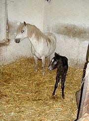
Horse behavior is best understood from the perspective that horses are prey animals with a well-developed fight-or-flight instinct. Their first response to a threat is to flee, although they are known to stand their ground and defend themselves or their offspring in cases where flight is...
and the fact that the horse is a flight animal. Both the strengths and weaknesses of the horse's visual abilities should be taken into consideration when training the animal, as an understanding of the horse's eye can help to discover why the animal behaves the way he does in various situations.
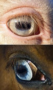
The anatomy of the equine eye
The equine eye includes the eyeball and the surrounding muscles and structures around the eyeball, termed the adnexaAdnexa
In anatomy, adnexa refers to the appendages of an organ. The term adnexa stems from a Latin word meaning appendages.More specifically, it can refer to:* Adnexa of eye * Adnexa of uterus...
.
The eyeball
The eyeball of the horse is not perfectly spherical, but rather is flattened anterior to posterior. However, research has found that the horse does not have a ramped retina, as was once thought.The wall of the eye is made up of three layers: the internal or nervous tunic, the vascular tunic, and the fibrous tunic.
- The nervous tunic (or retinaRetinaThe vertebrate retina is a light-sensitive tissue lining the inner surface of the eye. The optics of the eye create an image of the visual world on the retina, which serves much the same function as the film in a camera. Light striking the retina initiates a cascade of chemical and electrical...
) is made up of cells which are extensions of the brain, coming off the optic nerve. These receptors are light-sensitive, and include conesCone cellCone cells, or cones, are photoreceptor cells in the retina of the eye that are responsible for color vision; they function best in relatively bright light, as opposed to rod cells that work better in dim light. If the retina is exposed to an intense visual stimulus, a negative afterimage will be...
, which are less light-sensitive, but allow the eye to see color and provide visual acuity, and rod cells, which are more light sensitive, providing night vision but only seeing light and dark differences. Since only 2/3 of the eye can receive light, the receptor cells do not need to cover the entire interior of the eye, and line only the area from pupilPupilThe pupil is a hole located in the center of the iris of the eye that allows light to enter the retina. It appears black because most of the light entering the pupil is absorbed by the tissues inside the eye. In humans the pupil is round, but other species, such as some cats, have slit pupils. In...
to the optic disk. The part of the retina covered by light-sensitive cells is therefore termed the pars-optica retinae, and the blind part of the eye is termed the pars-ceaca retinae. The optic disk of the eye, however, does not contain any of these light-sensitive cells, as it is where the optic nerve leaves to the brain. It is therefore a blind spot within the eye. - The vascular tunic (or uveaUveaThe uvea , also called the uveal layer, uveal coat, uveal tract, or vascular tunic, is the pigmented middle of the three concentric layers that make up an eye. The name is possibly a reference to its reddish-blue or almost black colour, wrinkled appearance and grape-like size and shape when...
) is made up of the choroidChoroidThe choroid, also known as the choroidea or choroid coat, is the vascular layer of the eye, containing connective tissue, and lying between the retina and the sclera. The human choroid is thickest at the far extreme rear of the eye , while in the outlying areas it narrows to 0.1 mm...
, the ciliary bodyCiliary bodyThe ciliary body is the circumferential tissue inside the eye composed of the ciliary muscle and ciliary processes. It is triangular in horizontal section and is coated by a double layer, the ciliary epithelium. This epithelium produces the aqueous humor. The inner layer is transparent and covers...
, and the irisIris (anatomy)The iris is a thin, circular structure in the eye, responsible for controlling the diameter and size of the pupils and thus the amount of light reaching the retina. "Eye color" is the color of the iris, which can be green, blue, or brown. In some cases it can be hazel , grey, violet, or even pink...
. The choroid has a great deal of pigment, and is almost entirely made of blood vessels. It forms the tapetum lucidumTapetum lucidumThe tapetum lucidum is a layer of tissue in the eye of many vertebrate animals....
when it crosses over the fundusFundus (eye)The fundus of the eye is the interior surface of the eye, opposite the lens, and includes the retina, optic disc, macula and fovea, and posterior pole. The fundus can be viewed with an ophthalmoscope. The term may also be inclusive of Bruch's membrane and the choroid.The color of the fundus varies...
of the eye, causing the yellowish-green eye shine when light is directed into the animal's eyes at night. The tapetum lucidum reflects light back onto the retina, allowing for greater absorption in dark conditions. The iris lies between the cornes and the lensLens (anatomy)The crystalline lens is a transparent, biconvex structure in the eye that, along with the cornea, helps to refract light to be focused on the retina. The lens, by changing shape, functions to change the focal distance of the eye so that it can focus on objects at various distances, thus allowing a...
, and not only gives the eye its color, (see "eye color," below) but also allows for varying amounts of light to pass through its center hole, the pupilPupilThe pupil is a hole located in the center of the iris of the eye that allows light to enter the retina. It appears black because most of the light entering the pupil is absorbed by the tissues inside the eye. In humans the pupil is round, but other species, such as some cats, have slit pupils. In...
. - The fibrous tunic consists of the scleraScleraThe sclera , also known as the white or white of the eye, is the opaque , fibrous, protective, outer layer of the eye containing collagen and elastic fiber. In the development of the embryo, the sclera is derived from the neural crest...
and corneaCorneaThe cornea is the transparent front part of the eye that covers the iris, pupil, and anterior chamber. Together with the lens, the cornea refracts light, with the cornea accounting for approximately two-thirds of the eye's total optical power. In humans, the refractive power of the cornea is...
and protects the eye. The sclera (white of the eye) is made up of elastinElastinElastin is a protein in connective tissue that is elastic and allows many tissues in the body to resume their shape after stretching or contracting. Elastin helps skin to return to its original position when it is poked or pinched. Elastin is also an important load-bearing tissue in the bodies of...
and collagenCollagenCollagen is a group of naturally occurring proteins found in animals, especially in the flesh and connective tissues of mammals. It is the main component of connective tissue, and is the most abundant protein in mammals, making up about 25% to 35% of the whole-body protein content...
. The cornea (clear covering on the front of the eye) is made up of connective tissue and bathed in lacrimal fluid and aqueous humor. which provides it nutrition, as it does not have access to blood vessels. - The lensLens (anatomy)The crystalline lens is a transparent, biconvex structure in the eye that, along with the cornea, helps to refract light to be focused on the retina. The lens, by changing shape, functions to change the focal distance of the eye so that it can focus on objects at various distances, thus allowing a...
of the eye lies posterior to the iris, and is held suspended by the ciliary suspensory ligament and the ciliary muscle, which allows for "accommodation" of the eye: it allows the lens to change shape to focus on different objects. The lens is made up of onion-like layers of tissue.
Eye color
Horse markings
Markings on horses usually are distinctive white areas on an otherwise dark base coat color. Most horses have some markings, and they help to identify the horse as a unique individual. Markings are present at birth and do not change over the course of the horse's life...
or patterns
Pinto horse
A pinto horse has a coat color that consists of large patches of white and any other color. The distinction between "pinto" and "solid" can be tenuous, as so-called "solid" horses frequently have areas of white hair. Various cultures throughout history appear to have selectively bred for pinto...
. The white spotting patterns most often linked to blue eyes are splashed white
Splashed white
Splashed white or splash is a horse coat color pattern that produces pink-skinned, white markings. Many splashed whites have very modest markings, while others have the distinctive "dipped in white paint" pattern. Blue eyes are a hallmark of the pattern, and splash may account for otherwise "solid"...
, frame overo, and sometimes sabino
Sabino horse
Sabino is a group of white spotting patterns in horses that affect the skin and hair. A wide variety of irregular color patterns are accepted as sabino. In the strictest sense, "sabino" refers to the white patterns produced by the Sabino 1 gene, for which there is a DNA test...
. In the case of horses with white markings, one or both eyes may be blue, or part-blue
Heterochromia
In anatomy, heterochromia refers to a difference in coloration, usually of the iris but also of hair or skin. Heterochromia is a result of the relative excess or lack of melanin...
.
Homozygous cream dilutes
Cream gene
The cream gene is responsible for a number of horse coat colors. Horses that have the cream gene in addition to a base coat color that is chestnut will become palomino if they are heterozygous, having one copy of the cream gene, or cremello, if they are homozygous. Similarly, horses with a bay...
, sometimes called double-dilutes
Dilution gene
Dilution gene is a popular term for any one of a number of genes that act to create a lighter coat color in living creatures. There are many examples of such genes:-General:...
, always have light blue eyes to match their pale, cream-colored coats. Heterozygous or single-dilute creams, such as palomino
Palomino
Palomino is a coat color in horses, consisting of a gold coat and white mane and tail. Genetically, the palomino color is created by a single allele of a dilution gene called the cream gene working on a "red" base coat...
s and buckskins, often have light brown eyes. The eyes of horses with the Champagne gene
Champagne gene
The champagne gene is a simple dominant allele responsible for a number of rare horse coat colors. The most distinctive traits of horses with the champagne gene are the hazel eyes and pinkish, freckled skin, which are bright blue and bright pink at birth, respectively...
are typically greenish shades: aqua at birth, darkening to hazel with maturity.
As in humans, much of the genetics
Genetics
Genetics , a discipline of biology, is the science of genes, heredity, and variation in living organisms....
and etiology
Etiology
Etiology is the study of causation, or origination. The word is derived from the Greek , aitiologia, "giving a reason for" ....
behind eye color
Eye color
Eye color is a polygenic phenotypic character and is determined by two distinct factors: the pigmentation of the eye's iris and the frequency-dependence of the scattering of light by the turbid medium in the stroma of the iris....
are not yet fully understood.
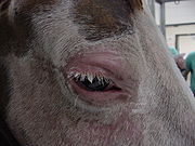
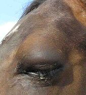
The adnexa
Eyelid
An eyelid is a thin fold of skin that covers and protects an eye. With the exception of the prepuce and the labia minora, it has the thinnest skin of the whole body. The levator palpebrae superioris muscle retracts the eyelid to "open" the eye. This can be either voluntarily or involuntarily...
s are made up of three layers of tissue: a thin layer of skin, which is covered in hair, a layer of muscles which allow the lid to open and close, and the palpebral conjunctiva, which lies against the eyeball. The opening between the two lids forms the palpebral fissue. The upper eyelid is larger and can move more than the lower lid. Unlike humans, horses also have a third eyelid (nictitating membrane
Nictitating membrane
The nictitating membrane is a transparent or translucent third eyelid present in some animals that can be drawn across the eye for protection and to moisten it while maintaining visibility. Some reptiles, birds, and sharks have a full nictitating membrane; in many mammals, there is a small...
) to protect the cornea. It lies on the inside corner of the eye, and closes diagonally over it.
The lacrimal apparatus
Lacrimal apparatus
The lacrimal apparatus is the physiologic system containing the orbital structures for tear production and drainage.It consists of:* the lacrimal gland, which secretes the tears, and its excretory ducts, which convey the fluid to the surface of the eye;...
produces tears, providing nutrition and moisture to the eye as well as helping to remove any debris that may have entered. The apparatus includes the lacrimal gland and the accessory lacrimal gland, which produce the tears. Blinking spreads the fluid over the eye, before it drains via the nasolacrimal duct
Nasolacrimal duct
The nasolacrimal duct carries tears from the lacrimal sac into the nasal cavity. Excess tears flow through nasolacrimal duct which drains into the inferior nasal meatus...
, which carries the lacrimal fluid into the nostril of the horse.
The ocular muscles allow the eye to move within the skull.
Visual field

Monocular vision
Monocular vision is vision in which each eye is used separately. By using the eyes in this way, as opposed by binocular vision, the field of view is increased, while depth perception is limited. The eyes are usually positioned on opposite sides of the animal's head giving it the ability to see two...
. Horses have the largest eyes of any land mammal, and are lateral-eyed, meaning that their eyes are positioned on the sides of their heads. This means that horses have a range of vision of more than 350°, with approximately 65° of this being binocular vision
Binocular vision
Binocular vision is vision in which both eyes are used together. The word binocular comes from two Latin roots, bini for double, and oculus for eye. Having two eyes confers at least four advantages over having one. First, it gives a creature a spare eye in case one is damaged. Second, it gives a...
and the remaining 285° monocular vision
Monocular vision
Monocular vision is vision in which each eye is used separately. By using the eyes in this way, as opposed by binocular vision, the field of view is increased, while depth perception is limited. The eyes are usually positioned on opposite sides of the animal's head giving it the ability to see two...
.
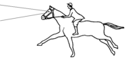
This provides a horse with the best chance to spot predators. The horse's wide range of monocular vision has two "blind spots," or areas where the animal can not see: in front of the face (making a cone that comes to a point at about 3–4 feet in front of the horse) and right behind his head, which extends over the back and behind the tail when standing with the head facing straight forward. Therefore, as a horse jumps an obstacle, it briefly disappears from sight right before the horse takes off.
There is a trade-off to a wide range of monocular vision: The placement of the horse's eyes decreases the possible range of binocular vision
Binocular vision
Binocular vision is vision in which both eyes are used together. The word binocular comes from two Latin roots, bini for double, and oculus for eye. Having two eyes confers at least four advantages over having one. First, it gives a creature a spare eye in case one is damaged. Second, it gives a...
(vision using both eyes at the same time) to around 65 degrees on a horizontal plane, occurring in a triangular shape primarily in front of the horse's face. Therefore the horse has a smaller field of depth perception than a human. The horse uses its binocular vision by looking straight at an object, raising its head when a horse looks at a distant predator or focuses on an obstacle to jump. To use binocular vision on a closer object near the ground, such as a snake or threat to its feet, the horse drops its nose and looks downward with neck somewhat arched.
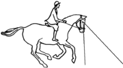
A horse will raise or lower its head to increase its range of binocular vision. A horse's visual field is lowered when it is asked to go "on the bit" with the head held perpendicular to the ground. This makes the horse's binocular vision focus less on distant objects and more on the immediate ground in front of the horse, suitable for arena distances, but less adaptive to a cross-country setting. Riders who ride with their horses "deep," "behind the vertical," or in a rollkur
Rollkur
Rollkur or hyperflexion of the horse's neck is a practice in equestrianism defined as "flexion of the horse's neck achieved through aggressive force" and is banned by the world governing body, the International Federation for Equestrian Sports...
frame decrease the range of the horse's distance vision even more, focusing only a few feet ahead of the front feet. Riders of Jumpers
Show jumping
Show jumping, also known as "stadium jumping," "open jumping," or "jumpers," is a member of a family of English riding equestrian events that also includes dressage, eventing, hunters, and equitation. Jumping classes commonly are seen at horse shows throughout the world, including the Olympics...
take the horse's use of distance vision into consideration, allowing their horse to raise the head a few strides before a jump, so that the animal is able to assess the jump and the proper take-off spot.
Visual acuity and sensitivity to motion
The visual acuityVisual acuity
Visual acuity is acuteness or clearness of vision, which is dependent on the sharpness of the retinal focus within the eye and the sensitivity of the interpretative faculty of the brain....
of the horse, or how well he is able to see details, is around 20/33. This is slightly worse than the usual 20/20 in humans, but much better than the visual acuity of dogs (20/50), cats (20/75), and rats (20/300). However, it is difficult to test an animal's visual acuity, and therefore the results may vary between studies.
The horse also has a "visual streak," or an area within the retina, linear in shape, with a high concentration of ganglion cells (up to 6100 cells/mm² in the visual streak compared to the 150 and 200 cells/mm² in the peripheral area). Horses have better acuity when the object they are looking at falls in this region. They therefore will tilt or raise their head, to help place the object within the area of the visual streak.
The horse is very sensitive to motion, as motion is usually the first alert that a predator is approaching. Such motion is usually first detected in their periphery, where they have poor visual acuity, and horses will usually act defensive and run if something suddenly moves into their peripheral field of vision.
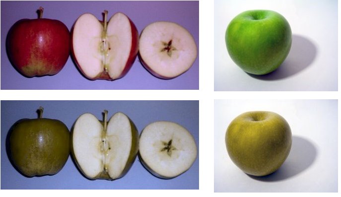
Color vision
Color blindness
Color blindness or color vision deficiency is the inability or decreased ability to see color, or perceive color differences, under lighting conditions when color vision is not normally impaired...
, but have two-color, or dichromatic vision. This means that they see two of the basic three wavelengths of visible light, compared to the three-color trichromic vision of most humans. In other words, horses naturally see the blue and green colors of the spectrum and the color variations based upon them, but cannot distinguish red. Research indicates that their color vision is somewhat like red-green color blindness in humans. This means that certain colors, especially red and related colors, appear more green.
Dichromatic vision is the result of the animal having two types of cones in their eyes: a short-wavelength sensitive cone (S) that is optimal at 428 nm (pastel bluish-gray), and a middle-to-long wavelength sensitive cone (M/L) which sees optimally at 539 nm, more of a yellowish color. This structure may be because horses are most active at dawn and dusk, a time when the rods
Rod cell
Rod cells, or rods, are photoreceptor cells in the retina of the eye that can function in less intense light than can the other type of visual photoreceptor, cone cells. Named for their cylindrical shape, rods are concentrated at the outer edges of the retina and are used in peripheral vision. On...
of the eye are especially useful.
The horse's limited ability to see color is sometimes taken into consideration when designing obstacles for the horse to jump, since the animal will have a harder time distinguishing between the obstacle and the ground if the two are only a few shades off. Therefore, most people paint their jump rails a different color from the footing or the surrounding landscape so that the horse may better judge the obstacle on the approach. Studies have shown that horses are less likely to have a rail down when the jump is painted with two or more contrasting colors, rather than one single color It is especially difficult for horses to distinguish between yellows and greens.
Sensitivity to light
Tapetum lucidum
The tapetum lucidum is a layer of tissue in the eye of many vertebrate animals....
, giving them superior night vision. This also gives them better vision on slightly cloudy days, relative to bright, sunny days. In a 2009 laboratory study, horses were able to distinguish different shapes in low light, including levels mimicking dark, moonless nights in wooded areas. When light dropped to nearly dark, horses could not discriminate between different shapes, but remained able to negotiate around the enclosure and testing equipment in conditions where humans in the same enclosure "stumbled into walls, apparatus, pylons, and even the horse itself. "
However, horses are less able to adjust to sudden changes of light than are humans, such as when moving from a bright day into a dark barn. This is a consideration during training, as certain tasks, such as loading into a trailer, may frighten a horse simply because it cannot see adequately. It is also important in riding, as quickly moving from light to dark or vice-versa will temporarily make it difficult for the animal to judge what is in front of it.
Near- and Far-sightedness
Many domestic horses (about 1/3) tend to have myopiaMyopia
Myopia , "shortsightedness" ) is a refractive defect of the eye in which collimated light produces image focus in front of the retina under conditions of accommodation. In simpler terms, myopia is a condition of the eye where the light that comes in does not directly focus on the retina but in...
(near-sightness), with few being far-sighted. Wild horses, however, are usually far-sighted.
Accommodation
Horses have relatively poor "accommodation" (the ability to turn the eye within the socket to focus on an object), as they have weak ciliary muscles. However, this does not usually place them at a disadvantage, as accommodation is often used when focusing with high acuity on things up close, and horses rarely need to do so. Instead, the horse often tilts its head slightly to focus on things without the benefit of a high degree of accommodation.Disorders of the eye
- Corneal abrasion
- Corneal ulcerCorneal ulcerA corneal ulcer, or ulcerative keratitis, is an inflammatory condition of the cornea involving loss of its outer layer. It is very common in dogs and is sometimes seen in cats...
- KeratitisKeratitisKeratitis is a condition in which the eye's cornea, the front part of the eye, becomes inflamed. The condition is often marked by moderate to intense pain and usually involves impaired eyesight.-Types:...
- ConjunctivitisConjunctivitisConjunctivitis refers to inflammation of the conjunctiva...
- UveitisUveitisUveitis specifically refers to inflammation of the middle layer of the eye, termed the "uvea" but in common usage may refer to any inflammatory process involving the interior of the eye....
: including recurrent uveitis and periodic ophthalmia ("moon blindness"). Spontaneous Equine Recurrent UveitisEquine Recurrent UveitisEquine Recurrent Uveitis, is an acute, non-granulomatous inflammation of the uveal tract of the eye, occurring commonly in horses of all breeds, worldwide. The causative factor is not known, but several pathogeneses have been suggested. It is the most common cause of blindness in horses...
(ERU) occurs in 10-15% of the equine population, with the AppaloosaAppaloosaThe Appaloosa is a horse breed best known for its colorful leopard-spotted coat pattern. There is a wide range of body types within the breed, stemming from the influence of multiple breeds of horses throughout its history. Each horse's color pattern is genetically the result of various spotting...
breed having an eightfold higher risk than the general horse population. - HabronemaHabronemaHabronema muscae is an internal stomach parasite that is most commonly found in Horses.-Eggs:The adult worms lay eggs within the horse's stomach. The eggs are later excreted through the feces...
- Keratoconjunctivitis siccaKeratoconjunctivitis siccaKeratoconjunctivitis sicca , also called keratitis sicca, xerophthalmia or dry eye syndrome is an eye disease caused by eye dryness, which, in turn, is caused by either decreased tear production or increased tear film evaporation. It is found in humans and some animals...