

Visual acuity
Encyclopedia
Visual perception
Visual perception is the ability to interpret information and surroundings from the effects of visible light reaching the eye. The resulting perception is also known as eyesight, sight, or vision...
, which is dependent on the sharpness of the retinal focus within the eye
Human eye
The human eye is an organ which reacts to light for several purposes. As a conscious sense organ, the eye allows vision. Rod and cone cells in the retina allow conscious light perception and vision including color differentiation and the perception of depth...
and the sensitivity of the interpretative faculty of the brain.
Visual acuity is a measure of the spatial resolution of the visual processing system and is usually tested in a manner to optimise and standardise the conditions. To this end, black symbols on a white background are used (for maximum contrast
Contrast (vision)
Contrast is the difference in visual properties that makes an object distinguishable from other objects and the background. In visual perception of the real world, contrast is determined by the difference in the color and brightness of the object and other objects within the same field of view...
) and a sufficient distance allowed to approximate infinity in the way the lens
Lens (anatomy)
The crystalline lens is a transparent, biconvex structure in the eye that, along with the cornea, helps to refract light to be focused on the retina. The lens, by changing shape, functions to change the focal distance of the eye so that it can focus on objects at various distances, thus allowing a...
attempts to focus. Twenty feet, or six metres, is essentially infinity
Infinity
Infinity is a concept in many fields, most predominantly mathematics and physics, that refers to a quantity without bound or end. People have developed various ideas throughout history about the nature of infinity...
from an optical perspective (the difference in optical power required to focus at 20 feet versus infinity is only 0.164 diopters). Whilst in an eye exam, lenses of varying powers are used to precisely correct for refractive error
Refractive error
A refractive error, or refraction error, is an error in the focusing of light by the eye and a frequent reason for reduced visual acuity.-Classification:...
s, using a pinhole will largely correct for refractive errors and allow VA to be tested in other circumstances. Letters are normally used (as in the classic Snellen chart
Snellen chart
A Snellen chart is an eye chart used by eye care professionals and others to measure visual acuity. Snellen charts are named after the Dutch ophthalmologist Herman Snellen who developed the chart during 1862...
) as most people will recognise them but other symbols (such as a letter E
E Chart
An E Chart, also known as a Tumbling E Chart, is an ophthalmological chart used to measure a patient's acuity for distant vision.-Uses:This chart is useful for patients that are illiterate or too young to read but who can speak....
facing in different directions) can be used instead.
In the term "20/20 vision", the numerator
Fraction (mathematics)
A fraction represents a part of a whole or, more generally, any number of equal parts. When spoken in everyday English, we specify how many parts of a certain size there are, for example, one-half, five-eighths and three-quarters.A common or "vulgar" fraction, such as 1/2, 5/8, 3/4, etc., consists...
refers to the distance in feet between the subject and the chart. The denominator indicates the size of the letters, specifically it denotes the separation at which the lines that make up those letters would be separated by a visual angle
Visual angle
The visual angle is the angle a viewed object subtends at the eye, usually stated in degrees of arc.It also is called the object's angular size....
of 1 arc minute, which for the lowest line that is read by an eye with no refractive error (or the errors corrected) is usually 20 feet. The metric equivalent is 6/6 vision where the distance is 6 metres. This means that at 20 feet or 6 metres, a typical human eye, able to separate 1 arc minute, can resolve lines with a spacing of about 1.75mm. 20/20 or 6/6 vision can be considered nominal
Real versus nominal value
In economics, nominal value refers to a value expressed in money terms in a given year or series of years. By contrast, real value adjusts nominal value to remove effects of price changes over time...
performance for human distance vision; 20/40 or 6/12 vision can be considered half that acuity for distance vision and 20/10 or 6/3 vision would be twice normal acuity. The 20/x number does not directly relate to the eyeglass prescription
Eyeglass prescription
An eyeglass prescription is an order written by an eyewear prescriber, such as an optometrist or ophthalmologist, that specifies the value of all parameters the prescriber has deemed necessary to construct and/or dispense corrective lenses appropriate for a patient.If an examination indicates that...
required to correct vision, because it does not specify the nature of the problem corrected by the lens, only the resulting performance. Instead an eye exam seeks to find the prescription that will provide the best corrected visual performance achievable. This may be greater or lesser than 6/6 for many reasons.
History
| Year | Event | |
|---|---|---|
| 1843 | German treatise advocating the need for standardized vision tests and developed a set of three charts. | |
| 1854 | Eduard von Jaeger published a set of reading samples to document functional vision. He published samples in German, French, English and other languages. He used fonts that were available in the State Printing House in Vienna in 1854 and labeled them with the numbers from that printing house catalogue. | |
| 1861 | Franciscus Donders Franciscus Donders -External links:* B. Theunissen. , F.C. Donders: turning refracting into science, @ History of science and scholarship in the Netherlands.* in the Virtual Laboratory of the Max Planck Institute for the History of Science* P. Eling, , Geneeskundige en fysioloog.... coined the term visual acuity to describe the “sharpness of vision” and defined it as the ratio between a subject's VA and a standard VA. |
|
| 1862 | Hermann Snellen Hermann Snellen Herman Snellen was a Dutch ophthalmologist who introduced the Snellen chart to study visual acuity . He took over directorship of the Netherlands Hospital for Eye Patients after Dr... published his famous letter chart Snellen chart A Snellen chart is an eye chart used by eye care professionals and others to measure visual acuity. Snellen charts are named after the Dutch ophthalmologist Herman Snellen who developed the chart during 1862... . His most significant decision was not to use existing typefaces but to design special targets, which he called optotype Optotype An optotype is a standardized symbol for testing vision. Optotypes can be specially shaped letters, numbers, or geometric symbols. For instance, to determine visual acuity, optotypes of different sizes are presented to a person and the smallest size is determined at which the person can reliably... s. He based it on a 5x5 grid. This was crucial because it was a physical standard measure to reproduce the chart. Snellen defined “standard vision” as the ability to recognize one of his optotypes when it subtended 5 minutes of arc, thus the optotype can be recognized only if the person viewing it can discriminate a spatial pattern separated by a visual angle Visual angle The visual angle is the angle a viewed object subtends at the eye, usually stated in degrees of arc.It also is called the object's angular size.... of 1 minute of arc (one element of the grid). |
|
| 1868 | John Green of St. Louis, who had worked with Donders and Snellen, proposed a chart with a geometric progression of letter sizes and proportional spacing between letters. At that time, Green’s proposals were not accepted. A century later, his principles would be incorporated in international standards. | |
| 1875 |
|
|
| 1888 | Edmund Landolt proposed the Landolt C Landolt C A Landolt C, also known as a Japanese Vision Test, Landolt ring or Landolt broken ring, is an optotype, i.e. a standardized symbol used for testing vision. It was developed by the Swiss-born ophthalmologist Edmund Landolt.... , a symbol that has only one element of detail and varies only in its orientation. The broken ring symbol is made with a "C" like figure in a 5 x 5 grid that, in the 20/20 optotype, subtends 5 minutes of arc and has an opening (oriented in the top, bottom, right or left) measuring 1 minute of arc. This proposal was based in the fact that not all of Snellen's optotypes were equally recognizable. This chart is actually the preferred visual acuity measurement symbol for laboratory experiments but gained only limited acceptance in clinical use. |
|
| 1898 | Marius Tscherning Marius Tscherning Marius Hans Erik Tscherning was a Danish ophthalmologist.He studied ophthalmology under Edmund Hansen Grut in Copenhagen, later becoming an adjunct director at the ophthalmological laboratory at the Sorbonne in Paris. Tscherning spent 25 years at the Sorbonne, where he worked closely with Louis... reported the inadequacy of 20/20 (1 minute of arc) as a norm value of VA and explained the Snellen’s mistake who referred to a normal observer using this wrong value. Tscherning’s opinion is echoed by many modern investigators who have found that Snellen’s criterion does not represent the normal limits of vision. Many observers are capable of producing results that surpass the limit of the supposed 20/20 standard for visual acuity. Surprisingly, the 20/20 myth still continues today. |
|
| 1923 | Soviet Soviet Union The Soviet Union , officially the Union of Soviet Socialist Republics , was a constitutionally socialist state that existed in Eurasia between 1922 and 1991.... ophthalmologists Sergei Golovin and D. A. Sivtsev developed the table for testing visual acuity. Later this table became known as Golovin-Sivtsev Table Golovin-Sivtsev Table In 1923, Soviet ophthalmologists Sergei Golovin and D. A. Sivtsev developed the table for testing visual acuity, that was used in the USSR and is still used in some post-Soviet states... . |
|
| 1959 | Louise Sloan designed a new optotype Sloan letters Sloan letters, designed by Louise Sloan in 1959, are a set of optotypes used to test visual acuity generally used in Snellen charts and logMAR charts.This set of optotypes consists of ten specially formed "letters", "C, D, H, K, N, O, R, S, V and Z"... set of 10 letters, all to be shown in each and every line tested, in order to avoid the problem that not all letters are equally recognizable. The larger letter sizes thus required more than one physical line. Louise Sloan also proposed a new letter size notation using the SI system stating that standard acuity (1.0, 20/20) represents the ability to recognize a standard letter size (1 M-bunit) at a standard distance (1 meter). |
|
| 1976 |
Lea test A Lea test is a visual acuity test tailored for children who do not know the alphabet normally used in eye charts. The optotypes are outlines of an apple, a house, a square, and a circle. This test was developed by Finnish ophthalmologist Lea Hyvärinen in 1976 and has been calibrated against... , using outlines of figures (an apple, a house, a circle and a square) to measure visual acuity in preschool children. |
|
| 1982 |
Rick Ferris et al. of the National Eye Institute chose the Bailey-Lovie layout, implemented with Sloan letters, to establish a standardized method of visual acuity measurement for the Early Treatment of Diabetic Retinopathy Study (ETDRS). These charts were used in all subsequent clinical studies, and did much to familiarize the profession with the new layout and progression. Data from the ETDRS were used to select letter combinations that give each line the same average difficulty, without using all letters on each line. |
|
| 1984 | The International Council of Ophthalmology approved a new 'Visual Acuity Measurement Standard', also incorporating the above features. |
Physiology of visual acuity
In low lightScotopic vision
Scotopic vision is the vision of the eye under low light conditions. The term comes from Greek skotos meaning darkness and -opia meaning a condition of sight...
vision, there is low resolution despite the high sensitivity thereof. This is due to spatial summation of rods, so 100 rods could merge into many bipolars, in turn converging on ganglion cells, and the unit for resolution is very large, thus acuity being small. The farther a pattern of white and black lines is presented to a person, the less he can distinguish the lines, culminating to a distance when the pattern is seen as a uniform gray. The angle subtended by the detail at minimum acuity is the resolving power, and its reciprocal is the visual acuity. For example, a visual acuity of 1 subtends 1 minute on the retina, that of 2 is 1/2 minutes (30 seconds) of arc. Visual acuity is much better in bright light than dim light, the former reaching 2 with a bright center and surrounding, the latter perhaps having visual acuity of 0.04 (25 minutes on eye). In this case, the stimulus is 1.7 inches (4.4 cm) and a distance of 20 ft (6 m).
Thus, visual acuity, or resolving power, is the property of cones.
To resolve detail, the eye's optical system has to project a focused image on the fovea
Fovea
The fovea centralis, also generally known as the fovea , is a part of the eye, located in the center of the macula region of the retina....
, a region inside the macula
Macula
The macula or macula lutea is an oval-shaped highly pigmented yellow spot near the center of the retina of the human eye. It has a diameter of around 5 mm and is often histologically defined as having two or more layers of ganglion cells...
having the highest density of cone
Cone cell
Cone cells, or cones, are photoreceptor cells in the retina of the eye that are responsible for color vision; they function best in relatively bright light, as opposed to rod cells that work better in dim light. If the retina is exposed to an intense visual stimulus, a negative afterimage will be...
photoreceptor cells (the only kind of photoreceptors existing on the fovea), thus having the highest resolution and best color vision. Acuity and color vision, despite being mediated by the same cells, are different physiologic functions that do not interrelate except by position. Acuity and color vision can be affected independently.
The grain of a photographic mosaic has just as limited resolving power as the "grain" of the retinal mosaic. In order to see detail, two sets of receptors must be intervened by a middle set. The maximum resolution is that 30 seconds of arc, corresponding to the foveal cone diameter or the angle subtended at the nodal point of the eye. In order to get reception from each cone, as it would be if vision was on a mosaic basis, the "local sign" must be obtained from a single cone via a chain of one bipolar, ganglion, and lateral geniculate cell each. A key factor of obtaining detailed vision, however, is inhibition. This is mediated by neurons such as the amacrine and horizontal cells, which functionally render the spread or convergence of signals inactive. This tendency to one-to-one shuttle of signals is powered by brightening of the center and its surroundings, which triggers the inhibition leading to a one-to-one wiring. This scenario, however, is rare, as cones may connect to both midget and flat (diffuse) bipolars, and amacrine and horizontal cells can merge messages just as easily as inhibit them.
Light
Light
Light or visible light is electromagnetic radiation that is visible to the human eye, and is responsible for the sense of sight. Visible light has wavelength in a range from about 380 nanometres to about 740 nm, with a frequency range of about 405 THz to 790 THz...
travels from the fixation object to the fovea through an imaginary path called the visual axis. The eye's tissues and structures that are in the visual axis (and also the tissues adjacent to it) affect the quality of the image. These structures are: tear film, cornea, anterior chamber, pupil, lens, vitreous, and finally the retina. The posterior part of the retina, called the retinal pigment epithelium (RPE) is responsible for, among many other things, absorbing light that crosses the retina so it cannot bounce to other parts of the retina. Interestingly, in many vertebrates, such as cats, where high visual acuity is not a priority, there is a reflecting tapetum
Tapetum lucidum
The tapetum lucidum is a layer of tissue in the eye of many vertebrate animals....
layer that gives the photoreceptors a "second chance" to absorb the light, thus improving the ability to see in the dark. This is what causes an animal's eyes to seemingly glow in the dark when a light is shone on them. The RPE also has a vital function of recycling the chemicals used by the rods and cones in photon detection. If the RPE is damaged and does not clean up this "shed" blindness can result.
As in a photographic lens
Photographic lens
A camera lens is an optical lens or assembly of lenses used in conjunction with a camera body and mechanism to make images of objects either on photographic film or on other media capable of storing an image chemically or electronically.While in principle a simple convex lens will suffice, in...
, visual acuity is affected by the size of the pupil. Optical aberrations of the eye that decrease visual acuity are at a maximum when the pupil is largest (about 8 mm), which occurs in low-light conditions. When the pupil is small (1–2 mm), image sharpness may be limited by diffraction
Diffraction
Diffraction refers to various phenomena which occur when a wave encounters an obstacle. Italian scientist Francesco Maria Grimaldi coined the word "diffraction" and was the first to record accurate observations of the phenomenon in 1665...
of light by the pupil (see diffraction limit). Between these extremes is the pupil diameter that is generally best for visual acuity in normal, healthy eyes; this tends to be around 3 or 4 mm.
If the optics of the eye were otherwise perfect, theoretically, acuity would be limited by pupil diffraction, which would be a diffraction-limited acuity of 0.4 minutes of arc (minarc) or 20/8 acuity. The smallest cone cells in the fovea have sizes corresponding to 0.4 minarc of the visual field, which also places a lower limit on acuity. The optimal acuity of 0.4 minarc or 20/8 can be demonstrated using a laser interferometer
Interferometry
Interferometry refers to a family of techniques in which electromagnetic waves are superimposed in order to extract information about the waves. An instrument used to interfere waves is called an interferometer. Interferometry is an important investigative technique in the fields of astronomy,...
that bypasses any defects in the eye's optics and projects a pattern of dark and light bands directly on the retina. Laser interferometers are now used routinely in patients with optical problems, such as cataract
Cataract
A cataract is a clouding that develops in the crystalline lens of the eye or in its envelope, varying in degree from slight to complete opacity and obstructing the passage of light...
s, to assess the health of the retina before subjecting them to surgery.
The visual cortex
Visual cortex
The visual cortex of the brain is the part of the cerebral cortex responsible for processing visual information. It is located in the occipital lobe, in the back of the brain....
is the part of the cerebral cortex
Cerebral cortex
The cerebral cortex is a sheet of neural tissue that is outermost to the cerebrum of the mammalian brain. It plays a key role in memory, attention, perceptual awareness, thought, language, and consciousness. It is constituted of up to six horizontal layers, each of which has a different...
in the posterior part of the brain
Brain
The brain is the center of the nervous system in all vertebrate and most invertebrate animals—only a few primitive invertebrates such as sponges, jellyfish, sea squirts and starfishes do not have one. It is located in the head, usually close to primary sensory apparatus such as vision, hearing,...
responsible for processing visual stimuli, called the occipital lobe
Occipital lobe
The occipital lobe is the visual processing center of the mammalian brain containing most of the anatomical region of the visual cortex. The primary visual cortex is Brodmann area 17, commonly called V1...
. The central 10° of field (approximately the extension of the macula
Macula
The macula or macula lutea is an oval-shaped highly pigmented yellow spot near the center of the retina of the human eye. It has a diameter of around 5 mm and is often histologically defined as having two or more layers of ganglion cells...
) is represented by at least 60% of the visual cortex. Many of these neurons are believed to be involved directly in visual acuity processing.
Proper development of normal visual acuity depends on an animal having normal visual input when it is very young. Any visual deprivation, that is, anything interfering with such input over a prolonged period, such as a cataract
Cataract
A cataract is a clouding that develops in the crystalline lens of the eye or in its envelope, varying in degree from slight to complete opacity and obstructing the passage of light...
, severe eye turn or strabismus
Strabismus
Strabismus is a condition in which the eyes are not properly aligned with each other. It typically involves a lack of coordination between the extraocular muscles, which prevents bringing the gaze of each eye to the same point in space and preventing proper binocular vision, which may adversely...
, or covering or patching the eye during medical treatment, will usually result in a severe and permanent decrease in visual acuity in the affected eye if not treated early in life. The decreased acuity is reflected in various abnormalities in cell properties in the visual cortex. These changes include a marked decrease in the number of cells connected to the affected eye as well as few cells connected to both eyes, resulting in a loss of binocular vision
Binocular vision
Binocular vision is vision in which both eyes are used together. The word binocular comes from two Latin roots, bini for double, and oculus for eye. Having two eyes confers at least four advantages over having one. First, it gives a creature a spare eye in case one is damaged. Second, it gives a...
and depth perception
Depth perception
Depth perception is the visual ability to perceive the world in three dimensions and the distance of an object. Depth sensation is the ability to move accurately, or to respond consistently, based on the distances of objects in an environment....
, or stereopsis
Stereopsis
Stereopsis refers to impression of depth that is perceived when a scene is viewed with both eyes by someone with normal binocular vision. Binocular viewing of a scene creates two slightly different images of the scene in the two eyes due the the eyes' different positions on the head...
. The period of time over which an animal is highly sensitive to such visual deprivation is referred to as the critical period
Critical period
This article is about a critical period in an organism's or person's development. See also America's Critical Period.In general, a critical period is a limited time in which an event can occur, usually to result in some kind of transformation...
.
The eye is connected to the visual cortex by the optic nerve
Optic nerve
The optic nerve, also called cranial nerve 2, transmits visual information from the retina to the brain. Derived from the embryonic retinal ganglion cell, a diverticulum located in the diencephalon, the optic nerve doesn't regenerate after transection.-Anatomy:The optic nerve is the second of...
coming out of the back of the eye. The two optic nerves come together behind the eyes at the optic chiasm
Optic chiasm
The optic chiasm or optic chiasma is the part of the brain where the optic nerves partially cross...
, where about half of the fibers from each eye cross over to the opposite side and join fibers from the other eye representing the corresponding visual field, the combined nerve fibers from both eyes forming the optic tract
Optic tract
The optic tract is a part of the visual system in the brain.It is a continuation of the optic nerve and runs from the optic chiasm to the lateral geniculate nucleus....
. This ultimately forms the physiological basis of binocular vision
Binocular vision
Binocular vision is vision in which both eyes are used together. The word binocular comes from two Latin roots, bini for double, and oculus for eye. Having two eyes confers at least four advantages over having one. First, it gives a creature a spare eye in case one is damaged. Second, it gives a...
. The tracts project to a relay station in the midbrain called the lateral geniculate nucleus
Lateral geniculate nucleus
The lateral geniculate nucleus is the primary relay center for visual information received from the retina of the eye. The LGN is found inside the thalamus of the brain....
, part of the thalamus
Thalamus
The thalamus is a midline paired symmetrical structure within the brains of vertebrates, including humans. It is situated between the cerebral cortex and midbrain, both in terms of location and neurological connections...
, and then to the visual cortex along a collection of nerve fibers called the optic radiation
Optic radiation
The optic radiation is a collection of axons from relay neurons in the lateral geniculate nucleus of the thalamus carrying visual information to the visual cortex along the calcarine fissure.There is one such tract on each side of the brain.-Parts:A distinctive...
s.
Any pathological process in the visual system, even in older humans beyond the critical period, will often cause decreases in visual acuity. Thus measuring visual acuity is a simple test in accessing the health of the eyes, the visual brain, or pathway to the brain. Any relatively sudden decrease in visual acuity is always a cause for concern. Common causes of decreases in visual acuity are cataract
Cataract
A cataract is a clouding that develops in the crystalline lens of the eye or in its envelope, varying in degree from slight to complete opacity and obstructing the passage of light...
s and scarred cornea
Cornea
The cornea is the transparent front part of the eye that covers the iris, pupil, and anterior chamber. Together with the lens, the cornea refracts light, with the cornea accounting for approximately two-thirds of the eye's total optical power. In humans, the refractive power of the cornea is...
s, which affect the optical path, diseases that affect the retina, such as macular degeneration
Macular degeneration
Age-related macular degeneration is a medical condition which usually affects older adults and results in a loss of vision in the center of the visual field because of damage to the retina. It occurs in “dry” and “wet” forms. It is a major cause of blindness and visual impairment in older adults...
and diabetes, diseases affecting the optic pathway to the brain such as tumor
Tumor
A tumor or tumour is commonly used as a synonym for a neoplasm that appears enlarged in size. Tumor is not synonymous with cancer...
s and multiple sclerosis
Multiple sclerosis
Multiple sclerosis is an inflammatory disease in which the fatty myelin sheaths around the axons of the brain and spinal cord are damaged, leading to demyelination and scarring as well as a broad spectrum of signs and symptoms...
, and diseases affecting the visual cortex such as tumors and stroke
Stroke
A stroke, previously known medically as a cerebrovascular accident , is the rapidly developing loss of brain function due to disturbance in the blood supply to the brain. This can be due to ischemia caused by blockage , or a hemorrhage...
s.
Though the resolving power depends on size and packing density of the photoreceptors, the neural system of receptors must interpret this resolving power. As determined from various experiments on the cat, different ganglion cells are tuned to different frequencies of detail, as from a grating
Grating
A grating is any regularly spaced collection of essentially identical, parallel, elongated elements. Gratings usually consist of a single set of elongated elements, but can consist of two sets, in which case the second set is usually perpendicular to the first...
, so some ganglion cells have better acuity than others. In humans the results are the same, this time utilizing the same method as well as a device to read electrical changes in the scalp.
Optical aspects
Besides the neural connections of the receptors, the optical system is an equally key player in retinal resolution. In the ideal eye, the image of a diffraction gratingDiffraction grating
In optics, a diffraction grating is an optical component with a periodic structure, which splits and diffracts light into several beams travelling in different directions. The directions of these beams depend on the spacing of the grating and the wavelength of the light so that the grating acts as...
, can subtend 0.5 micrometre on the retina. This is certainly not the case, however, and furthermore the pupil can cause diffraction
Diffraction
Diffraction refers to various phenomena which occur when a wave encounters an obstacle. Italian scientist Francesco Maria Grimaldi coined the word "diffraction" and was the first to record accurate observations of the phenomenon in 1665...
of the light. Thus, black lines on a grating will be mixed with the intervening white lines to make a gray appearance. Defective optical issues (such as myopia) can render it worse, but suitable lenses can help. Images (such as gratings) can be sharpened by lateral inhibition, i.e., more highly excited cells inhibiting the less excited cells. A similar reaction is in the case of chromatic aberrations, in which the color fringes around black-and-white objects are inhibited similarly.
Visual acuity expression
| Foot | Metre | Decimal | LogMAR |
|---|---|---|---|
| 20/200 | 6/60 | 0.10 | 1.00 |
| 20/160 | 6/48 | 0.125 | 0.90 |
| 20/125 | 6/38 | 0.16 | 0.80 |
| 20/100 | 6/30 | 0.20 | 0.70 |
| 20/80 | 6/24 | 0.25 | 0.60 |
| 20/63 | 6/19 | 0.32 | 0.50 |
| 20/50 | 6/15 | 0.40 | 0.40 |
| 20/40 | 6/12 | 0.50 | 0.30 |
| 20/32 | 6/9.5 | 0.63 | 0.20 |
| 20/25 | 6/7.5 | 0.80 | 0.10 |
| 20/20 | 6/6 | 1.00 | 0.00 |
| 20/16 | 6/4.8 | 1.25 | -0.10 |
| 20/12.5 | 6/3.8 | 1.60 | -0.20 |
| 20/10 | 6/3 | 2.00 | -0.30 |
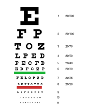
Visual acuity is often measured according to the size of letters viewed on a Snellen chart
Snellen chart
A Snellen chart is an eye chart used by eye care professionals and others to measure visual acuity. Snellen charts are named after the Dutch ophthalmologist Herman Snellen who developed the chart during 1862...
or the size of other symbols, such as Landolt C
Landolt C
A Landolt C, also known as a Japanese Vision Test, Landolt ring or Landolt broken ring, is an optotype, i.e. a standardized symbol used for testing vision. It was developed by the Swiss-born ophthalmologist Edmund Landolt....
s or Tumbling E
E Chart
An E Chart, also known as a Tumbling E Chart, is an ophthalmological chart used to measure a patient's acuity for distant vision.-Uses:This chart is useful for patients that are illiterate or too young to read but who can speak....
.
In some countries, acuity is expressed as a vulgar fraction, and in some as a decimal number
Decimal
The decimal numeral system has ten as its base. It is the numerical base most widely used by modern civilizations....
.
Using the foot as a unit of measurement, (fractional) visual acuity is expressed relative to 20/20. Otherwise, using the metre
Metre
The metre , symbol m, is the base unit of length in the International System of Units . Originally intended to be one ten-millionth of the distance from the Earth's equator to the North Pole , its definition has been periodically refined to reflect growing knowledge of metrology...
, visual acuity is expressed relative to 6/6. For all intents and purposes, 6/6 vision is equivalent to 20/20. In the decimal system, the acuity is defined as the reciprocal value of the size of the gap (measured in arc minutes) of the smallest Landolt C
Landolt C
A Landolt C, also known as a Japanese Vision Test, Landolt ring or Landolt broken ring, is an optotype, i.e. a standardized symbol used for testing vision. It was developed by the Swiss-born ophthalmologist Edmund Landolt....
that can be reliably identified. A value of 1.0 is equal to 20/20.
LogMAR is another commonly used scale, expressed as the (decadic) logarithm of the minimum angle of resolution. LogMAR scale converts the geometric sequence of a traditional chart to a linear scale. It measures visual acuity loss; positive values indicate vision loss, while negative values denote normal or better visual acuity. This scale is rarely used clinically; it is more frequently used in statistical calculations because it provides a more scientific equivalent for the traditional clinical statement of “lines lost” or “lines gained”, which is valid only when all steps between lines are equal, which is not usually the case.
A visual acuity of 20/20 is frequently described as meaning that a person can see detail from 20 feet away the same as a person with normal eyesight would see from 20 feet. If a person has a visual acuity of 20/40, he is said to see detail from 20 feet away the same as a person with normal eyesight would see it from 40 feet away. It is possible to have vision superior to 20/20: the maximum acuity of the human eye without visual aids (such as binoculars
Binoculars
Binoculars, field glasses or binocular telescopes are a pair of identical or mirror-symmetrical telescopes mounted side-by-side and aligned to point accurately in the same direction, allowing the viewer to use both eyes when viewing distant objects...
) is generally thought to be around 20/10 (6/3) however, recent test subjects have exceeded 20/8 vision. Some birds of prey, such as hawk
Hawk
The term hawk can be used in several ways:* In strict usage in Australia and Africa, to mean any of the species in the subfamily Accipitrinae, which comprises the genera Accipiter, Micronisus, Melierax, Urotriorchis and Megatriorchis. The large and widespread Accipiter genus includes goshawks,...
s, are believed to have an acuity of around 20/2; in this respect, their vision is much better than human eyesight.
When visual acuity is below the largest optotype on the chart, the reading distance is reduced until the patient can read it. Once the patient is able to read the chart, the letter size and test distance are noted. If the patient is unable to read the chart at any distance, he or she is tested as follows:
| Name | Abbreviation | Definition |
|---|---|---|
| Counting Fingers | CF | Ability to count fingers at a given distance. |
| Hand Motion | HM | Ability to distinguish a hand if it is moving or not in front of the patient's face. |
| Light Perception | LP | Ability to perceive any light. |
| No Light Perception | NLP | Inability to see any light. Total blindness. |
Many humans have one eye that has superior visual acuity over the other.
Legal Definitions
Various countries have defined statutory limits for poor visual acuity that qualifies as a disability. For example, in Australia, the Social Security Act defines blindness as:In the USA, the relevant federal statute defines blindness as follows:
[T]he term "blindness" means central visual acuity of 20/200 or less in the better eye with the use of a correcting lens. An eye that is accompanied by a limitation in the fields of vision such that the widest diameter of the visual field subtends an angle no greater than 20 degrees shall be considered for purposes in this paragraph as having a central visual acuity of 20/200 or less.
A person's visual acuity is registered documenting the following: whether the test was for distant or near vision, the eye(s) evaluated and whether corrective lenses (i.e. glasses
Glasses
Glasses, also known as eyeglasses , spectacles or simply specs , are frames bearing lenses worn in front of the eyes. They are normally used for vision correction or eye protection. Safety glasses are a kind of eye protection against flying debris or against visible and near visible light or...
or contact lens
Contact lens
A contact lens, or simply contact, is a lens placed on the eye. They are considered medical devices and can be worn to correct vision, for cosmetic or therapeutic reasons. In 2004, it was estimated that 125 million people use contact lenses worldwide, including 28 to 38 million in the United...
es) were used:
- Distance from the chart
- D (distant) for the evaluation done at 20 feet (or 6 meters).
- N (near) for the evaluation done at 15.7 inches (or 40 cm).
- Eye evaluated
- OD (Latin oculus dexter) for the right eye.
- OS (Latin oculus sinister) for the left eye.
- OU (Latin oculi uterque) for both eyes.
- Usage of spectacles during the test
- cc (Latin cum correctore) with correctors.
- sc: (Latin sine correctore) without correctors.
- Pinhole occluderPinhole occluderA pinhole occluder is an opaque disk with one or more small holes through it, used by ophthalmologists and optometrists to test visual acuity. The occluder is a simple way to focus light, as in a pinhole camera, temporarily removing the effects of refractive errors such as myopia...
- The abbreviation PH is followed by the visual acuity as measured with a pinhole occluder, which temporarily corrects for refractive errorRefractive errorA refractive error, or refraction error, is an error in the focusing of light by the eye and a frequent reason for reduced visual acuity.-Classification:...
s such as myopia or astigmatism.
- The abbreviation PH is followed by the visual acuity as measured with a pinhole occluder, which temporarily corrects for refractive error
So, distant visual acuity of 20/60 and 20/25 with pinhole in the right eye will be:
DscOD 20/60 PH 20/25
Distant visual acuity of count fingers and 20/50 with pinhole in the left eye will be:
DscOS CF PH 20/50
Near visual acuity of 20/25 with pinhole remaining at 20/25 in both eyes with spectacles will be:
NccOU 20/25 PH 20/25
"Dynamic visual acuity" defines the ability of the eye to visually discern fine detail in a moving object.
Measurement considerations
Visual acuity measurement involves more than being able to see the optotypes. The patient should be cooperative, understand the optotypes, be able to communicate with the physician, and many more factors. If any of these factors is missing, then the measurement will not represent the patient's real visual acuity.Visual acuity is a subjective test meaning that if the patient is unwilling or unable to cooperate, the test cannot be done. A patient being sleepy, intoxicated, or having any disease that can alter the patient's consciousness or his mental status can make the measured visual acuity worse than it actually is.
Illiterate patients who cannot read letters and/or numbers will be registered as having very low visual acuity if this is not known. Some of the patients will not tell the physician that they don't know the optotypes unless asked directly about it. Brain damage can result in a patient not being able to recognize printed letters, or being unable to spell them.
A motor inability can make a person respond incorrectly to the optotype shown and negatively affect the visual acuity measurement.
Variables such as pupil
Pupil
The pupil is a hole located in the center of the iris of the eye that allows light to enter the retina. It appears black because most of the light entering the pupil is absorbed by the tissues inside the eye. In humans the pupil is round, but other species, such as some cats, have slit pupils. In...
size, background adaptation luminance, duration of presentation, type of optotype used, interaction effects from adjacent visual contours (or “crowding") can all affect visual acuity measurement.
Visual acuity testing in children
The newborn’s visual acuity is approximately 20/400, developing to 20/20 well after the age of six in most children, according to a study published in 2009.The measurement of visual acuity in infants, pre-verbal children and special populations (for instance, handicapped individuals) is not always possible with a letter chart. For these populations, specialised testing is necessary. As a basic examination step, one must check whether visual stimuli can be fixed, centered and followed.
More formal testing using preferential looking
Preferential looking
Preferential looking is an experimental method in developmental psychology used to gain insight into the young mind/brain. The method as used today was developed by the developmental psychologist Robert L. Frantz in the 1960s.-General account:...
techniques use Teller acuity cards (presented by a technician from behind a window in the wall) to check if the child is more visually attentive to a random presentation of vertical or horizontal bars on one side compared with a blank page on the other side — the bars become progressively finer or closer together, and the endpoint is noted when the child in its adult carer's lap equally prefers the two sides.
Another popular technique is electro-physiologic testing using visual evoked potentials (VEP), which can be used to estimate visual acuity in doubtful cases and expected severe vision loss cases like Leber's congenital amaurosis
Leber's congenital amaurosis
Leber's congenital amaurosis is a rare inherited eye disease that appears at birth or in the first few months of life, and affects around 1 in 80,000 of the population.It was first described by Theodor Leber in the 19th century...
.
VEP testing of acuity is somewhat similar to preferential looking in using a series of black and white stripes or checkerboard patterns (which produce larger responses than stripes). However, behaviorial responses are not required. Instead brain waves created by the presentation of the patterns are recorded. The patterns become finer and finer until the evoked brain wave just disappears, which is considered to be the endpoint measure of visual acuity. In adults and older, verbal children capable of paying attention and following instructions, the endpoint provided by the VEP corresponds very well to the perceptual endpoint determined by asking the subject when they can no longer see the pattern. There is an assumption that this correspondence also applies to much younger children and infants, though this does not necessarily have to be the case. Studies do show the evoked brain waves, as well as derived acuities, are very adult-like by one year of age.
For reasons not totally understood, until a child is several years old, visual acuities from behavioral preferential looking techniques typically lag behind those determined using the VEP, a direct physiological measure of early visual processing in the brain. Possibly it takes longer for more complex behavioral and attentional responses, involving brain areas not directly involved in processing vision, to mature. Thus the visual brain may detect the presence of a finer pattern (reflected in the evoked brain wave), but the "behavioral brain" of a small child may not find it salient enough to pay special attention to.
A simple but less-used technique is checking oculomotor responses with an optokinetic nystagmus drum, where the subject is placed inside the drum and surrounded by rotating black and white stripes. This creates an involuntary flicking or nystagumus of the eyes as they attempt to track the moving stripes. There is a good correspondence between the optikinetic and usual eye-chart acuities in adults. A potentially serious problem with this technique is that the process is reflexive and mediated in the low-level brain stem
Brain stem
In vertebrate anatomy the brainstem is the posterior part of the brain, adjoining and structurally continuous with the spinal cord. The brain stem provides the main motor and sensory innervation to the face and neck via the cranial nerves...
, not in the visual cortex. Thus someone can have a normal optokinetic response and yet be cortically blind with no conscious visual sensation.
Normal vision
Visual acuity depends upon how accurately light is focused on the retina (mostly the maculaMacula
The macula or macula lutea is an oval-shaped highly pigmented yellow spot near the center of the retina of the human eye. It has a diameter of around 5 mm and is often histologically defined as having two or more layers of ganglion cells...
r region), the integrity of the eye's neural elements, and the interpretative faculty of the brain. Normal visual acuity is frequently considered to be what was defined by Snellen as the ability to recognize an optotype
Optotype
An optotype is a standardized symbol for testing vision. Optotypes can be specially shaped letters, numbers, or geometric symbols. For instance, to determine visual acuity, optotypes of different sizes are presented to a person and the smallest size is determined at which the person can reliably...
when it subtended 5 minutes of arc
Minute of arc
A minute of arc, arcminute, or minute of angle , is a unit of angular measurement equal to one sixtieth of one degree. In turn, a second of arc or arcsecond is one sixtieth of one minute of arc....
, that is Snellen's chart 20/20 feet, 6/6 meter, 1.00 decimal or 0.0 logMAR. In humans, the maximum acuity of a healthy, emmetropic eye (and even ametropic eyes with correctors) is approximately 20/16 to 20/12, so it is inaccurate to refer to 20/20 visual acuity as "perfect" vision. 20/20 is the visual acuity needed to discriminate two points separated by 1 arc minute—about 1/16 of an inch at 20 feet. This is because a 20/20 letter, E for example, has three limbs and two spaces in between them, giving 5 different detailed areas. The ability to resolve this therefore requires 1/5 of the letter's total arc, which in this case would be 1 minute. The significance of the 20/20 standard can best be thought of as the lower limit of normal or as a screening cutoff. When used as a screening test subjects that reach this level need no further investigation, even though the average visual acuity of healthy eyes is 20/16 to 20/12.
Some people may suffer from other visual problems, such as color blindness
Color blindness
Color blindness or color vision deficiency is the inability or decreased ability to see color, or perceive color differences, under lighting conditions when color vision is not normally impaired...
, reduced contrast
Contrast (vision)
Contrast is the difference in visual properties that makes an object distinguishable from other objects and the background. In visual perception of the real world, contrast is determined by the difference in the color and brightness of the object and other objects within the same field of view...
, or inability to track fast-moving objects and still have normal visual acuity. Thus, normal visual acuity does not mean normal vision. The reason visual acuity is very widely used is that it is a test that corresponds very well with the normal daily activities a person can handle, and evaluate their impairment to do them.
Other measures of visual acuity
Normally visual acuity refers to the ability to resolve two separated points or lines, but there are other measures of the ability of the visual system to discern spatial differences.Vernier acuity measures the ability to align two line segments. Humans can do this with remarkable accuracy, it is a hyperacuity (scientific term)
Hyperacuity (scientific term)
Hyperacuity is the term used when a sensory performance is better than the limits set by its underlying anatomical apparatus.-Hyperacuity in Various Sense Modalities:...
. Under optimal conditions of good illumination, high contrast, and long line segments, the limit to vernier
Vernier scale
A vernier scale is an additional scale which allows a distance or angle measurement to be read more precisely than directly reading a uniformly-divided straight or circular measurement scale...
acuity is about 8 arc seconds or 0.13 arc minutes, compared to about 0.6 arc minutes (20/12) for normal visual acuity or the 0.4 arc minute diameter of a foveal cone. Because the limit of vernier acuity is well below that imposed on regular visual acuity by the "retinal grain" or size of the foveal cones, it is thought to be a process of the visual cortex
Visual cortex
The visual cortex of the brain is the part of the cerebral cortex responsible for processing visual information. It is located in the occipital lobe, in the back of the brain....
rather than the retina. Supporting this idea, vernier acuity seems to correspond very closely (and may have the same underlying mechanism) enabling one to discern very slight differences in the orientations of two lines, where orientation is known to be processed in the visual cortex.
The smallest detectable visual angle produced by a single fine dark line against a uniformally illuminated background is also much less than foveal cone size or regular visual acuity. In this case, under optimal conditions, the limit is about 0.5 arc seconds or only about 2% of the diameter of a foveal cone. This produces a contrast of about 1% with the illumination
Lighting
Lighting or illumination is the deliberate application of light to achieve some practical or aesthetic effect. Lighting includes the use of both artificial light sources such as lamps and light fixtures, as well as natural illumination by capturing daylight...
of surrounding cones. The mechanism of detection is the ability to detect such small differences in contrast or illumination, and does not depend on the angular width of the bar, which cannot be discerned. Thus as the line gets finer, it appears to get fainter but not thinner.
Stereoscopic acuity
Stereoscopic acuity
Stereoscopic acuity, also stereoacuity, is the smallest detectable depth difference that can be seen in binocular vision.-Specification and Measurement:...
is the ability to detect tiny differences in depth
Depth perception
Depth perception is the visual ability to perceive the world in three dimensions and the distance of an object. Depth sensation is the ability to move accurately, or to respond consistently, based on the distances of objects in an environment....
with the two eyes. For more complex targets, stereoacuity is similar to normal monocular visual acuity, or around 0.6-1.0 arc minutes, but for much simpler targets, such as vertical rods, may be as low as only 2 arc seconds. Although stereoacuity normally corresponds very well with monocular acuity, it may be very poor or even absent even with normal monocular acuities. Such individuals typically have abnormal visual development when they are very young, such as an alternating strabismus
Strabismus
Strabismus is a condition in which the eyes are not properly aligned with each other. It typically involves a lack of coordination between the extraocular muscles, which prevents bringing the gaze of each eye to the same point in space and preventing proper binocular vision, which may adversely...
or eye turn, where both eyes rarely or never point in the same direction and therefore do not function together.
See also
- DioptreDioptreA dioptre, or diopter, is a unit of measurement of the optical power of a lens or curved mirror, which is equal to the reciprocal of the focal length measured in metres . It is thus a unit of reciprocal length. For example, a 3-dioptre lens brings parallel rays of light to focus at metre...
- Eye examinationEye examinationAn eye examination is a battery of tests performed by an ophthalmologist, optometrist, or orthoptist assessing vision and ability to focus on and discern objects, as well as other tests and examinations pertaining to the eyes....
- Optical resolutionOptical resolutionOptical resolution describes the ability of an imaging system to resolve detail in the object that is being imaged.An imaging system may have many individual components including a lens and recording and display components...
- Pediatric ophthalmologyPediatric ophthalmologyPediatric ophthalmology is a sub-speciality of ophthalmology concerned with eye diseases, visual development, and vision care in children.-Training:...
- Refractive errorRefractive errorA refractive error, or refraction error, is an error in the focusing of light by the eye and a frequent reason for reduced visual acuity.-Classification:...
- StrabismusStrabismusStrabismus is a condition in which the eyes are not properly aligned with each other. It typically involves a lack of coordination between the extraocular muscles, which prevents bringing the gaze of each eye to the same point in space and preventing proper binocular vision, which may adversely...
- Troxler's fadingTroxler's fadingTroxler's fading or Troxler's effect is a phenomenon of visual perception. When one fixates a particular point, after about 20 seconds or so, a stimulus away from the fixation point, in peripheral vision, will fade away and disappear. The effect is enhanced if the stimulus is small, is of low...
- Retinal summationRetinal summationRetinal summation describes the relationship between different types of cells in the retina: cone photoreceptor cells, bipolar cells, and ganglion cells. With high retinal summation, a large number of photoreceptor cells converge on a smaller number of bipolar cells in transferring their signals...
- Hyperacuity (scientific term)Hyperacuity (scientific term)Hyperacuity is the term used when a sensory performance is better than the limits set by its underlying anatomical apparatus.-Hyperacuity in Various Sense Modalities:...
Further reading
- Duane's Clinical Ophthalmology, V.1 C.5, V.1 C.33, V.2 C.2, V.2 C.4, V.5 C.49, V.5 C.51, V.8 C.17, Lippincott Williams & Wilkins, 2004. Головин С.С. Сивцев Д.А. Таблица для исследования остроты зрения. 3 изд. М., 1927
External links
- How Visual Acuity is Measured
- Visual Acuity of the Human Eye
- Golovin-Sivtsev Table for testing visual acuity - used in the USSR and post-Soviet states in different formats (as pdf or cdr files)
- Visual Acuity Chapter from the Webvision reference, University of Utah