

Hippocampus anatomy
Encyclopedia
Hippocampus
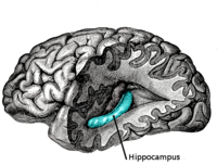
The hippocampus is a major component of the brains of humans and other vertebrates. It belongs to the limbic system and plays important roles in the consolidation of information from short-term memory to long-term memory and spatial navigation. Humans and other mammals have two hippocampi, one in...
, a neural structure in the medial temporal lobe
Temporal lobe
The temporal lobe is a region of the cerebral cortex that is located beneath the Sylvian fissure on both cerebral hemispheres of the mammalian brain....
of the brain
Brain
The brain is the center of the nervous system in all vertebrate and most invertebrate animals—only a few primitive invertebrates such as sponges, jellyfish, sea squirts and starfishes do not have one. It is located in the head, usually close to primary sensory apparatus such as vision, hearing,...
that has a distinctive, curved shape that has been likened to the sea horse
Hippocamp
The hippocamp or hippocampus , often called a sea-horse in English, is a mythological creature shared by Phoenician and Greek mythology, though the name by which it is recognised is purely Greek; it became part of Etruscan mythology...
monster of Greek mythology
Greek mythology
Greek mythology is the body of myths and legends belonging to the ancient Greeks, concerning their gods and heroes, the nature of the world, and the origins and significance of their own cult and ritual practices. They were a part of religion in ancient Greece...
and the ram's horns of Amun
Amun
Amun, reconstructed Egyptian Yamānu , was a god in Egyptian mythology who in the form of Amun-Ra became the focus of the most complex system of theology in Ancient Egypt...
in Egyptian mythology
Egyptian mythology
Ancient Egyptian religion was a complex system of polytheistic beliefs and rituals which were an integral part of ancient Egyptian society. It centered on the Egyptians' interaction with a multitude of deities who were believed to be present in, and in control of, the forces and elements of nature...
. This general layout holds across the full range of mammalian
Mammal
Mammals are members of a class of air-breathing vertebrate animals characterised by the possession of endothermy, hair, three middle ear bones, and mammary glands functional in mothers with young...
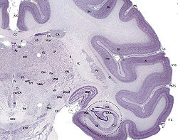
species, from hedgehog to human, although the details vary. For example, in the rat
Rat
Rats are various medium-sized, long-tailed rodents of the superfamily Muroidea. "True rats" are members of the genus Rattus, the most important of which to humans are the black rat, Rattus rattus, and the brown rat, Rattus norvegicus...
, the two hippocampi look similar to a pair of bananas, joined at the stems. In primate
Primate
A primate is a mammal of the order Primates , which contains prosimians and simians. Primates arose from ancestors that lived in the trees of tropical forests; many primate characteristics represent adaptations to life in this challenging three-dimensional environment...
brains, including humans, the portion of the hippocampus near the base of the temporal lobe is much broader than the part at the top. Due to the three-dimensional curvature of this structure, two-dimensional sections such as shown are commonly seen. Neuroimaging
Neuroimaging
Neuroimaging includes the use of various techniques to either directly or indirectly image the structure, function/pharmacology of the brain...
pictures can show a number of different shapes, depending on the angle and location of the cut.
Limbic system
The limbic system is a set of brain structures including the hippocampus, amygdala, anterior thalamic nuclei, septum, limbic cortex and fornix, which seemingly support a variety of functions including emotion, behavior, long term memory, and olfaction. The term "limbic" comes from the Latin...
(Latin limbus =
border), with the hippocampus lining the posterior edge of this hole. These limbic structures include the hippocampus, cingulate cortex
Cingulate cortex
The cingulate cortex is a part of the brain situated in the medial aspect of the cortex. It includes the cortex of the cingulate gyrus, which lies immediately above the corpus callosum, and the continuation of this in the cingulate sulcus...
, olfactory cortex
Olfactory system
The olfactory system is the sensory system used for olfaction, or the sense of smell. Most mammals and reptiles have two distinct parts to their olfactory system: a main olfactory system and an accessory olfactory system. The main olfactory system detects volatile, airborne substances, while the...
, and amygdala
Amygdala
The ' are almond-shaped groups of nuclei located deep within the medial temporal lobes of the brain in complex vertebrates, including humans. Shown in research to perform a primary role in the processing and memory of emotional reactions, the amygdalae are considered part of the limbic system.-...
. Paul MacLean once suggested, as part of his triune brain
Triune brain
The triune brain is a model of the evolution of the vertebrate forebrain and behavior proposed by the American physician and neuroscientist Paul D. MacLean. MacLean originally formulated his model in the 1960s and propounded it at length in his 1990 book The Triune Brain in Evolution...
theory, that the limbic structures comprise the neural basis of emotion
Emotion
Emotion is a complex psychophysiological experience of an individual's state of mind as interacting with biochemical and environmental influences. In humans, emotion fundamentally involves "physiological arousal, expressive behaviors, and conscious experience." Emotion is associated with mood,...
. While most neuroscientists no longer believe in the concept of a unified "limbic system", these regions are highly interconnected and do interact with one another.

Basic Hippocampal Circuit
.png)
series of narrow zones. The first of these, the dentate gyrus
Dentate gyrus
The dentate gyrus is part of the hippocampal formation. It is thought to contribute to new memories as well as other functional roles. It is notable as being one of a select few brain structures currently known to have high rates of neurogenesis in adult rats, .The dentate gyrus cells receive...
(DG), is actually a separate
structure, a tightly packed layer of small granule cells wrapped around the end of the hippocampus
proper, forming a pointed wedge in some cross-sections, a semicircle in others. Next
come a series of Cornu Ammonis areas: first CA4 (which underlies the dentate gyrus), then CA3, then
a very small zone called CA2, then CA1. The CA areas are all filled with densely packed pyramidal cells similar to those found in the neocortex
Neocortex
The neocortex , also called the neopallium and isocortex , is a part of the brain of mammals. It is the outer layer of the cerebral hemispheres, and made up of six layers, labelled I to VI...
. After CA1 comes an area called the subiculum
Subiculum
The subiculum is the most inferior component of the hippocampal formation. It lies between the entorhinal cortex and the CA1 subfield of the hippocampus proper.-Paths:...
. After this comes a pair of ill-defined areas called the presubiculum and parasubiculum, then a
transition to the cortex proper (mostly the entorhinal
Entorhinal cortex
The entorhinal cortex is located in the medial temporal lobe and functions as a hub in a widespread network for memory and navigation. The EC is the main interface between the hippocampus and neocortex...
area of the cortex). Most anatomists
use the term "hippocampus proper" to refer to the four CA fields, and "hippocampal formation"
to refer to the hippocampus proper plus dentate gyrus and subiculum.
The major pathways of signal flow through the hippocampus combine to form a loop. Most external input comes from the adjoining entorhinal cortex, via the axons of the so-called perforant path
Perforant path
In the brain, the perforant pathway provides a connectional route from the entorhinal cortex to all fields of the hippocampal formation, including the dentate gyrus, all CA fields , and the subiculum....
. These axons arise from layer 2 of the entorhinal cortex (EC), and terminate in the dentate gyrus and CA3. There is also a distinct pathway from layer 3 of the EC directly to CA1. Granule cells of the DG send their axons (called "mossy fibers") to CA3. Pyramidal cells of CA3 send their axons to CA1. Pyramidal cells of CA1 send their axons to the subiculum and deep layers of the EC. Subicular neurons send their axons mainly to the EC. The perforant path-to-dentate gyrus-to-CA3-to-CA1 was called the trisynaptic circuit
Trisynaptic loop
The trisynaptic loop is a relay of synaptic transmission in the hippocampus, which is made up of three major cell groups: granule cells, CA3 pyramidal neurons, and CA1 pyramidal cells. The hippocampal relay involves 3 main regions within the hippocampus which are classified according to their...
by Per Andersen, who noted that thin slices could be cut out of the hippocampus perpendicular to its long axis, in a way that preserves all of these connections. This observation was the basis of his lamellar hypothesis, which proposed that the hippocampus can be thought of as a series of parallel strips, operating in a functionally independent way. The lamellar concept is still sometimes considered to be a useful organizing principle, but more recent data, showing extensive longitudinal connections within the hippocampal system, have required it to be substantially modified.
Perforant path input from EC layer II enters the dentate gyrus and is relayed to region CA3 (and to mossy cells, located in the hilus of the dentate gyrus, which then send information to distant portions of the dentate gyrus where the cycle is repeated). Region CA3 combines this input with signals from EC layer II and sends extensive connections within the region and also sends connections to region CA1 through a set of fibers called the Schaffer collateral
Schaffer collateral
Schaffer collaterals are axon collaterals given off by CA3 pyramidal cells in the hippocampus. These collaterals project to area CA1 of the hippocampus and are an integral part of memory formation and the emotional network of the Papez circuit, and of the hippocampal trisynaptic loop...
s. Region CA1 receives input from the CA3 subfield, EC layer III and the nucleus reuniens of the thalamus (which project only to the terminal apical dendritic tufts in the stratum lacunosum-moleculare). In turn, CA1 projects to the subiculum as well as sending information along the aforementioned output paths of the hippocampus. The subiculum is the final stage in the pathway, combining information from the CA1 projection and EC layer III to also send information along the output pathways of the hippocampus.
The hippocampus also receives a number of subcortical inputs. In Macaca fascicularis, these inputs include the amygdala
Amygdala
The ' are almond-shaped groups of nuclei located deep within the medial temporal lobes of the brain in complex vertebrates, including humans. Shown in research to perform a primary role in the processing and memory of emotional reactions, the amygdalae are considered part of the limbic system.-...
(specifically the anterior amygdaloid area, the basolateral nucleus, and the periamygdaloid cortex), the medial septum and the diagonal band of Broca
Diagonal band of Broca
The diagonal band of Broca forms the medial margin of the anterior perforated substance.-Contents:It consists of fibers that are said to arise in the parolfactory area, the gyrus subcallosus and the anterior perforated substance, and course backward in the longitudinal striae to the dentate gyrus...
, the claustrum
Claustrum
The claustrum, which is suspected to be present in all mammals, is a fairly thin vertical curved sheet of subcortical gray matter...
, the substantia innominata
Substantia innominata
The substantia innominata of Meynert is a stratum in the human brain consisting partly of gray and partly of white substance, which lies below the anterior part of the thalamus and lentiform nucleus...
and the basal nucleus of Meynert, the thalamus
Thalamus
The thalamus is a midline paired symmetrical structure within the brains of vertebrates, including humans. It is situated between the cerebral cortex and midbrain, both in terms of location and neurological connections...
(including the anterior nuclear complex, the laterodorsal nucleus, the paraventricular and parataenial nuclei, the nucleus reuniens, and the nucleus centralis medialis), the lateral preoptic and lateral hypothalamic
Hypothalamus
The Hypothalamus is a portion of the brain that contains a number of small nuclei with a variety of functions...
areas, the supramammillary and retromammillary regions, the ventral tegmental area, the tegmental reticular fields, the raphe nuclei
Raphe nuclei
The raphe nuclei are a moderate-size cluster of nuclei found in the brain stem. Their main function is to release serotonin to the rest of the brain...
(the nucleus centralis superior and the dorsal raphe nucleus), the nucleus reticularis tegementi pontis
Reticular formation
The reticular formation is a part of the brain that is involved in actions such as awaking/sleeping cycle, and filtering incoming stimuli to discriminate irrelevant background stimuli...
, the central gray, the dorsal tegmental nucleus, and the locus coeruleus.
The hippocampus also receives direct monosynaptic projections from the cerebellar fastigial nucleus.
Hippocampal Cells and Layers

Hippocampus proper
The hippocampusHippocampus
The hippocampus is a major component of the brains of humans and other vertebrates. It belongs to the limbic system and plays important roles in the consolidation of information from short-term memory to long-term memory and spatial navigation. Humans and other mammals have two hippocampi, one in...
is composed of multiple subfields. Though terminology varies among authors, the terms most frequently used are dentate gyrus
Dentate gyrus
The dentate gyrus is part of the hippocampal formation. It is thought to contribute to new memories as well as other functional roles. It is notable as being one of a select few brain structures currently known to have high rates of neurogenesis in adult rats, .The dentate gyrus cells receive...
and the cornu ammonis (literally "Amun
Amun
Amun, reconstructed Egyptian Yamānu , was a god in Egyptian mythology who in the form of Amun-Ra became the focus of the most complex system of theology in Ancient Egypt...
's horns", abbreviated CA). The dentate gyrus contains the fascia dentata and the hilus, while CA is differentiated into fields CA1, CA2, and CA3.
Cut in cross section
Cross section (geometry)
In geometry, a cross-section is the intersection of a figure in 2-dimensional space with a line, or of a body in 3-dimensional space with a plane, etc...
, the hippocampus is a C-shaped structure that resembles a ram's
Ovis
Ovis is a genus of mammals, part of the goat-antelope subfamily of the ruminant family Bovidae. Its five or more highly gregarious species are known as sheep...
horns
Horn (anatomy)
A horn is a pointed projection of the skin on the head of various animals, consisting of a covering of horn surrounding a core of living bone. True horns are found mainly among the ruminant artiodactyls, in the families Antilocapridae and Bovidae...
. The name cornu ammonis refers to the Egypt
Egypt
Egypt , officially the Arab Republic of Egypt, Arabic: , is a country mainly in North Africa, with the Sinai Peninsula forming a land bridge in Southwest Asia. Egypt is thus a transcontinental country, and a major power in Africa, the Mediterranean Basin, the Middle East and the Muslim world...
ian deity Amun
Amun
Amun, reconstructed Egyptian Yamānu , was a god in Egyptian mythology who in the form of Amun-Ra became the focus of the most complex system of theology in Ancient Egypt...
, who has the head of a ram. The horned appearance of the hippocampus is caused by cell density differentials and the existence of varying degrees of neuron
Neuron
A neuron is an electrically excitable cell that processes and transmits information by electrical and chemical signaling. Chemical signaling occurs via synapses, specialized connections with other cells. Neurons connect to each other to form networks. Neurons are the core components of the nervous...
al fibers.
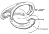
In rodents, the hippocampus is positioned so that, roughly, one end is near the top of the head (the dorsal or septal end) and one end near the bottom of the head (the ventral or temporal end). As shown in the figure, the structure itself is curved and subfields or regions are defined along the curve, from CA4 through CA1 (only CA3 and CA1 are labeled). The CA regions are also structured depthwise in clearly defined strata (or layers):
- The alveus is the deepest layer and contains the axons from pyramidal neurons, passing on toward the fimbria/fornix, one of the major outputs of the hippocampus.
- Stratum oriens (str. oriens) is the next layer superficial to the alveus. The cell bodies of inhibitory basket cells and horizontal trilaminar cells, named for their axons innervating three layers-- the oriens, pyramidal, and radiatum are located in this stratum. The basal dendrites of pyramidal neurons are also found here, where they receive input from other pyramidal cells, septalSeptumIn anatomy, a septum is a wall, dividing a cavity or structure into smaller ones.-In human anatomy:...
fibers and commissural fibers from the contralateral hippocampus (usually recurrent connections, especially in CA3 and CA2.) In rodents the two hippocampi are highly connected, but in primates this commissural connection is much sparser.
- Stratum pyramidale (str. pyr.) contains the cell bodies of the pyramidal neurons, which are the principal excitatory neurons of the hippocampus. This stratum tends to be one of the more visible strata to the naked eye. In region CA3, this stratum contains synapses from the mossy fibers that course through stratum lucidum. This stratum also contains the cell bodies of many interneurons, including axo-axonic cells, bistratified cells, and radial trilaminar cells.
- Stratum lucidum (str. luc.) is one of the thinnest strata in the hippocampus and only found in the CA3 region. Mossy fibers from the dentate gyrus granule cells course through this stratum in CA3, though synapses from these fibers can be found in str. pyr.
- Stratum radiatum (str. rad.), like str. oriens, contains septal and commissural fibers. It also contains Schaffer collateral fibers, which are the projection forward from CA3 to CA1. Some interneurons that can be found in more superficial layers can also be found here, including basket cells, bistratified cells, and radial trilaminar cells.
- Stratum lacunosum (str. lac.) is a thin stratum that too contains Schaffer collateral fibers, but it also contains perforant pathPerforant pathIn the brain, the perforant pathway provides a connectional route from the entorhinal cortex to all fields of the hippocampal formation, including the dentate gyrus, all CA fields , and the subiculum....
fibers from the superficial layers of entorhinal cortex. Due to its small size, it is often grouped together with stratum moleculare into a single stratum called stratum lacunosum-moleculare (str. l-m.).
- Stratum moleculare (str. mol.) is the most superficial stratum in the hippocampus. Here the perforant path fibers form synapses onto the distal, apical dendrites of pyramidal cells.
- The hippocampal sulcusHippocampal sulcusThe hippocampal sulcus, also known as the hippocampal fissure, is a sulcus that separates the dentate gyrus from the subiculum and the CA1 field in the hippocampus.-Development in humans:...
(sulc.) or fissure is a cell-free region that separates the CA1 field from the dentate gyrus. Because the phase of recorded theta rhythmTheta rhythmA theta rhythm is an oscillatory pattern in EEG signals recorded either from inside the brain or from electrodes glued to the scalp. Two types of theta rhythm have been described...
varies systematically through the strata, the fissure is often used as a fixed reference point for recording EEG as it is easily identifiable.
Dentate gyrus
The dentate gyrus is composed of a similar series of strata:- The polymorphic layer (poly. lay.) is the most superficial layer of the dentate gyrus and is often considered a separate subfield (see CA4/hilus below). This layer contains many interneurons, and the axons of the dentate granule cells pass through this stratum on the way to CA3.
- Stratum granulosum (str. gr.) contains the cell bodies of the dentate granule cells.
- Stratum moleculare, inner third (str. mol. 1/3) is where both commissural fibers from the contralateral dentate gyrus run and form synapses as well as where inputs from the medial septum terminate, both on the proximal dendrites of the granule cells.
- Stratum moleculare, external two thirds (str. mol. 2/3) is the deepest of the strata, sitting just superficial to the hippocampal fissure across from stratum moleculare in the CA fields. The perforant path fibers run through this strata, making excitatory synapses onto the distal apical dendrites of granule cells.
Subfields
- Fascia dentataFascia dentataThe fascia dentata is the earliest stage of the hippocampal circuit. Its primary input is the perforant path from the superficial layers of entorhinal cortex. Its principal neurons are tiny granule cells which give rise to unmyelinated axons called the mossy fibers which project to the hilus and...
- Region IV of hippocampus properRegion IV of hippocampus properRegion IV of hippocampus proper is a portion of the hippocampal formation.Region CA4 Region IV of hippocampus proper is a portion of the hippocampal formation.Region CA4 Region IV of hippocampus proper is a portion of the hippocampal formation.Region CA4 (often called the hilus or hilar region when...
- Region III of hippocampus properRegion III of hippocampus properRegion III of hippocampus proper is a portion of the hippocampal formation.Region CA3 receives input along the mossy fibers from granule cells in the dentate gyrus and from projection cells in entorhinal cortex along the perforant path...
- Region II of hippocampus properRegion II of hippocampus properRegion II of hippocampus proper is a portion of the hippocampal formation.Region CA2 is a small region located between CA3 and CA1. Anatomically, in the rat, it is best defined as the strip of cells that receive perforant path input from EC layer II but do not receive mossy fiber connections from DG...
- Region I of hippocampus properRegion I of hippocampus properRegion I of hippocampus proper is a portion of the hippocampal formation.Region CA1 is the first region in the hippocampal circuit that yields a significant output pathway, which goes to entorhinal cortex layer V. It also sends significant output forward to the subiculum...
- SubiculumSubiculumThe subiculum is the most inferior component of the hippocampal formation. It lies between the entorhinal cortex and the CA1 subfield of the hippocampus proper.-Paths:...