
Arrhythmogenic right ventricular dysplasia
Encyclopedia
Arrhythmogenic right ventricular dysplasia (ARVD), also called arrhythmogenic right ventricular cardiomyopathy (ARVC) or arrhythmogenic right ventricular dysplasia/cardiomyopathy (ARVD/C), is an inherited heart disease
.
ARVD is caused by genetic defects
of the parts of heart muscle (also called myocardium or cardiac muscle) known as desmosome
s, areas on the surface of heart muscle cells which link the cells together. The desmosomes are composed of several protein
s, and many of those proteins can have harmful mutation
s.
The disease is a type of nonischemic cardiomyopathy
that involves primarily the right ventricle
. It is characterized by hypokinetic
areas involving the free wall of the right ventricle, with fibrofatty replacement of the right ventricular myocardium, with associated arrhythmias originating in the right ventricle.
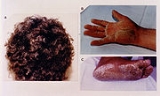
ARVD is often found in association with diffuse palmoplantar keratoderma, and woolly hair, because their genes are nearby and often inherited together
ARVC/D is an important cause of ventricular arrhythmias in children and young adults. It is seen predominantly in males, and 30-50% of cases have a familial distribution.
) are the main causatives for the development of this disease. Recently it could be shown, that mutations in the desmin
-gene could cause ARVC. Desmin
is a intermediate filament protein, which is linked to the desmosomes. The penetrance
is 20-35% in general, but significantly higher in Italy. Seven gene loci have been implicated in ARVD. However, about 50% of families that express ARVD that undergo genetic screening do not show linkage with any of the known chromosomal
loci
. It is unclear whether the pathogenesis varies with the different loci involved. Standard genetic screening test are currently tested and evaluated in different state of the art cardiovascular research centres and hospitals.
Types include:
, although some studies have suggested that it may be as common as 1/1,000. Recently, 1/200 were found to be carriers of mutations that predispose to ARVC It accounts for up to 17% of all sudden cardiac deaths in the young. In Italy
, the incidence is 40/10,000, making it the most common cause of sudden cardiac death in the young population.
Symptoms are usually exercise-related. In populations where hypertrophic cardiomyopathy
is screened out prior to involvement in competitive athletics, it is a common cause of sudden cardiac death.
The first clinical signs of ARVD are usually during adolescence
. However, signs of ARVD have been demonstrated in infants.
(programmed cell death) appears to play a large role. It is unclear why only the right ventricle is involved. The disease process starts in the subepicardial region and works its way towards the endocardial surface, leading to transmural involvement (possibly accounting for the aneurysmal dilatation of the RV). Residual myocardium is confined to the subendocardial region and the trabeculae of the RV. These trabeculae may become hypertrophied.
Aneurysmal dilatation is seen in 50% of cases at autopsy. It usually occurs in the diaphragmatic, apical, and infundibular regions (known as the triangle of dysplasia). The left ventricle is involved in 50-67% of individuals. If the left ventricle is involved, it is usually late in the course of disease, and confers a poor prognosis.
There are two pathological patterns seen in ARVD, Fatty infiltration and fibro-fatty infiltration.
(myocardial cell) degeneration and death seen in 50% of cases of fatty infiltration.
s) seen on microscopy. Myocardial atrophy is due to injury and apoptosis
. This leads to thinning of the RV free wall (to < 3 mm thickness) Myocytes are replaced with fibrofatty tissue. The regions preferentially involved include the RV inflow tract, the RV outflow tract, and the RV apex. However, the LV free wall may be involved in some cases. Involvement of the ventricular septum is rare. The areas involved are prone to aneurysm formation.
Ventricular arrhythmias due to ARVD typically arise from the diseased right ventricle. The type of arrhythmia ranges from frequent premature ventricular complexes
(PVCs) to ventricular tachycardia
(VT) to ventricular fibrillation
(VF).
While the initiating factor of the ventricular arrhythmias is unclear, it may be due to triggered activity or reentry.
Ventricular arrhythmias are usually exercise-related, suggesting that they are sensitive to catecholamines. The ventricular beats typically have a right axis deviation. Multiple morphologies of ventricular tachycardia may be present in the same individual, suggesting multiple arrhythmogenic foci or pathways.
Right ventricular outflow tract (RVOT) tachycardia is the most common VT seen in individuals with ARVD. In this case, the EKG shows a left bundle branch block
(LBBB) morphology with an inferior axis.
(EKG), echocardiography
, right ventricular angiography, cardiac MRI, and genetic testing
.
(RBBB), women, and children under 12 years old.
RBBB itself is seen frequently in individuals with ARVD. This may be due to delayed activation of the right ventricle, rather than any intrinsic abnormality in the right bundle branch.
The epsilon wave is found in about 50% of those with ARVD. This is described as a terminal notch in the QRS complex. It is due to slowed intraventricular conduction. The epsilon wave may be seen on a surface EKG; however, it is more commonly seen on signal averaged EKGs.
Ventricular ectopy
seen on a surface EKG in the setting of ARVD is typically of left bundle branch block
(LBBB) morphology, with a QRS axis of -90 to +110 degrees. The origin of the ectopic beats is usually from one of the three regions of fatty degeneration (the "triangle of dysplasia"): the RV outflow tract, the RV inflow tract, and the RV apex.
) is used to detect late potentials and epsilon waves in individuals with ARVD.
Cardiac MRI can visualize the extreme thinning and akinesis of the RV free wall. However, the normal RV free wall may be about 3 mm thick, making the test less sensitive.
for the diagnosis of ARVD. Findings consistent with ARVD are an akinetic or dyskinetic bulging localized to the infundibular, apical, and subtricuspid regions of the RV. The specificity is 90%; however, the test is observer dependent.
False negatives are common, however, because the disease progresses typically from the epicardium to the endocardium (with the biopsy sample coming from the endocardium), and the segmental nature of the disease. Also, due to the paper-thin right ventricular free wall that is common in this disease process, most biopsy samples are taken from the ventricular septum, which is commonly not involved in the disease process.
A biopsy sample that is consistent with ARVD would have > 3% fat, >40% fibrous tissue, and <45% myocytes.
. Approximately 40-50% of ARVD patients have a mutation identified in one of several genes encoding components of the desmosome
, which can help confirm a diagnosis of ARVD. Since ARVD is an autosomal dominant trait, children of an ARVD patient have a 50% chance of inheriting the disease causing mutation. Whenever a mutation is identified by genetic testing, family-specific genetic testing can be used to differentiate between relatives who are at-risk for the disease and those who are not. ARVD genetic testing is clinically available.
Major Criteria
Minor Criteria
Many individuals have symptoms associated with ventricular tachycardia, such as palpitations, light-headedness, or syncope. Others may have symptoms and signs related to right ventricular failure, such as lower extremity edema, liver congestion with elevated hepatic enzymes. Unfortunately, sudden death may be the first manifestation of disease.
ARVD is a progressive disease. Over time, the right ventricle becomes more involved, leading to right ventricular failure. The right ventricle will fail before there is left ventricular dysfunction. However, by the time the individual has signs of overt right ventricular failure, there will be histological involvement of the left ventricle. Eventually, the left ventricle will also become involved, leading to bi-ventricular failure. Signs and symptoms of left ventricular failure may become evident, including congestive heart failure, atrial fibrillation, and an increased incidence of thromboembolic events.
A certain subgroup of individuals with ARVD are considered at high risk for sudden cardiac death. Characteristics associated with high risk of sudden cardiac death include:
Management options include pharmacological, surgical, catheter ablation, and placement of an implantable cardioverter-defibrillator
.
Prior to the decision of the treatment option, programmed electrical stimulation in the electrophysiology
laboratory may be performed for additional prognostic information. Goals of programmed stimulation include:
Regardless of the management option chosen, the individual is typically suggested to undergo lifestyle modification, including avoidance of strenuous exercise, cardiac stimulants (i.e.: caffeine, nicotine, pseudoephedrine) and alcohol. If the individual wishes to begin an exercise regimen, an exercise stress test may have added benefit.
Sotalol
, a beta blocker
and a class III antiarrhythmic agent
, is the most effective antiarrhythmic agent in ARVD. Other antiarrhythmic agents used include amiodarone
and conventional beta blockers (i.e.: metoprolol). If antiarrhythmic agents are used, their efficacy should be guided by series ambulatory holter monitoring, to show a reduction in arrhythmic events.
While angiotensin converting enzyme inhibitors (ACE Inhibitors) are well known for slowing progression in other cardiomyopathies, they have not been proven to be helpful in ARVD.
Individuals with decreased RV ejection fraction with dyskinetic portions of the right ventricle may benefit from long term anticoagulation with warfarin
to prevent thrombus formation and subsequent pulmonary embolism
.
placement, causing frequent discharges of the ICD.
is the most effective prevention against sudden cardiac death. Due to the prohibitive cost of ICDs, they are not routinely placed in all individuals with ARVD.
Indications for ICD placement in the setting of ARVD include:
Since ICDs are typically placed via a transvenous approach into the right ventricle, there are complications associated with ICD placement and follow-up.
Due to the extreme thinning of the RV free wall, it is possible to perforate the RV during implantation, potentially causing pericardial tamponade
. Because of this, every attempt is made at placing the defibrillator lead on the ventricular septum.
After a successful implantation, the progressive nature of the disease may lead to fibro-fatty replacement of the myocardium at the site of lead placement. This may lead to undersensing of the individual's electrical activity (potentially causing inability to sense VT or VF), and inability to pace the ventricle.
and Spanish international left wing-back Antonio Puerta
died from the condition, at the age of 22, on 28 August 2007, three days after suffering several cardiac arrests, while disputing a La Liga
game against Getafe CF
. Englishman Matt Gadsby
also died from the condition after collapsing on the pitch on 9 September 2006, while playing with his team Hinckley United in a Conference North
game against Harrogate Town.
Model Krissy Taylor
, sister of Niki Taylor
, died from the disease at age 17 in 1995.
Heart disease
Heart disease, cardiac disease or cardiopathy is an umbrella term for a variety of diseases affecting the heart. , it is the leading cause of death in the United States, England, Canada and Wales, accounting for 25.4% of the total deaths in the United States.-Types:-Coronary heart disease:Coronary...
.
ARVD is caused by genetic defects
Genetic disorder
A genetic disorder is an illness caused by abnormalities in genes or chromosomes, especially a condition that is present from before birth. Most genetic disorders are quite rare and affect one person in every several thousands or millions....
of the parts of heart muscle (also called myocardium or cardiac muscle) known as desmosome
Desmosome
A desmosome , also known as macula adherens , is a cell structure specialized for cell-to-cell adhesion...
s, areas on the surface of heart muscle cells which link the cells together. The desmosomes are composed of several protein
Protein
Proteins are biochemical compounds consisting of one or more polypeptides typically folded into a globular or fibrous form, facilitating a biological function. A polypeptide is a single linear polymer chain of amino acids bonded together by peptide bonds between the carboxyl and amino groups of...
s, and many of those proteins can have harmful mutation
Mutation
In molecular biology and genetics, mutations are changes in a genomic sequence: the DNA sequence of a cell's genome or the DNA or RNA sequence of a virus. They can be defined as sudden and spontaneous changes in the cell. Mutations are caused by radiation, viruses, transposons and mutagenic...
s.
The disease is a type of nonischemic cardiomyopathy
Cardiomyopathy
Cardiomyopathy, which literally means "heart muscle disease," is the deterioration of the function of the myocardium for any reason. People with cardiomyopathy are often at risk of arrhythmia or sudden cardiac death or both. Cardiomyopathy can often go undetected, making it especially dangerous to...
that involves primarily the right ventricle
Right ventricle
The right ventricle is one of four chambers in the human heart. It receives deoxygenated blood from the right atrium via the tricuspid valve, and pumps it into the pulmonary artery via the pulmonary valve and pulmonary trunk....
. It is characterized by hypokinetic
Hypokinesia
Hypokinesia refers to decreased bodily movement. It is associated with basal ganglia diseases , mental health disorders and prolonged inactivity due to illness, amongst other diseases.Hypokinesia describes a spectrum of disorders:...
areas involving the free wall of the right ventricle, with fibrofatty replacement of the right ventricular myocardium, with associated arrhythmias originating in the right ventricle.
ARVD is often found in association with diffuse palmoplantar keratoderma, and woolly hair, because their genes are nearby and often inherited together
ARVC/D is an important cause of ventricular arrhythmias in children and young adults. It is seen predominantly in males, and 30-50% of cases have a familial distribution.
Genetics
It is usually inherited in an autosomal dominant pattern, with variable expression. Novel studies showed that mutations (point mutations) in genes encoding for desmosomal proteins (see Intercalated DiscIntercalated disc
When observing cardiac tissue through a microscope, intercalated discs are an identifying feature of cardiac muscle. Cardiac muscle consists of single heart muscle cells which have to be connected by intercalated discs to work as a functional organ. By contrast, skeletal muscle consists of...
) are the main causatives for the development of this disease. Recently it could be shown, that mutations in the desmin
Desmin
Desmin is a protein that in humans is encoded by the DES gene.Desmin is a type III intermediate filament found near the Z line in sarcomeres. It was first described in 1976, first purified in 1977, the gene was cloned in 1989, and the first knock-out mouse was created in 1996. Desmin is only...
-gene could cause ARVC. Desmin
Desmin
Desmin is a protein that in humans is encoded by the DES gene.Desmin is a type III intermediate filament found near the Z line in sarcomeres. It was first described in 1976, first purified in 1977, the gene was cloned in 1989, and the first knock-out mouse was created in 1996. Desmin is only...
is a intermediate filament protein, which is linked to the desmosomes. The penetrance
Penetrance
Penetrance in genetics is the proportion of individuals carrying a particular variant of a gene that also express an associated trait . In medical genetics, the penetrance of a disease-causing mutation is the proportion of individuals with the mutation who exhibit clinical symptoms...
is 20-35% in general, but significantly higher in Italy. Seven gene loci have been implicated in ARVD. However, about 50% of families that express ARVD that undergo genetic screening do not show linkage with any of the known chromosomal
Chromosome
A chromosome is an organized structure of DNA and protein found in cells. It is a single piece of coiled DNA containing many genes, regulatory elements and other nucleotide sequences. Chromosomes also contain DNA-bound proteins, which serve to package the DNA and control its functions.Chromosomes...
loci
Allele
An allele is one of two or more forms of a gene or a genetic locus . "Allel" is an abbreviation of allelomorph. Sometimes, different alleles can result in different observable phenotypic traits, such as different pigmentation...
. It is unclear whether the pathogenesis varies with the different loci involved. Standard genetic screening test are currently tested and evaluated in different state of the art cardiovascular research centres and hospitals.
Types include:
| Type | OMIM | Gene | Locus |
|---|---|---|---|
| ARVD1 | TGFB3 | 14q23-q24 | |
| ARVD2 | RYR2 | 1q42-q43 | |
| ARVD3 | ? | 14q12-q22 | |
| ARVD4 | ? | 2q32.1-q32.3 | |
| ARVD5 | TMEM43 TMEM43 Transmembrane protein 43 is a protein that in humans is encoded by the TMEM43 gene.. TMEM43 may have an important role in maintaining nuclear envelope structure by organizing protein complexes at the inner nuclear membrane. Required for retaining emerin at the inner nuclear membrane. Mutations in... |
3p23 | |
| ARVD6 | ? | 10p14-p12 | |
| ARVD7 | ? | 10q22.3 | |
| ARVD8 | DSP Desmoplakin Desmoplakin is a protein associated with desmosomes.Desmoplakin is a protein that in humans is encoded by the DSP gene. The C-terminus of desmoplakin binds with intermediate filaments. These are further sub divided to three homologous Plakin repeat domains . In the mid-region of desmoplakin, a... |
6p24 | |
| ARVD9 | PKP2 PKP2 Plakophilin-2 is a protein that in humans is encoded by the PKP2 gene.-Interactions:PKP2 has been shown to interact with Desmoplakin, Plakoglobin and Desmoglein 1.-External links:* *... |
12p11 | |
| ARVD10 | DSG2 | 18q12.1-q12 | |
| ARVD11 | DSC2 DSC2 Desmocollin-2 is a protein that in humans is encoded by the DSC2 gene.-External links:* *... |
18q12.1 | |
| ARVD12 | JUP | 17q21 |
Incidence
The incidence of ARVD is about 1/10,000 in the general population in the United StatesUnited States
The United States of America is a federal constitutional republic comprising fifty states and a federal district...
, although some studies have suggested that it may be as common as 1/1,000. Recently, 1/200 were found to be carriers of mutations that predispose to ARVC It accounts for up to 17% of all sudden cardiac deaths in the young. In Italy
Italy
Italy , officially the Italian Republic languages]] under the European Charter for Regional or Minority Languages. In each of these, Italy's official name is as follows:;;;;;;;;), is a unitary parliamentary republic in South-Central Europe. To the north it borders France, Switzerland, Austria and...
, the incidence is 40/10,000, making it the most common cause of sudden cardiac death in the young population.
Presentation
Up to 80% of individuals with ARVD present with syncope or sudden cardiac death. The remainder frequently present with palpitations or other symptoms due to right ventricular outflow tract (RVOT) tachycardia (a type of monomorphic ventricular tachycardia).Symptoms are usually exercise-related. In populations where hypertrophic cardiomyopathy
Hypertrophic cardiomyopathy
Hypertrophic cardiomyopathy is a disease of the myocardium in which a portion of the myocardium is hypertrophied without any obvious cause...
is screened out prior to involvement in competitive athletics, it is a common cause of sudden cardiac death.
The first clinical signs of ARVD are usually during adolescence
Adolescence
Adolescence is a transitional stage of physical and mental human development generally occurring between puberty and legal adulthood , but largely characterized as beginning and ending with the teenage stage...
. However, signs of ARVD have been demonstrated in infants.
Pathogenesis
The pathogenesis of ARVD is largely unknown. ApoptosisApoptosis
Apoptosis is the process of programmed cell death that may occur in multicellular organisms. Biochemical events lead to characteristic cell changes and death. These changes include blebbing, cell shrinkage, nuclear fragmentation, chromatin condensation, and chromosomal DNA fragmentation...
(programmed cell death) appears to play a large role. It is unclear why only the right ventricle is involved. The disease process starts in the subepicardial region and works its way towards the endocardial surface, leading to transmural involvement (possibly accounting for the aneurysmal dilatation of the RV). Residual myocardium is confined to the subendocardial region and the trabeculae of the RV. These trabeculae may become hypertrophied.
Aneurysmal dilatation is seen in 50% of cases at autopsy. It usually occurs in the diaphragmatic, apical, and infundibular regions (known as the triangle of dysplasia). The left ventricle is involved in 50-67% of individuals. If the left ventricle is involved, it is usually late in the course of disease, and confers a poor prognosis.
There are two pathological patterns seen in ARVD, Fatty infiltration and fibro-fatty infiltration.
Fatty infiltration
The first, fatty infiltration, is confined to the right ventricle. This involves a partial or near-complete substitution of myocardium with fatty tissue without wall thinning. It involves predominantly the apical and infundibular regions of the RV. The left ventricle and ventricular septum are usually spared. No inflammatory infiltrates are seen in fatty infiltration. There is evidence of myocyteMyocyte
A myocyte is the type of cell found in muscles. They arise from myoblasts.Each myocyte contains myofibrils, which are long, long chains of sarcomeres, the contractile units of the cell....
(myocardial cell) degeneration and death seen in 50% of cases of fatty infiltration.
Fibro-fatty infiltration
The second, fibro-fatty infiltration, involves replacement of myocytes with fibrofatty tissue. A patchy myocarditis is involved in up to 2/3 of cases, with inflammatory infiltrates (mostly T cellT cell
T cells or T lymphocytes belong to a group of white blood cells known as lymphocytes, and play a central role in cell-mediated immunity. They can be distinguished from other lymphocytes, such as B cells and natural killer cells , by the presence of a T cell receptor on the cell surface. They are...
s) seen on microscopy. Myocardial atrophy is due to injury and apoptosis
Apoptosis
Apoptosis is the process of programmed cell death that may occur in multicellular organisms. Biochemical events lead to characteristic cell changes and death. These changes include blebbing, cell shrinkage, nuclear fragmentation, chromatin condensation, and chromosomal DNA fragmentation...
. This leads to thinning of the RV free wall (to < 3 mm thickness) Myocytes are replaced with fibrofatty tissue. The regions preferentially involved include the RV inflow tract, the RV outflow tract, and the RV apex. However, the LV free wall may be involved in some cases. Involvement of the ventricular septum is rare. The areas involved are prone to aneurysm formation.
Ventricular arrhythmias
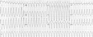 |
| Monomorphic ventricular tachycardia originating from the right ventricular outflow tract. |
Ventricular arrhythmias due to ARVD typically arise from the diseased right ventricle. The type of arrhythmia ranges from frequent premature ventricular complexes
Premature ventricular contraction
A premature ventricular contraction , also known as a premature ventricular complex, ventricular premature contraction , ventricular premature beat , or extrasystole, is a relatively common event where the heartbeat is initiated by the heart ventricles rather than by the sinoatrial node, the...
(PVCs) to ventricular tachycardia
Ventricular tachycardia
Ventricular tachycardia is a tachycardia, or fast heart rhythm, that originates in one of the ventricles of the heart...
(VT) to ventricular fibrillation
Ventricular fibrillation
Ventricular fibrillation is a condition in which there is uncoordinated contraction of the cardiac muscle of the ventricles in the heart, making them quiver rather than contract properly. Ventricular fibrillation is a medical emergency and most commonly identified arrythmia in cardiac arrest...
(VF).
While the initiating factor of the ventricular arrhythmias is unclear, it may be due to triggered activity or reentry.
Ventricular arrhythmias are usually exercise-related, suggesting that they are sensitive to catecholamines. The ventricular beats typically have a right axis deviation. Multiple morphologies of ventricular tachycardia may be present in the same individual, suggesting multiple arrhythmogenic foci or pathways.
Right ventricular outflow tract (RVOT) tachycardia is the most common VT seen in individuals with ARVD. In this case, the EKG shows a left bundle branch block
Left bundle branch block
Left bundle branch block is a cardiac conduction abnormality seen on the electrocardiogram . In this condition, activation of the left ventricle is delayed, which results in the left ventricle contracting later than the right ventricle....
(LBBB) morphology with an inferior axis.
Diagnosis
The differential diagnosis for the ventricular tachycardia due to ARVD include:- Congenital heart disease
- Repaired tetralogy of FallotTetralogy of FallotTetralogy of Fallot is a congenital heart defect which is classically understood to involve four anatomical abnormalities...
- Ebstein's anomalyEbstein's anomalyEbstein anomaly is a congenital heart defect in which the opening of the tricuspid valve is displaced towards the apex of the right ventricle of the heart.-Presentation:...
- Uhl's anomaly
- Atrial septal defectAtrial septal defectAtrial septal defect is a form of congenital heart defect that enables blood flow between the left and right atria via the interatrial septum. The interatrial septum is the tissue that divides the right and left atria...
- Partial anomalous venous return
- Repaired tetralogy of Fallot
- Acquired heart disease
- Tricuspid valve disease
- Pulmonary hypertension
- Right ventricular infarction
- Bundle-branch re-entrant tachycardia
- Miscellaneous
- Pre-excited AV re-entry tachycardia
- Idiopathic RVOT tachycardia
- Sarcoidosis
Clinical testing
In order to make the diagnosis of ARVD, a number of clinical tests are employed, including the electrocardiogramElectrocardiogram
Electrocardiography is a transthoracic interpretation of the electrical activity of the heart over a period of time, as detected by electrodes attached to the outer surface of the skin and recorded by a device external to the body...
(EKG), echocardiography
Echocardiography
An echocardiogram, often referred to in the medical community as a cardiac ECHO or simply an ECHO, is a sonogram of the heart . Also known as a cardiac ultrasound, it uses standard ultrasound techniques to image two-dimensional slices of the heart...
, right ventricular angiography, cardiac MRI, and genetic testing
Genetic testing
Genetic testing is among the newest and most sophisticated of techniques used to test for genetic disorders which involves direct examination of the DNA molecule itself. Other genetic tests include biochemical tests for such gene products as enzymes and other proteins and for microscopic...
.
Electrocardiogram
90% of individuals with ARVD have some EKG abnormality. The most common EKG abnormality seen in ARVD is T wave inversion in leads V1 to V3. However, this is a non-specific finding, and may be considered a normal variant in right bundle branch blockRight bundle branch block
A right bundle branch block is a defect in the heart's electrical conduction system.During a right bundle branch block, the right ventricle is not directly activated by impulses travelling through the right bundle branch. The left ventricle however, is still normally activated by the left bundle...
(RBBB), women, and children under 12 years old.
RBBB itself is seen frequently in individuals with ARVD. This may be due to delayed activation of the right ventricle, rather than any intrinsic abnormality in the right bundle branch.
 |
|
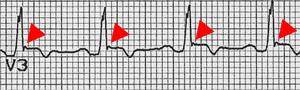
| The epsilon wave (marked by red triangle), seen in ARVD. |
The epsilon wave is found in about 50% of those with ARVD. This is described as a terminal notch in the QRS complex. It is due to slowed intraventricular conduction. The epsilon wave may be seen on a surface EKG; however, it is more commonly seen on signal averaged EKGs.
Ventricular ectopy
Cardiac ectopy
Ectopic beat is a disturbance of the cardiac rhythm frequently related to the electrical conduction system of the heart, in which beats arise from fiber or group of fibers outside the region in the heart muscle ordinarily responsible for impulse formation, i.e., the Sinus node...
seen on a surface EKG in the setting of ARVD is typically of left bundle branch block
Left bundle branch block
Left bundle branch block is a cardiac conduction abnormality seen on the electrocardiogram . In this condition, activation of the left ventricle is delayed, which results in the left ventricle contracting later than the right ventricle....
(LBBB) morphology, with a QRS axis of -90 to +110 degrees. The origin of the ectopic beats is usually from one of the three regions of fatty degeneration (the "triangle of dysplasia"): the RV outflow tract, the RV inflow tract, and the RV apex.
Signal averaged ECG
Signal averaged ECG (SAECGSignal-averaged electrocardiogram
Signal-averaged electrocardiography is a special electrocardiographic technique, in which multiple electric signals from the heart are averaged to remove interference and reveal small variations in the QRS complex, usually the so-called "late potentials"...
) is used to detect late potentials and epsilon waves in individuals with ARVD.
Echocardiography
Echocardiography may reveal an enlarged, hypokinetic right ventricle with a paper-thin RV free wall. The dilatation of the RV will cause dilatation of the tricuspid valve annulus, with subsequent tricuspid regurgitation. Paradoxical septal motion may also be present.Cardiac MRI
Fatty infiltration of the RV free wall can be visible on cardiac MRI. Fat has increased intensity in T1-weighted images. However, it may be difficult to differentiate intramyocardial fat and the epicardial fat that is commonly seen adjacent to the normal heart. Also, the sub-tricuspid region may be difficult to distinguish from the atrioventricular sulcus, which is rich in fat.Cardiac MRI can visualize the extreme thinning and akinesis of the RV free wall. However, the normal RV free wall may be about 3 mm thick, making the test less sensitive.
Right ventricular angiography
Right ventricular angiography is considered the gold standardGold standard (test)
In medicine and statistics, gold standard test refers to a diagnostic test or benchmark that is the best available under reasonable conditions. It does not have to be necessarily the best possible test for the condition in absolute terms...
for the diagnosis of ARVD. Findings consistent with ARVD are an akinetic or dyskinetic bulging localized to the infundibular, apical, and subtricuspid regions of the RV. The specificity is 90%; however, the test is observer dependent.
Right ventricular biopsy
Transvenous biopsy of the right ventricle can be highly specific for ARVD, but it has low sensitivity. False positives include other conditions with fatty infiltration of the ventricle, such as chronic alcohol abuse and Duchenne/Becker muscular dystrophy.False negatives are common, however, because the disease progresses typically from the epicardium to the endocardium (with the biopsy sample coming from the endocardium), and the segmental nature of the disease. Also, due to the paper-thin right ventricular free wall that is common in this disease process, most biopsy samples are taken from the ventricular septum, which is commonly not involved in the disease process.
A biopsy sample that is consistent with ARVD would have > 3% fat, >40% fibrous tissue, and <45% myocytes.
Autopsy
A post mortem histological demonstration of full thickness substitution of the RV myocardium by fatty or fibro-fatty tissue is consistent with ARVD.Genetic Testing
ARVD is an autosomal dominant trait with reduced penetrancePenetrance
Penetrance in genetics is the proportion of individuals carrying a particular variant of a gene that also express an associated trait . In medical genetics, the penetrance of a disease-causing mutation is the proportion of individuals with the mutation who exhibit clinical symptoms...
. Approximately 40-50% of ARVD patients have a mutation identified in one of several genes encoding components of the desmosome
Desmosome
A desmosome , also known as macula adherens , is a cell structure specialized for cell-to-cell adhesion...
, which can help confirm a diagnosis of ARVD. Since ARVD is an autosomal dominant trait, children of an ARVD patient have a 50% chance of inheriting the disease causing mutation. Whenever a mutation is identified by genetic testing, family-specific genetic testing can be used to differentiate between relatives who are at-risk for the disease and those who are not. ARVD genetic testing is clinically available.
Diagnostic Criteria
There is no pathognomonic feature of ARVD. The diagnosis of ARVD is based on a combination of major and minor criteria. To make a diagnosis of ARVD requires either 2 major criteria or 1 major and 2 minor criteria or 4 minor criteria.Major Criteria
- Right ventricular dysfunction
- Severe dilatation and reduction of RV ejection fractionEjection fractionIn cardiovascular physiology, ejection fraction is the fraction of Blood pumped out of the Right Ventricle of the heart to the Pulmonary Circulation and Left Ventricle of the heart to the Systemic Circulation with each Heart beat or Cardiac cycle...
with little or no LV impairment - Localized RV aneurysms
- Severe segmental dilatation of the RV
- Severe dilatation and reduction of RV ejection fraction
- Tissue characterization
- Fibrofatty replacement of myocardium on endomyocardial biopsy
- Conduction abnormalities
- Epsilon waves in V1 - V3.
- Localized prolongation (>110 ms) of QRS in V1 - V3
- Family history
- Familial disease confirmed on autopsy or surgery
Minor Criteria
- Right ventricular dysfunction
- Mild global RV dilatation and/or reduced ejection fraction with normal LV.
- Mild segmental dilatation of the RV
- Regional RV hypokinesis
- Tissue characterization
- Conduction abnormalities
- Inverted T waves in V2 and V3 in an individual over 12 years old, in the absence of a right bundle branch blockRight bundle branch blockA right bundle branch block is a defect in the heart's electrical conduction system.During a right bundle branch block, the right ventricle is not directly activated by impulses travelling through the right bundle branch. The left ventricle however, is still normally activated by the left bundle...
(RBBB) - Late potentials on signal averaged EKG.
- Ventricular tachycardia with a left bundle branch blockLeft bundle branch blockLeft bundle branch block is a cardiac conduction abnormality seen on the electrocardiogram . In this condition, activation of the left ventricle is delayed, which results in the left ventricle contracting later than the right ventricle....
(LBBB) morphology - Frequent PVCs (> 1000 PVCs / 24 hours)
- Inverted T waves in V2 and V3 in an individual over 12 years old, in the absence of a right bundle branch block
- Family history
- Family history of sudden cardiac death before age 35
- Family history of ARVD
Natural history
There is a long asymptomatic lead-time in individuals with ARVD. While this is a genetically transmitted disease, individuals in their teens may not have any characteristics of ARVD on screening tests.Many individuals have symptoms associated with ventricular tachycardia, such as palpitations, light-headedness, or syncope. Others may have symptoms and signs related to right ventricular failure, such as lower extremity edema, liver congestion with elevated hepatic enzymes. Unfortunately, sudden death may be the first manifestation of disease.
ARVD is a progressive disease. Over time, the right ventricle becomes more involved, leading to right ventricular failure. The right ventricle will fail before there is left ventricular dysfunction. However, by the time the individual has signs of overt right ventricular failure, there will be histological involvement of the left ventricle. Eventually, the left ventricle will also become involved, leading to bi-ventricular failure. Signs and symptoms of left ventricular failure may become evident, including congestive heart failure, atrial fibrillation, and an increased incidence of thromboembolic events.
Management
The goal of management of ARVD is to decrease the incidence of sudden cardiac death. This raises a clinical dilemma: How to prophylactically treat the asymptomatic patient who was diagnosed during family screening.A certain subgroup of individuals with ARVD are considered at high risk for sudden cardiac death. Characteristics associated with high risk of sudden cardiac death include:
- Young age
- Competitive sports activity
- Malignant familial history
- Extensive RV disease with decreased right ventricular ejection fraction.
- Left ventricular involvement
- Syncope
- Episode of ventricular arrhythmia
Management options include pharmacological, surgical, catheter ablation, and placement of an implantable cardioverter-defibrillator
Implantable cardioverter-defibrillator
An implantable cardioverter-defibrillator is a small battery-powered electrical impulse generator which is implanted in patients who are at risk of sudden cardiac death due to ventricular fibrillation and ventricular tachycardia. The device is programmed to detect cardiac arrhythmia and correct it...
.
Prior to the decision of the treatment option, programmed electrical stimulation in the electrophysiology
Cardiac electrophysiology
Cardiac electrophysiology is the science of elucidating, diagnosing, and treating the electrical activities of the heart. The term is usually used to describe studies of such phenomena by invasive catheter recording of spontaneous activity as well as of cardiac responses to programmed electrical...
laboratory may be performed for additional prognostic information. Goals of programmed stimulation include:
- Assessment of the disease's arrhythmogenic potential
- Evaluate the hemodynamic consequences of sustained VT
- Determine whether the VT can be interrupted via antitachycardia pacing.
Regardless of the management option chosen, the individual is typically suggested to undergo lifestyle modification, including avoidance of strenuous exercise, cardiac stimulants (i.e.: caffeine, nicotine, pseudoephedrine) and alcohol. If the individual wishes to begin an exercise regimen, an exercise stress test may have added benefit.
Pharmacologic management
Pharmacologic management of ARVD involves arrhythmia suppression and prevention of thrombus formation.Sotalol
Sotalol
Sotalol is a drug used in individuals with rhythm disturbances of the heart, and to treat hypertension in some individuals. It is a non-selective competitive β-adrenergic receptor blocker that also exhibits Class III antiarrhythmic properties by its inhibition of potassium channels...
, a beta blocker
Beta blocker
Beta blockers or beta-adrenergic blocking agents, beta-adrenergic antagonists, beta-adrenoreceptor antagonists or beta antagonists, are a class of drugs used for various indications. They are particularly for the management of cardiac arrhythmias, cardioprotection after myocardial infarction ,...
and a class III antiarrhythmic agent
Antiarrhythmic agent
Antiarrhythmic agents are a group of pharmaceuticals that are used to suppress abnormal rhythms of the heart , such as atrial fibrillation, atrial flutter, ventricular tachycardia, and ventricular fibrillation....
, is the most effective antiarrhythmic agent in ARVD. Other antiarrhythmic agents used include amiodarone
Amiodarone
Amiodarone is an antiarrhythmic agent used for various types of tachyarrhythmias , both ventricular and supraventricular arrhythmias. Discovered in 1961, it was not approved for use in the United States until 1985...
and conventional beta blockers (i.e.: metoprolol). If antiarrhythmic agents are used, their efficacy should be guided by series ambulatory holter monitoring, to show a reduction in arrhythmic events.
While angiotensin converting enzyme inhibitors (ACE Inhibitors) are well known for slowing progression in other cardiomyopathies, they have not been proven to be helpful in ARVD.
Individuals with decreased RV ejection fraction with dyskinetic portions of the right ventricle may benefit from long term anticoagulation with warfarin
Warfarin
Warfarin is an anticoagulant. It is most likely to be the drug popularly referred to as a "blood thinner," yet this is a misnomer, since it does not affect the thickness or viscosity of blood...
to prevent thrombus formation and subsequent pulmonary embolism
Pulmonary embolism
Pulmonary embolism is a blockage of the main artery of the lung or one of its branches by a substance that has travelled from elsewhere in the body through the bloodstream . Usually this is due to embolism of a thrombus from the deep veins in the legs, a process termed venous thromboembolism...
.
Catheter ablation
Catheter ablation may be used to treat intractable ventricular tachycardia. It has a 60-90% success rate. Unfortunately, due to the progressive nature of the disease, recurrence is common (60% recurrence rate), with the creation of new arrhythmogenic foci. Indications for catheter ablation include drug-refractory VT and frequent recurrence of VT after ICDImplantable cardioverter-defibrillator
An implantable cardioverter-defibrillator is a small battery-powered electrical impulse generator which is implanted in patients who are at risk of sudden cardiac death due to ventricular fibrillation and ventricular tachycardia. The device is programmed to detect cardiac arrhythmia and correct it...
placement, causing frequent discharges of the ICD.
Implantable cardioverter-defibrillator
An ICDImplantable cardioverter-defibrillator
An implantable cardioverter-defibrillator is a small battery-powered electrical impulse generator which is implanted in patients who are at risk of sudden cardiac death due to ventricular fibrillation and ventricular tachycardia. The device is programmed to detect cardiac arrhythmia and correct it...
is the most effective prevention against sudden cardiac death. Due to the prohibitive cost of ICDs, they are not routinely placed in all individuals with ARVD.
Indications for ICD placement in the setting of ARVD include:
- Cardiac arrest due to VT or VF
- Symptomatic VT that is not inducible during programmed stimulation
- Failed programmed stimulation-guided drug therapy
- Severe RV involvement with poor tolerance of VT
- Sudden death of immediate family member
Since ICDs are typically placed via a transvenous approach into the right ventricle, there are complications associated with ICD placement and follow-up.
Due to the extreme thinning of the RV free wall, it is possible to perforate the RV during implantation, potentially causing pericardial tamponade
Cardiac tamponade
Cardiac tamponade, also known as pericardial tamponade, is an emergency condition in which fluid accumulates in the pericardium ....
. Because of this, every attempt is made at placing the defibrillator lead on the ventricular septum.
After a successful implantation, the progressive nature of the disease may lead to fibro-fatty replacement of the myocardium at the site of lead placement. This may lead to undersensing of the individual's electrical activity (potentially causing inability to sense VT or VF), and inability to pace the ventricle.
Cardiac transplant surgery
Cardiac transplant surgery may be performed in ARVD. It may be indicated if the arrhythmias associated with the disease are uncontrollable or if there is severe bi-ventricular heart failure that is not manageable with pharmacological therapy.Family screening
All first degree family members of the affected individual should be screened for ARVD. This is used to establish the pattern of inheritance. Screening should begin during the teenage years unless otherwise indicated. Screening tests include:- Echocardiogram
- EKG
- Signal averaged EKGSignal-averaged electrocardiogramSignal-averaged electrocardiography is a special electrocardiographic technique, in which multiple electric signals from the heart are averaged to remove interference and reveal small variations in the QRS complex, usually the so-called "late potentials"...
- Holter monitorHolter monitorIn medicine, a Holter monitor is a portable device for continuously monitoring various electrical activity of the cardiovascular system for at least 24 hours...
ing - Cardiac MRICardiovascular Magnetic ResonanceCardiovascular magnetic resonance imaging , sometimes known as cardiac MRI, is a medical imaging technology for the non-invasive assessment of the function and structure of the cardiovascular system. It is derived from and based on the same basic principles as magnetic resonance imaging but with...
- Exercise stress test
Highly-publicised deaths from arrhythmogenic right ventricular dysplasia
Sevilla FCSevilla FC
Sevilla Fútbol Club S.A.D. is a Spanish professional football club based in Seville, Spain that plays in the Spanish La Liga championship.They are one of the most successful clubs in Spanish football having won a 1 La Liga title, 5 Spanish "Copa del Rey" Cups, 1 Spanish Super Cup and 2 UEFA...
and Spanish international left wing-back Antonio Puerta
Antonio Puerta
Antonio José Puerta Pérez was a Spanish footballer who played solely for Sevilla.Mainly a left midfielder who could also operate as an offensive left back, he died on 28 August 2007, affected with arrhythmogenic right ventricular cardiomyopathy, three days after suffering a series of cardiac...
died from the condition, at the age of 22, on 28 August 2007, three days after suffering several cardiac arrests, while disputing a La Liga
La Liga
The Primera División of the Liga Nacional de Fútbol Profesional , commonly known as La Liga or, for sponsorship reasons, Liga BBVA since 2008, is the top professional association football division of the Spanish football league system...
game against Getafe CF
Getafe CF
Getafe Club de Fútbol is a Spanish La Liga football club based in Getafe, a city in the Madrid metropolitan area, founded in 1946 and refounded in 1983.In the top level since 2004-05, it holds home games at the Coliseum Alfonso Pérez....
. Englishman Matt Gadsby
Matt Gadsby
Matthew John Gadsby was an English professional footballer. Born in Sutton Coldfield, he played for Walsall, Mansfield Town, Kidderminster Harriers, Forest Green and Hinckley United as a defender and midfielder....
also died from the condition after collapsing on the pitch on 9 September 2006, while playing with his team Hinckley United in a Conference North
Conference North
The Conference North also known as Blue Square Bet North for sponsorship reasons, is a division of the Football Conference in England, taking its place immediately below the Conference National. Along with Conference South it is at Step 2 of the National League System and the sixth overall tier of...
game against Harrogate Town.
Model Krissy Taylor
Krissy Taylor
-Early life and career:Taylor was born in Miami and lived in South Florida with her parents and two older sisters. Taylor’s foray into the modeling world was largely due to being supermodel Niki Taylor's younger sister. At age 11, Taylor began going to shoots with Niki, and photographers began to...
, sister of Niki Taylor
Niki Taylor
Nicole Renee "Niki" Taylor is an American model.-Early life:Taylor was born in Fort Lauderdale, Florida, to Ken and Barbara Taylor, a highway patrol lieutenant and a photographer, respectively. She was raised in Pembroke Pines, Florida, and attended Cooper City High School...
, died from the disease at age 17 in 1995.
External links
- GeneReviews/NCBI/NIH/UW entry on Arrhythmogenic Right Ventricular Dysplasia/Cardiomyopathy, Autosomal Dominant
- http://www.ncbi.nlm.nih.gov/omim/604772,605676,601214,107970,125645,125647,125671,173325,180902,190230,600996,602086,602087,602861,604400,604401,607450,609040,609160,610193,610476,611528,612048,107970,125645,125647,125671,173325,180902,190230,600996,602086,602087,602861,604400,604401,607450,609040,609160,610193,610476,611528,612048 OMIM entries on Arrhythmogenic Right Ventricular Dysplasia/Cardiomyopathy, Autosomal Dominant]
- http://www.arvd.com
- http://www.arvd.org
- http://www.arvd-arvc-info.com
- http://ourworld.compuserve.com/homepages/drmarknorman/
- http://telethon.bio.unipd.it/ARVDnet/
- http://www.cardiomyopathy.org/html/which_card_arvc.htm