
Trochlear nerve
Encyclopedia
The trochlear nerve is a motor nerve (a “somatic efferent” nerve) that innervates a single muscle: the superior oblique
muscle of the eye.
The trochlear nerve is unique among the cranial nerves in several respects. It is the smallest nerve in terms of the number of axons it contains. It has the greatest intracranial length. Other than the optic nerve (cranial nerve II), it is the only cranial nerve that decussates (crosses to the other side) before innervating its target. Finally, it is the only cranial nerve that exits from the dorsal aspect of the brainstem.
Homologous trochlear nerves are found in all jawed vertebrates. The unique features of the trochlear nerve, including its dorsal exit from the brainstem and its contralateral innervation, are seen in the primitive brains of sharks.
The human trochlear nerve is derived from the basal plate
of the embryo
nic midbrain.
 The trochlear nerve emerges from the dorsal aspect of the brainstem at the level of the caudal mesencephalon
, just below the inferior colliculus
. It circles anteriorly around the brainstem and runs forward toward the eye in the subarachnoid space
. It passes between the posterior cerebral artery
and the superior cerebellar artery
, and then pierces the dura
just under free margin of the tentorium cerebelli
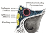
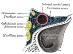
, close to the crossing of the attached margin of the tentorium and within millimeters of the posterior clinoid process. It runs on the lateral wall of the cavernous sinus
, where it is joined by the other two extraocular nerves (III) and the first two branches of the trigeminal nerve
(V), Ophthalmic V1 and Maxillary V2. The internal carotid artery
and Abducens nerve (VI) run within the cavernous sinus. Finally, it enters the orbit through the superior orbital fissure and innervates the superior oblique muscle
.
The superior oblique muscle ends in a tendon that passes through a fibrous loop, the trochlea, located anteriorly on the medial aspect of the orbit. Trochlea means “pulley” in Latin; the fourth nerve is named after this structure.
The body of the superior oblique muscle is located behind the eyeball, but the tendon (which is redirected by the trochlea) approaches the eyeball from the front. The tendon attaches to the top (superior aspect) of the eyeball at an angle of 51 degrees with respect to the primary position of the eye (looking straight forward). The force of the tendon’s pull therefore has two components: a forward component that tends to pull the eyeball downward (depression), and a medial component that tends to rotate the top of the eyeball toward the nose (intorsion).
The relative strength of these two forces depends on which way the eye is looking. When the eye is adducted (looking toward the nose), the force of depression increases. When the eye is abducted (looking away from the nose), the force of intorsion increases, while the force of depression decreases. When the eye is in the primary position (looking straight ahead), contraction of the superior oblique produces depression and intorsion in roughly equal amounts.
To summarize, the actions of the superior oblique muscle are (1) depression of the eyeball, especially when the eye is adducted; and (2) intorsion of the eyeball, especially when the eye is abducted. The clinical consequences of weakness in the superior oblique (caused, for example, by fourth nerve palsies) are discussed below.
This summary of the superior oblique muscle describes its most important functions. However, it is an oversimplification of the actual situation. For example, the tendon of the superior oblique inserts behind the equator of the eyeball in the frontal plane, so contraction of the muscle also tends to abduct the eyeball (turn it outward). In fact, each of the six extraocular muscles exerts rotational forces in all three planes (elevation-depression, adduction-abduction, intorsion-extorsion) to varying degrees, depending on which way the eye is looking. The relative forces change every time the eyeball moves – every time the direction of gaze changes. The central control of this process, which involves the continuous, precise adjustment of forces on twelve different tendons in order to point both eyes in exactly the same direction, is truly remarkable.
The recent discovery of soft tissue pulleys in the orbit – similar to the trochlea, but anatomically more subtle and previously missed – has completely changed (and greatly simplified) our understanding of the actions of the extraocular muscles. Perhaps the most important finding is that a 2-dimensional representation of the visual field is sufficient for most purposes.
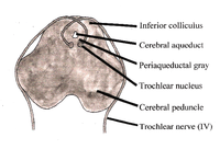 The nucleus of the trochlear nerve
is located in the caudal mesencephalon
beneath the cerebral aqueduct
. It is immediately below the nucleus of the oculomotor nerve
(III) in the rostral mesencephalon.
The trochlear nucleus is unique in that its axons run dorsally and cross the midline before emerging from the brainstem. Thus a lesion of the trochlear nucleus affects the contralateral eye. Lesions of all other cranial nuclei affect the ipsilateral side (except of course the optic nerves - cranial nerves II - which innervate both eyes).
(double vision). The affected eye drifts upward relative to the normal eye, due to the unopposed actions of the remaining extraocular muscles. The patient sees two visual fields (one from each eye), separated vertically. To compensate for this, patients learn to tilt the head forward (tuck the chin in) in order to bring the fields back together – to fuse the two images into a single visual field. This accounts for the “dejected” appearance of patients with “pathetic nerve” palsies.
As would be expected, the diplopia gets worse when the affected eye looks toward the nose – the contribution of the superior oblique muscle to downward gaze is greater in this position. Common activities requiring this type of convergent gaze are reading the newspaper and walking down stairs. Diplopia associated with these activities may be the initial symptom of a fourth nerve palsy.
Alfred Bielschowsky
's head tilt test
is a test for palsy of the superior oblique muscle caused by damage to cranial nerve IV (trochlear nerve).
Weakness of intorsion results in torsional diplopia, in which two different visual fields, tilted with respect to each other, are seen at the same time. To compensate for this, patients with trochlear nerve palsies tilt their heads to the opposite side, in order to fuse the two images into a single visual field.
The characteristic appearance of patients with fourth nerve palsies (head tilted to one side, chin tucked in) suggests the diagnosis, but other causes must be ruled out. For example, torticollis
can produce a similar appearance.
The most common cause of acute fourth nerve palsy is head trauma. Even relatively minor trauma can transiently stretch the fourth nerve (by transiently displacing the brainstem relative to the posterior clinoid process). Patients with minor damage to the fourth nerve will complain of “blurry” vision. Patients with more extensive damage will notice frank diplopia and rotational (torsional) disturbances of the visual fields. The usual clinical course is complete recovery within weeks to months.
Isolated injury to the fourth nerve can be caused by any process that stretches or compresses the nerve. A generalized increase in intracranial pressure – hydrocephalus
, pseudotumor cerebri, hemorrhage, edema
– will affect the fourth nerve, but the abducens nerve (VI) is usually affected first (producing horizontal diplopia, not vertical diplopia). Infections (meningitis
, herpes zoster
), demyelination (multiple sclerosis
), diabetic neuropathy
and cavernous sinus
disease can affect the fourth nerve, as can orbital tumors and Tolosa-Hunt syndrome
. In general, these diseases affect other cranial nerves as well. Isolated damage to the fourth nerve is uncommon in these settings.
The most common cause of chronic fourth nerve palsy is a congenital defect, in which the development of the fourth nerve (or its nucleus) is abnormal or incomplete. Congenital defects may be noticed in childhood, but minor defects may not become evident until adult life, when compensatory mechanisms begin to fail. Congenital fourth nerve palsies are amenable to surgical treatment.
The trochlear nucleus and its axons within the brainstem can be damaged by infarctions, hemorrhage, arteriovenous malformation
s, tumors and demyelination. Collateral damage to other structures will usually dominate the clinical picture.
The fourth nerve is one of the final common pathways for cortical systems that control eye movement in general. Cortical control of eye movement (saccades, smooth pursuit, accommodation
) involves conjugate gaze
, not unilateral eye movement.
Superior oblique muscle
For the abdominal muscle see: Abdominal external oblique muscleThe superior oblique muscle, or obliquus oculi superior, is a fusiform muscle originating in the upper, medial side of the orbit which abducts, depresses and internally rotates the eye...
muscle of the eye.
The trochlear nerve is unique among the cranial nerves in several respects. It is the smallest nerve in terms of the number of axons it contains. It has the greatest intracranial length. Other than the optic nerve (cranial nerve II), it is the only cranial nerve that decussates (crosses to the other side) before innervating its target. Finally, it is the only cranial nerve that exits from the dorsal aspect of the brainstem.
Homologous trochlear nerves are found in all jawed vertebrates. The unique features of the trochlear nerve, including its dorsal exit from the brainstem and its contralateral innervation, are seen in the primitive brains of sharks.
The human trochlear nerve is derived from the basal plate
Basal plate (neural tube)
In the developing nervous system, the basal plate is the region of the neural tube ventral to the sulcus limitans. It extends from the rostral mesencephalon to the end of the spinal cord and contains primarily motor neurons, whereas neurons found in the alar plate are primarily associated with...
of the embryo
Embryo
An embryo is a multicellular diploid eukaryote in its earliest stage of development, from the time of first cell division until birth, hatching, or germination...
nic midbrain.
Peripheral anatomy
Mesencephalon
The midbrain or mesencephalon is a portion of the central nervous system associated with vision, hearing, motor control, sleep/wake, arousal , and temperature regulation....
, just below the inferior colliculus
Inferior colliculus
The inferior colliculus is the principal midbrain nucleus of the auditory pathway and receives input from several more peripheral brainstem nuclei in the auditory pathway, as well as inputs from the auditory cortex...
. It circles anteriorly around the brainstem and runs forward toward the eye in the subarachnoid space
Subarachnoid space
In the central nervous system, the subarachnoid cavity is the interval between the arachnoid membrane and pia mater....
. It passes between the posterior cerebral artery
Posterior cerebral artery
-External links: - Posterior Cerebral Artery Stroke* at strokecenter.org* at State University of New York Upstate Medical University* at psyweb.com* at neuropat.dote.hu...
and the superior cerebellar artery
Superior cerebellar artery
The superior cerebellar artery arises near the termination of the basilar artery.It passes lateralward, immediately below the oculomotor nerve, which separates it from the posterior cerebral artery, winds around the cerebral peduncle, close to the trochlear nerve, and, arriving at the upper...
, and then pierces the dura
Dura
Dura may refer to:* Dura , a Palestinian town in the southern West Bank located eleven kilometers southwest of Hebron in the Hebron Governorate* Dura language, a critically endangered language of Nepal...
just under free margin of the tentorium cerebelli
Tentorium cerebelli
The tentorium cerebelli or cerebellar tentorium is an extension of the dura mater that separates the cerebellum from the inferior portion of the occipital lobes.-Anatomy:...
, close to the crossing of the attached margin of the tentorium and within millimeters of the posterior clinoid process. It runs on the lateral wall of the cavernous sinus
Cavernous sinus
The cavernous sinus , within the human head, is a large collection of thin-walled veins creating a cavity bordered by the temporal bone of the skull and the sphenoid bone, lateral to the sella turcica.-Contents:...
, where it is joined by the other two extraocular nerves (III) and the first two branches of the trigeminal nerve
Trigeminal nerve
The trigeminal nerve contains both sensory and motor fibres. It is responsible for sensation in the face and certain motor functions such as biting, chewing, and swallowing. Sensory information from the face and body is processed by parallel pathways in the central nervous system...
(V), Ophthalmic V1 and Maxillary V2. The internal carotid artery
Internal carotid artery
In human anatomy, the internal carotid arteries are two major arteries, one on each side of the head and neck. They arise from the common carotid arteries where these bifurcate into the internal and external carotid artery, and they supply the brain....
and Abducens nerve (VI) run within the cavernous sinus. Finally, it enters the orbit through the superior orbital fissure and innervates the superior oblique muscle
Superior oblique muscle
For the abdominal muscle see: Abdominal external oblique muscleThe superior oblique muscle, or obliquus oculi superior, is a fusiform muscle originating in the upper, medial side of the orbit which abducts, depresses and internally rotates the eye...
.
The superior oblique muscle ends in a tendon that passes through a fibrous loop, the trochlea, located anteriorly on the medial aspect of the orbit. Trochlea means “pulley” in Latin; the fourth nerve is named after this structure.
Actions of the superior oblique muscle
In order to understand the actions of the superior oblique muscle, it is useful to imagine the eyeball as a sphere that is constrained – like the trackball of a computer mouse – in such a way that only certain rotational movements are possible. Allowable movements for the superior oblique are (1) rotation in a vertical plane – looking down and up (depression and elevation of the eyeball) and (2) rotation in the plane of the face (intorsion and extorsion of the eyeball).The body of the superior oblique muscle is located behind the eyeball, but the tendon (which is redirected by the trochlea) approaches the eyeball from the front. The tendon attaches to the top (superior aspect) of the eyeball at an angle of 51 degrees with respect to the primary position of the eye (looking straight forward). The force of the tendon’s pull therefore has two components: a forward component that tends to pull the eyeball downward (depression), and a medial component that tends to rotate the top of the eyeball toward the nose (intorsion).
The relative strength of these two forces depends on which way the eye is looking. When the eye is adducted (looking toward the nose), the force of depression increases. When the eye is abducted (looking away from the nose), the force of intorsion increases, while the force of depression decreases. When the eye is in the primary position (looking straight ahead), contraction of the superior oblique produces depression and intorsion in roughly equal amounts.
To summarize, the actions of the superior oblique muscle are (1) depression of the eyeball, especially when the eye is adducted; and (2) intorsion of the eyeball, especially when the eye is abducted. The clinical consequences of weakness in the superior oblique (caused, for example, by fourth nerve palsies) are discussed below.
This summary of the superior oblique muscle describes its most important functions. However, it is an oversimplification of the actual situation. For example, the tendon of the superior oblique inserts behind the equator of the eyeball in the frontal plane, so contraction of the muscle also tends to abduct the eyeball (turn it outward). In fact, each of the six extraocular muscles exerts rotational forces in all three planes (elevation-depression, adduction-abduction, intorsion-extorsion) to varying degrees, depending on which way the eye is looking. The relative forces change every time the eyeball moves – every time the direction of gaze changes. The central control of this process, which involves the continuous, precise adjustment of forces on twelve different tendons in order to point both eyes in exactly the same direction, is truly remarkable.
The recent discovery of soft tissue pulleys in the orbit – similar to the trochlea, but anatomically more subtle and previously missed – has completely changed (and greatly simplified) our understanding of the actions of the extraocular muscles. Perhaps the most important finding is that a 2-dimensional representation of the visual field is sufficient for most purposes.
Central anatomy
Trochlear nucleus
The nucleus of the trochlear nerve is located in the midbrain, at the level of the inferior colliculus. It is a motor nucleus, so is located near the midline....
is located in the caudal mesencephalon
Mesencephalon
The midbrain or mesencephalon is a portion of the central nervous system associated with vision, hearing, motor control, sleep/wake, arousal , and temperature regulation....
beneath the cerebral aqueduct
Cerebral aqueduct
The mesencephalic duct, also known as the aqueductus mesencephali, aqueduct of Sylvius or the cerebral aqueduct, contains cerebrospinal fluid , is within the mesencephalon and connects the third ventricle in the diencephalon to the fourth ventricle in the mesencephalon, which is between the pons...
. It is immediately below the nucleus of the oculomotor nerve
Oculomotor nerve
The oculomotor nerve is the 3rd of 12 paired cranial nerves. It enters the orbit via the superior orbital fissure and controls most of the eye's movements, including constriction of the pupil and maintaining an open eyelid by innervating the Levator palpebrae superiors muscle. The optic nerve is...
(III) in the rostral mesencephalon.
The trochlear nucleus is unique in that its axons run dorsally and cross the midline before emerging from the brainstem. Thus a lesion of the trochlear nucleus affects the contralateral eye. Lesions of all other cranial nuclei affect the ipsilateral side (except of course the optic nerves - cranial nerves II - which innervate both eyes).
Vertical diplopia
Injury to the trochlear nerve cause weakness of infromedial movement with consequent vertical diplopiaDiplopia
Diplopia, commonly known as double vision, is the simultaneous perception of two images of a single object that may be displaced horizontally, vertically, or diagonally in relation to each other...
(double vision). The affected eye drifts upward relative to the normal eye, due to the unopposed actions of the remaining extraocular muscles. The patient sees two visual fields (one from each eye), separated vertically. To compensate for this, patients learn to tilt the head forward (tuck the chin in) in order to bring the fields back together – to fuse the two images into a single visual field. This accounts for the “dejected” appearance of patients with “pathetic nerve” palsies.
As would be expected, the diplopia gets worse when the affected eye looks toward the nose – the contribution of the superior oblique muscle to downward gaze is greater in this position. Common activities requiring this type of convergent gaze are reading the newspaper and walking down stairs. Diplopia associated with these activities may be the initial symptom of a fourth nerve palsy.
Alfred Bielschowsky
Alfred Bielschowsky
Alfred Bielschowsky was a German ophthalmologist. His specialty was physiology and pathology of the eye, particularly in regards to research of eye movement, space perception and diagnosis of oculomotor anomalies....
's head tilt test
Bielschowsky's head tilt test
Bielschowsky's head tilt test is a medical test to detect damage to the IVth cranial nerve leading to a palsy of the superior oblique muscle of the eye....
is a test for palsy of the superior oblique muscle caused by damage to cranial nerve IV (trochlear nerve).
Torsional diplopia
Trochlear nerve palsy also affects torsion (rotation of the eyeball in the plane of the face). Torsion is a normal response to tilting the head sideways. The eyes automatically rotate in an equal and opposite direction, so that the orientation of the environment remains unchanged – vertical things remain vertical.Weakness of intorsion results in torsional diplopia, in which two different visual fields, tilted with respect to each other, are seen at the same time. To compensate for this, patients with trochlear nerve palsies tilt their heads to the opposite side, in order to fuse the two images into a single visual field.
The characteristic appearance of patients with fourth nerve palsies (head tilted to one side, chin tucked in) suggests the diagnosis, but other causes must be ruled out. For example, torticollis
Torticollis
Torticollis, or wryneck, is a stiff neck associated with muscle spasm, classically causing lateral flexion contracture of the cervical spine musculature...
can produce a similar appearance.
Peripheral lesions
A peripheral lesion is a damage to the bundle of nerves, in contrast to a central lesion, which is a damage to the trochlear nucleus. Acute symptoms are probably a result of a trauma or disease, while chronic symptoms probably are congenital.Acute palsy
The most common cause of acute fourth nerve palsy is head trauma. Even relatively minor trauma can transiently stretch the fourth nerve (by transiently displacing the brainstem relative to the posterior clinoid process). Patients with minor damage to the fourth nerve will complain of “blurry” vision. Patients with more extensive damage will notice frank diplopia and rotational (torsional) disturbances of the visual fields. The usual clinical course is complete recovery within weeks to months.
Isolated injury to the fourth nerve can be caused by any process that stretches or compresses the nerve. A generalized increase in intracranial pressure – hydrocephalus
Hydrocephalus
Hydrocephalus , also known as "water in the brain," is a medical condition in which there is an abnormal accumulation of cerebrospinal fluid in the ventricles, or cavities, of the brain. This may cause increased intracranial pressure inside the skull and progressive enlargement of the head,...
, pseudotumor cerebri, hemorrhage, edema
Edema
Edema or oedema ; both words from the Greek , oídēma "swelling"), formerly known as dropsy or hydropsy, is an abnormal accumulation of fluid beneath the skin or in one or more cavities of the body that produces swelling...
– will affect the fourth nerve, but the abducens nerve (VI) is usually affected first (producing horizontal diplopia, not vertical diplopia). Infections (meningitis
Meningitis
Meningitis is inflammation of the protective membranes covering the brain and spinal cord, known collectively as the meninges. The inflammation may be caused by infection with viruses, bacteria, or other microorganisms, and less commonly by certain drugs...
, herpes zoster
Herpes zoster
Herpes zoster , commonly known as shingles and also known as zona, is a viral disease characterized by a painful skin rash with blisters in a limited area on one side of the body, often in a stripe...
), demyelination (multiple sclerosis
Multiple sclerosis
Multiple sclerosis is an inflammatory disease in which the fatty myelin sheaths around the axons of the brain and spinal cord are damaged, leading to demyelination and scarring as well as a broad spectrum of signs and symptoms...
), diabetic neuropathy
Diabetic neuropathy
Diabetic neuropathies are neuropathic disorders that are associated with diabetes mellitus. These conditions are thought to result from diabetic microvascular injury involving small blood vessels that supply nerves in addition to macrovascular conditions that can culminate in diabetic neuropathy...
and cavernous sinus
Cavernous sinus
The cavernous sinus , within the human head, is a large collection of thin-walled veins creating a cavity bordered by the temporal bone of the skull and the sphenoid bone, lateral to the sella turcica.-Contents:...
disease can affect the fourth nerve, as can orbital tumors and Tolosa-Hunt syndrome
Tolosa-Hunt syndrome
Tolosa-Hunt syndrome is a rare disorder characterized by severe and unilateral headaches with extraocular palsies, usually involving the third, fourth, fifth, and sixth cranial nerves, and pain around the sides and back of the eye, along with weakness and paralysis of certain eye muscles.In 2004,...
. In general, these diseases affect other cranial nerves as well. Isolated damage to the fourth nerve is uncommon in these settings.
Chronic palsy
The most common cause of chronic fourth nerve palsy is a congenital defect, in which the development of the fourth nerve (or its nucleus) is abnormal or incomplete. Congenital defects may be noticed in childhood, but minor defects may not become evident until adult life, when compensatory mechanisms begin to fail. Congenital fourth nerve palsies are amenable to surgical treatment.
Central lesions
Central damage is a damage to the trochlear nucleus. It affects the contralateral eye. The nuclei of other cranial nerves affect ipsilateral structures (except of course the optic nerves - cranial nerves II - which innervate both eyes).The trochlear nucleus and its axons within the brainstem can be damaged by infarctions, hemorrhage, arteriovenous malformation
Arteriovenous malformation
Arteriovenous malformation or AVM is an abnormal connection between veins and arteries, usually congenital. This pathology is widely known because of its occurrence in the central nervous system, but can appear in any location. An arteriovenous malformation is a vascular anomaly. It is a...
s, tumors and demyelination. Collateral damage to other structures will usually dominate the clinical picture.
The fourth nerve is one of the final common pathways for cortical systems that control eye movement in general. Cortical control of eye movement (saccades, smooth pursuit, accommodation
Accommodation (eye)
Accommodation is the process by which the vertebrate eye changes optical power to maintain a clear image on an object as its distance changes....
) involves conjugate gaze
Conjugate gaze palsy
Conjugate gaze palsy refers to an inability of both eyes to move in the same direction at the same time.It can be associated with a lesion of the paramedian pontine reticular formation.-See also:* Internuclear ophthalmoplegia* One and a half syndrome...
, not unilateral eye movement.