
Internal carotid artery
Encyclopedia
In human anatomy
, the internal carotid arteries are two major arteries, one on each side of the head and neck. They arise from the common carotid arteries where these bifurcate into the internal and external carotid artery
, and they supply the brain.
in 1998 subdivided the artery into four parts: "cervical", "petrous", "cavernous", and "cerebral". However, in clinical settings, the classification system of the internal carotid artery follows the 1996 recommendations by Bouthillier, and describes seven anatomical segments of the internal carotid artery. The Bouthillier system is often used clinically by neurosurgeons, neuroradiologists and neurologists. This nomenclature system is a clinical one, based on the angiographic appearance of the artery and its relationship to surrounding anatomy, in contrast to an embryologic classification system. An older clinical classification is based on work by Fischer in 1938 is also commonly used, as well as classification schemes based on the embryologic anatomy of the carotid artery.
The segments of the internal carotid artery are as follows:
; it arises around the level of the third cervical vertebra when the common carotid bifurcates into this artery and its more superficial counterpart, the external carotid artery
.
 The cervical segment, or C1, of the internal carotid extends from the carotid bifurcation until it enters the carotid canal
in the skull anterior to the jugular foramen.
At its origin, the internal carotid artery is somewhat dilated. This part of the artery is known as the carotid sinus
or the carotid bulb. The ascending portion of the cervical segment occurs distal to the bulb, when the vessel walls are again parallel.
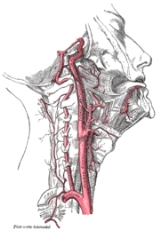
The internal carotid runs perpendicularly upward in the carotid sheath
, and enters the skull
through the carotid canal
. During this part of its course, it lies in front of the transverse processes of the upper three cervical vertebrae.
It is relatively superficial at its start, where it is contained in the carotid triangle of the neck, and lies behind and lateral to the external carotid, overlapped by the sternocleidomastoid muscle, and covered by the deep fascia, the platysma, and integument: it then passes beneath the parotid gland
, being crossed by the hypoglossal nerve
, the digastric muscle
and the stylohyoid muscle
, the occipital artery
and the posterior auricular artery
. Higher up, it is separated from the external carotid by the styloglossus
and stylopharyngeus muscles, the tip of the styloid process and the stylohyoid ligament
, the glossopharyngeal nerve
and the pharyngeal branch of the vagus nerve
. It is in relation, behind, with the longus capitis, the superior cervical ganglion
of the sympathetic trunk
, and the superior laryngeal nerve
; laterally, with the internal jugular vein
and vagus nerve, the nerve lying on a plane posterior to the artery; medially, with the pharynx
, superior laryngeal nerve, and ascending pharyngeal artery
. At the base of the skull the glossopharyngeal, vagus, accessory, and hypoglossal nerves lie between the artery and the internal jugular vein.
Unlike the external carotid artery
, the internal carotid normally has no branches in the neck
.
. This segment extends until the foramen lacerum
. The petrous portion classically has three sections: an ascending, or vertical portion; the genu, or bend; and the horizontal portion.
When the internal carotid artery enters the canal in the petrous portion of the temporal bone
, it first ascends a short distance, then curves anteriorly and medially. The artery lies at first in front of the cochlea
and tympanic cavity
; from the latter cavity it is separated by a thin, bony lamella, which is cribriform in the young subject, and often partly absorbed in old age. Farther forward it is separated from the trigeminal ganglion
by a thin plate of bone, which forms the floor of the fossa for the ganglion and the roof of the horizontal portion of the canal. Frequently this bony plate is more or less deficient, and then the ganglion is separated from the artery by fibrous membrane. The artery is separated from the bony wall of the carotid canal
by a prolongation of dura mater
, and is surrounded by a number of small veins and by filaments of the carotid plexus
, derived from the ascending branch of the superior cervical ganglion
of the sympathetic trunk.
The named branches of the petrous segment of the internal carotid artery are:
and ends at the petrolingual ligament, a reflection of periosteum
between the lingula and petrous apex (or petrosal process) of the sphenoid bone
. The lacerum portion is still considered to be 'extra-dural', as it is surrounded by periosteum and fibrocartilage along its course. The lacerum segment normally has no named branches, though the vidian artery may occasionally arise from this segment. It is erroneously stated in several anatomy text books that the internal carotid artery passes through the foramen lacerum. This at best has only ever been a partial truth in that it passes through the superior part of the foramen on its way to the cavernous sinus. As such it does not exit the skull through it. The inferior part of the foramen is actually filled with fibrocartilage. Recent consensus is that the Internal Carotid Artery should not be described as travelling through the foramen lacerum.
 The cavernous segment, or C4, of the internal carotid artery begins at the petrolingual ligament and extends to the proximal dural ring, which is formed by the medial and inferior periosteum of the anterior clinoid process. The cavernous segment is surrounded by the cavernous sinus
.
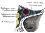
In this part of its course, the artery is situated between the layers of the dura mater forming the cavernous sinus, but covered by the lining membrane of the sinus. It at first ascends toward the posterior clinoid process, then passes forward by the side of the body of the sphenoid bone, and again curves upward on the medial side of the anterior clinoid process
, and perforates the dura mater forming the roof of the sinus. The curve in the cavernous segment is called the carotid siphon. This portion of the artery is surrounded by filaments of the sympathetic trunk, and on its lateral side is the abducent nerve
, or cranial nerve VI.
The named branches of the cavernous segment are:
The cavernous segment also gives rise to small capsular arteries that supply the wall of the cavernous sinus.
at the proximal dural ring and extends distally to the distal dural ring, after which the carotid artery is considered 'intra-dural' and has entered the subarachnoid space
.
The clinoid segment normally has no named branches, though the ophthalmic artery
may arise from the clinoid segment.
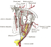 The ophthalmic segment, or C6, extends from the distal dural ring, which is continuous with the falciform ligament, and extends distally to the origin of the posterior communicating artery
. The ophthalmic segment courses roughly horizontally, parallel to the optic nerve
which runs superomedially to the carotid at this point.
The named branches of the ophthalmic segment are:
to the bifurcation of the internal carotid artery.
The named branches of the communicating segment are:
The internal carotid then divides to form the anterior cerebral artery and middle cerebral artery
. The internal carotid artery can receive blood flow via an important collateral pathway
supplying the brain, the cerebral arterial circle, which is more commonly known as the Circle of Willis
.
forms a plexus of nerves around the artery known as the carotid plexus
. The internal carotid nerve arises from the superior cervical ganglion
, and forms this plexus, which follows the internal carotid into the skull.
Human anatomy
Human anatomy is primarily the scientific study of the morphology of the human body. Anatomy is subdivided into gross anatomy and microscopic anatomy. Gross anatomy is the study of anatomical structures that can be seen by the naked eye...
, the internal carotid arteries are two major arteries, one on each side of the head and neck. They arise from the common carotid arteries where these bifurcate into the internal and external carotid artery
External carotid artery
In human anatomy, the external carotid artery is a major artery of the head and neck. It arises from the common carotid artery when it bifurcates into the external and internal carotid artery.-Course:...
, and they supply the brain.
Classification
Terminologia AnatomicaTerminologia Anatomica
Terminologia Anatomica is the international standard on human anatomic terminology. It was developed by the Federative Committee on Anatomical Terminology and the International Federation of Associations of Anatomists and was released in 1998. It supersedes the previous standard, Nomina...
in 1998 subdivided the artery into four parts: "cervical", "petrous", "cavernous", and "cerebral". However, in clinical settings, the classification system of the internal carotid artery follows the 1996 recommendations by Bouthillier, and describes seven anatomical segments of the internal carotid artery. The Bouthillier system is often used clinically by neurosurgeons, neuroradiologists and neurologists. This nomenclature system is a clinical one, based on the angiographic appearance of the artery and its relationship to surrounding anatomy, in contrast to an embryologic classification system. An older clinical classification is based on work by Fischer in 1938 is also commonly used, as well as classification schemes based on the embryologic anatomy of the carotid artery.
The segments of the internal carotid artery are as follows:
- Cervical segment, or C1, identical to the commonly used Cervical portion
- Petrous segment, or C2
- Lacerum segment, or C3
- C2 and C3 comprise the commonly used Petrous portionPetrous portion of the internal carotid arteryPetrous portion of the internal carotid artery .—When the internal carotid artery enters the canal in the petrous portion of the temporal bone, it first ascends a short distance, then curves forward and medially, and again ascends as it leaves the canal to enter the cavity of the skull between the...
- C2 and C3 comprise the commonly used Petrous portion
- Cavernous segment, or C4, almost identical to the commonly used Cavernous portion
- Clinoid segment, or C5. This segment is not identified in some earlier classifications, and lies between the commonly used Cavernous portion and Cerebral or Supraclinoid portionCerebral portion of internal carotid arteryThe cerebral portion of internal carotid artery.—Having perforated the dura mater on the medial side of the anterior clinoid process, the internal carotid passes between the optic and oculomotor nerves...
- Ophthalmic, or supraclinoid segment, or C6
- Communicating, or terminal segment, or C7
- C6 and C7 together comprise the commonly used Cerebral or Supraclinoid portionCerebral portion of internal carotid arteryThe cerebral portion of internal carotid artery.—Having perforated the dura mater on the medial side of the anterior clinoid process, the internal carotid passes between the optic and oculomotor nerves...
- C6 and C7 together comprise the commonly used Cerebral or Supraclinoid portion
Course
The internal carotid artery is a terminal branch of the common carotid arteryCommon carotid artery
In human anatomy, the common carotid artery is an artery that supplies the head and neck with oxygenated blood; it divides in the neck to form the external and internal carotid arteries. - Structure :...
; it arises around the level of the third cervical vertebra when the common carotid bifurcates into this artery and its more superficial counterpart, the external carotid artery
External carotid artery
In human anatomy, the external carotid artery is a major artery of the head and neck. It arises from the common carotid artery when it bifurcates into the external and internal carotid artery.-Course:...
.
C1: Cervical segment
Carotid canal
On the interior surface of the temporal bone, behind the rough surface of the apex, is the large circular aperture of the carotid canal, which ascends at first vertically, and then, making a bend, runs horizontally forward and medialward.-Contents:...
in the skull anterior to the jugular foramen.
At its origin, the internal carotid artery is somewhat dilated. This part of the artery is known as the carotid sinus
Carotid sinus
In human anatomy, the carotid sinus is a localized dilation of the internal carotid artery at its origin, the common carotid artery.-Functions:...
or the carotid bulb. The ascending portion of the cervical segment occurs distal to the bulb, when the vessel walls are again parallel.
The internal carotid runs perpendicularly upward in the carotid sheath
Carotid sheath
The carotid sheath is an anatomical term for the fibrous connective tissue that surrounds the vascular compartment of the neck. It is part of the deep cervical fascia of the neck, below the superficial cervical fascia meaning the subcutaneous adipose tissue immediately beneath the skin.The deep...
, and enters the skull
Human skull
The human skull is a bony structure, skeleton, that is in the human head and which supports the structures of the face and forms a cavity for the brain.In humans, the adult skull is normally made up of 22 bones...
through the carotid canal
Carotid canal
On the interior surface of the temporal bone, behind the rough surface of the apex, is the large circular aperture of the carotid canal, which ascends at first vertically, and then, making a bend, runs horizontally forward and medialward.-Contents:...
. During this part of its course, it lies in front of the transverse processes of the upper three cervical vertebrae.
It is relatively superficial at its start, where it is contained in the carotid triangle of the neck, and lies behind and lateral to the external carotid, overlapped by the sternocleidomastoid muscle, and covered by the deep fascia, the platysma, and integument: it then passes beneath the parotid gland
Parotid gland
The paired parotid glands are the largest of the salivary glands. They are each found wrapped around the mandibular ramus, and secrete saliva through Stensen's ducts into the oral cavity, to facilitate mastication and swallowing and to begin the digestion of starches.-Location:The parotid glands...
, being crossed by the hypoglossal nerve
Hypoglossal nerve
The hypoglossal nerve is the twelfth cranial nerve , leading to the tongue. The nerve arises from the hypoglossal nucleus and emerges from the medulla oblongata in the preolivary sulcus separating the olive and the pyramid. It then passes through the hypoglossal canal...
, the digastric muscle
Digastric muscle
The digastric muscle is a small muscle located under the jaw. so digastric muscles are muscle fibers in ligament of treitz ,omohyoid , occipitofrontalis....
and the stylohyoid muscle
Stylohyoid muscle
The stylohyoid muscle is a slender muscle, lying anterior, and superior of the posterior belly of the digastric muscle. It shares this muscle's innervation by the facial nerve, and functions to draw the hyoid bone backwards and elevate the tongue....
, the occipital artery
Occipital artery
The occipital artery arises from the external carotid artery opposite the facial artery, its path is below the posterior belly of digastric to the occipital region. This artery supplies blood to the back of the scalp and sterno-mastoid muscles...
and the posterior auricular artery
Posterior auricular artery
The posterior auricular artery is a small artery and arises from the external carotid artery, above the Digastric muscle and Stylohyoid muscle, opposite the apex of the styloid process....
. Higher up, it is separated from the external carotid by the styloglossus
Styloglossus
The Styloglossus, the shortest and smallest of the three styloid muscles, arises from the anterior and lateral surfaces of the styloid process, near its apex, and from the stylomandibular ligament....
and stylopharyngeus muscles, the tip of the styloid process and the stylohyoid ligament
Stylohyoid ligament
In connection with the stylohyoideus muscle a ligamentous band, the stylohyoid ligament, may be described.It is a fibrous cord, which is attached to the tip of the styloid process of the temporal bone and the lesser cornu of the hyoid bone....
, the glossopharyngeal nerve
Glossopharyngeal nerve
The glossopharyngeal nerve is the ninth of twelve pairs of cranial nerves . It exits the brainstem out from the sides of the upper medulla, just rostral to the vagus nerve...
and the pharyngeal branch of the vagus nerve
Vagus nerve
The vagus nerve , also called pneumogastric nerve or cranial nerve X, is the tenth of twelve paired cranial nerves...
. It is in relation, behind, with the longus capitis, the superior cervical ganglion
Superior cervical ganglion
The superior cervical ganglion , the largest of the cervical ganglia, is placed opposite the second and third cervical vertebræ. It contains neurons that supply sympathetic innervation to the face....
of the sympathetic trunk
Sympathetic trunk
The sympathetic trunks are a paired bundle of nerve fibers that run from the base of the skull to the coccyx.-Structure:...
, and the superior laryngeal nerve
Superior laryngeal nerve
The superior laryngeal nerve is a branch of the vagus nerve. It arises from the middle of the ganglion nodosum and in its course receives a branch from the superior cervical ganglion of the sympathetic....
; laterally, with the internal jugular vein
Internal jugular vein
The two internal jugular veins collect the blood from the brain, the superficial parts of the face, and the neck.-Path:On both sides and at the base of the brain, the inferior petrosal sinus and the sigmoid sinus join to form the internal jugular vein...
and vagus nerve, the nerve lying on a plane posterior to the artery; medially, with the pharynx
Pharynx
The human pharynx is the part of the throat situated immediately posterior to the mouth and nasal cavity, and anterior to the esophagus and larynx. The human pharynx is conventionally divided into three sections: the nasopharynx , the oropharynx , and the laryngopharynx...
, superior laryngeal nerve, and ascending pharyngeal artery
Ascending pharyngeal artery
The ascending pharyngeal artery, the smallest branch of the external carotid, is a long, slender vessel, deeply seated in the neck, beneath the other branches of the external carotid and under the Stylopharyngeus...
. At the base of the skull the glossopharyngeal, vagus, accessory, and hypoglossal nerves lie between the artery and the internal jugular vein.
Unlike the external carotid artery
External carotid artery
In human anatomy, the external carotid artery is a major artery of the head and neck. It arises from the common carotid artery when it bifurcates into the external and internal carotid artery.-Course:...
, the internal carotid normally has no branches in the neck
Neck
The neck is the part of the body, on many terrestrial or secondarily aquatic vertebrates, that distinguishes the head from the torso or trunk. The adjective signifying "of the neck" is cervical .-Boner anatomy: The cervical spine:The cervical portion of the human spine comprises seven boney...
.
C2: Petrous segment
The petrous segment, or C2, of the internal carotid is that which is inside the petrous part of the temporal boneTemporal bone
The temporal bones are situated at the sides and base of the skull, and lateral to the temporal lobes of the cerebrum.The temporal bone supports that part of the face known as the temple.-Parts:The temporal bone consists of four parts:* Squama temporalis...
. This segment extends until the foramen lacerum
Foramen lacerum
The foramen lacerum is a triangular hole in the base of the skull located at the base of the medial pterygoid plate.-Transit through the foramen lacerum:...
. The petrous portion classically has three sections: an ascending, or vertical portion; the genu, or bend; and the horizontal portion.
When the internal carotid artery enters the canal in the petrous portion of the temporal bone
Petrous portion of the temporal bone
The petrous portion of the temporal bone or pyramid is pyramidal and is wedged in at the base of the skull between the sphenoid and occipital bones. Directed medially, forward, and a little upward, it presents for examination a base, an apex, three surfaces, and three angles, and contains, in its...
, it first ascends a short distance, then curves anteriorly and medially. The artery lies at first in front of the cochlea
Cochlea
The cochlea is the auditory portion of the inner ear. It is a spiral-shaped cavity in the bony labyrinth, making 2.5 turns around its axis, the modiolus....
and tympanic cavity
Tympanic cavity
The tympanic cavity is a small cavity surrounding the bones of the middle ear.It is formed from the tubotympanic recess, an expansion of the first pharyngeal pouch....
; from the latter cavity it is separated by a thin, bony lamella, which is cribriform in the young subject, and often partly absorbed in old age. Farther forward it is separated from the trigeminal ganglion
Trigeminal ganglion
The trigeminal ganglion is a sensory ganglion of the trigeminal nerve that occupies a cavity in the dura mater, covering the trigeminal impression near the apex of the petrous part of the temporal bone.-Relations:It is somewhat crescentic in shape, with its convexity...
by a thin plate of bone, which forms the floor of the fossa for the ganglion and the roof of the horizontal portion of the canal. Frequently this bony plate is more or less deficient, and then the ganglion is separated from the artery by fibrous membrane. The artery is separated from the bony wall of the carotid canal
Carotid canal
On the interior surface of the temporal bone, behind the rough surface of the apex, is the large circular aperture of the carotid canal, which ascends at first vertically, and then, making a bend, runs horizontally forward and medialward.-Contents:...
by a prolongation of dura mater
Dura mater
The dura mater , or dura, is the outermost of the three layers of the meninges surrounding the brain and spinal cord. It is derived from Mesoderm. The other two meningeal layers are the pia mater and the arachnoid mater. The dura surrounds the brain and the spinal cord and is responsible for...
, and is surrounded by a number of small veins and by filaments of the carotid plexus
Carotid plexus
Carotid plexus can refer to:* Common carotid plexus* Internal carotid plexus* External carotid plexus...
, derived from the ascending branch of the superior cervical ganglion
Superior cervical ganglion
The superior cervical ganglion , the largest of the cervical ganglia, is placed opposite the second and third cervical vertebræ. It contains neurons that supply sympathetic innervation to the face....
of the sympathetic trunk.
The named branches of the petrous segment of the internal carotid artery are:
- the vidian artery or artery of the pterygoid canal
- the caroticotympanic artery
C3: Lacerum segment
The lacerum segment, or C3, is a short segment that begins above the foramen lacerumForamen lacerum
The foramen lacerum is a triangular hole in the base of the skull located at the base of the medial pterygoid plate.-Transit through the foramen lacerum:...
and ends at the petrolingual ligament, a reflection of periosteum
Periosteum
Periosteum is a membrane that lines the outer surface of all bones, except at the joints of long bones. Endosteum lines the inner surface of all bones....
between the lingula and petrous apex (or petrosal process) of the sphenoid bone
Sphenoid bone
The sphenoid bone is an unpaired bone situated at the base of the skull in front of the temporal bone and basilar part of the occipital bone.The sphenoid bone is one of the seven bones that articulate to form the orbit...
. The lacerum portion is still considered to be 'extra-dural', as it is surrounded by periosteum and fibrocartilage along its course. The lacerum segment normally has no named branches, though the vidian artery may occasionally arise from this segment. It is erroneously stated in several anatomy text books that the internal carotid artery passes through the foramen lacerum. This at best has only ever been a partial truth in that it passes through the superior part of the foramen on its way to the cavernous sinus. As such it does not exit the skull through it. The inferior part of the foramen is actually filled with fibrocartilage. Recent consensus is that the Internal Carotid Artery should not be described as travelling through the foramen lacerum.
C4: Cavernous segment
Cavernous sinus
The cavernous sinus , within the human head, is a large collection of thin-walled veins creating a cavity bordered by the temporal bone of the skull and the sphenoid bone, lateral to the sella turcica.-Contents:...
.
In this part of its course, the artery is situated between the layers of the dura mater forming the cavernous sinus, but covered by the lining membrane of the sinus. It at first ascends toward the posterior clinoid process, then passes forward by the side of the body of the sphenoid bone, and again curves upward on the medial side of the anterior clinoid process
Anterior clinoid process
In the sphenoid bone, the posterior border, smooth and rounded, is received into the lateral fissure of the brain; the medial end of this border forms the anterior clinoid process, which gives attachment to the tentorium cerebelli; it is sometimes joined to the middle clinoid process by a spicule...
, and perforates the dura mater forming the roof of the sinus. The curve in the cavernous segment is called the carotid siphon. This portion of the artery is surrounded by filaments of the sympathetic trunk, and on its lateral side is the abducent nerve
Abducent nerve
The abducens nerve or abducent nerve is a somatic efferent nerve that controls the movement of a single muscle, the lateral rectus muscle of the eye, in humans. In most other mammals it also innervates the musculus retractor bulbi, which can retract the eye for protection...
, or cranial nerve VI.
The named branches of the cavernous segment are:
- the meningohypophyseal arteryMeningohypophyseal arteryThe meningohypophyseal artery, or meningohypophyseal trunk, is an inconstant branch of the cavernous segment of the internal carotid artery. Classically, the meningohypophyseal artery has three named branches:#Dorsal meningeal artery...
- the inferolateral trunk
The cavernous segment also gives rise to small capsular arteries that supply the wall of the cavernous sinus.
C5: Clinoid segment
The clinoid segment, or C5, is another short segment of the internal carotid that begins after the artery exits the cavernous sinusCavernous sinus
The cavernous sinus , within the human head, is a large collection of thin-walled veins creating a cavity bordered by the temporal bone of the skull and the sphenoid bone, lateral to the sella turcica.-Contents:...
at the proximal dural ring and extends distally to the distal dural ring, after which the carotid artery is considered 'intra-dural' and has entered the subarachnoid space
Subarachnoid space
In the central nervous system, the subarachnoid cavity is the interval between the arachnoid membrane and pia mater....
.
The clinoid segment normally has no named branches, though the ophthalmic artery
Ophthalmic artery
The ophthalmic artery is the first branch of the internal carotid artery distal to the cavernous sinus. Branches of the OA supply all the structures in the orbit as well as some structures in the nose, face and meninges...
may arise from the clinoid segment.
C6: Ophthalmic segment
Posterior communicating artery
In human anatomy, the posterior communicating artery is one of a pair of right-sided and left-sided blood vessels in the circle of Willis. It connects the three cerebral arteries of the same side. Anteriorly, it connects to the internal carotid artery prior the terminal bifurcation of the ICA...
. The ophthalmic segment courses roughly horizontally, parallel to the optic nerve
Optic nerve
The optic nerve, also called cranial nerve 2, transmits visual information from the retina to the brain. Derived from the embryonic retinal ganglion cell, a diverticulum located in the diencephalon, the optic nerve doesn't regenerate after transection.-Anatomy:The optic nerve is the second of...
which runs superomedially to the carotid at this point.
The named branches of the ophthalmic segment are:
- the ophthalmic arteryOphthalmic arteryThe ophthalmic artery is the first branch of the internal carotid artery distal to the cavernous sinus. Branches of the OA supply all the structures in the orbit as well as some structures in the nose, face and meninges...
- the superior hypophyseal artery
C7: Communicating segment
The communicating segment, or terminal segment, or C7, of the internal carotid artery passes between the optic and oculomotor nerves to the anterior perforated substance at the medial extremity of the lateral cerebral fissure. Angiographically, this segment extends from the origin of the posterior communicating arteryPosterior communicating artery
In human anatomy, the posterior communicating artery is one of a pair of right-sided and left-sided blood vessels in the circle of Willis. It connects the three cerebral arteries of the same side. Anteriorly, it connects to the internal carotid artery prior the terminal bifurcation of the ICA...
to the bifurcation of the internal carotid artery.
The named branches of the communicating segment are:
- the posterior communicating arteryPosterior communicating arteryIn human anatomy, the posterior communicating artery is one of a pair of right-sided and left-sided blood vessels in the circle of Willis. It connects the three cerebral arteries of the same side. Anteriorly, it connects to the internal carotid artery prior the terminal bifurcation of the ICA...
- the anterior choroidal arteryAnterior choroidal arteryThe anterior choroidal artery originates from the internal carotid artery, though it also rarely arises from the middle cerebral artery.-Structures supplied:The anterior choroidal artery serves many structures in the cerebrum:...
The internal carotid then divides to form the anterior cerebral artery and middle cerebral artery
Middle cerebral artery
-External links:*...
. The internal carotid artery can receive blood flow via an important collateral pathway
Anastomosis
An anastomosis is the reconnection of two streams that previously branched out, such as blood vessels or leaf veins. The term is used in medicine, biology, mycology and geology....
supplying the brain, the cerebral arterial circle, which is more commonly known as the Circle of Willis
Circle of Willis
The Circle of Willis is a circle of arteries that supply blood to the brain...
.
Branches
The following are the branches of the internal carotid artery, listed by segment:- C1: Branches from the cervical portion - none.
- C2: Branches from the petrous portion
- Caroticotympanic arteriesCaroticotympanic arteriesThe caroticotympanic branch is small; it enters the tympanic cavity through a minute foramen in the carotid canal, and anastomoses with the anterior tympanic branch of the internal maxillary, and with the stylomastoid artery....
- vidian artery
- Caroticotympanic arteries
- C3: Branches from the lacerum portion - none
- C4: Branches from the cavernous portion
- Branches of the meningohypophyseal trunk:
- Tentorial basal branch
- Tentorial marginal branch
- Meningeal branch - helps supply blood to the meninges of the anterior cranial fossa
- Clivus branches - tiny branches that supply the clivusClivus (anatomy)The clivus is a part of the cranium, a shallow depression behind the dorsum sellæ that slopes obliquely backward. It forms a gradual sloping process at the anterior most portion of the basilar occipital bone at its junction with the sphenoid bone. On axial planes, it sits just posterior to the...
- Inferior hypophyseal artery
- Capsular branches - supplies wall of cavernous sinusCavernous sinusThe cavernous sinus , within the human head, is a large collection of thin-walled veins creating a cavity bordered by the temporal bone of the skull and the sphenoid bone, lateral to the sella turcica.-Contents:...
- Branches of the inferolateral trunk:
- Branches to trigeminal ganglion - provide blood to trigeminal ganglionTrigeminal ganglionThe trigeminal ganglion is a sensory ganglion of the trigeminal nerve that occupies a cavity in the dura mater, covering the trigeminal impression near the apex of the petrous part of the temporal bone.-Relations:It is somewhat crescentic in shape, with its convexity...
- Artery of the foramen rotundumForamen rotundumThe foramen rotundum is a circular hole in the sphenoid bone that connects the middle cranial fossa and the pterygopalatine fossa.-Structure:...
- Branches to nerves
- Branches to trigeminal ganglion - provide blood to trigeminal ganglion
- Branches of the meningohypophyseal trunk:
- C5: Branches from the clinoid portion - none
- C6: Branches from the ophthalmic portion
- Ophthalmic arteryOphthalmic arteryThe ophthalmic artery is the first branch of the internal carotid artery distal to the cavernous sinus. Branches of the OA supply all the structures in the orbit as well as some structures in the nose, face and meninges...
- Superior hypophyseal artery
- Ophthalmic artery
- C7: Branches from the communicating portion
- Posterior communicating arteryPosterior communicating arteryIn human anatomy, the posterior communicating artery is one of a pair of right-sided and left-sided blood vessels in the circle of Willis. It connects the three cerebral arteries of the same side. Anteriorly, it connects to the internal carotid artery prior the terminal bifurcation of the ICA...
- Anterior choroidal arteryAnterior choroidal arteryThe anterior choroidal artery originates from the internal carotid artery, though it also rarely arises from the middle cerebral artery.-Structures supplied:The anterior choroidal artery serves many structures in the cerebrum:...
- Anterior cerebral artery (a terminal branch)
- Middle cerebral arteryMiddle cerebral artery-External links:*...
(a terminal branch)
- Posterior communicating artery
Carotid plexus
The sympathetic trunkSympathetic trunk
The sympathetic trunks are a paired bundle of nerve fibers that run from the base of the skull to the coccyx.-Structure:...
forms a plexus of nerves around the artery known as the carotid plexus
Carotid plexus
Carotid plexus can refer to:* Common carotid plexus* Internal carotid plexus* External carotid plexus...
. The internal carotid nerve arises from the superior cervical ganglion
Superior cervical ganglion
The superior cervical ganglion , the largest of the cervical ganglia, is placed opposite the second and third cervical vertebræ. It contains neurons that supply sympathetic innervation to the face....
, and forms this plexus, which follows the internal carotid into the skull.
See also
- External carotid arteryExternal carotid arteryIn human anatomy, the external carotid artery is a major artery of the head and neck. It arises from the common carotid artery when it bifurcates into the external and internal carotid artery.-Course:...
- Carotid endarterectomyCarotid endarterectomyCarotid endarterectomy is a surgical procedure used to prevent stroke, by correcting stenosis in the common carotid artery...
- Carotid sinusCarotid sinusIn human anatomy, the carotid sinus is a localized dilation of the internal carotid artery at its origin, the common carotid artery.-Functions:...
- Carotid bodyCarotid bodyThe carotid body is a small cluster of chemoreceptors and supporting cells located near the fork of the carotid artery ....
- Carotid sheathCarotid sheathThe carotid sheath is an anatomical term for the fibrous connective tissue that surrounds the vascular compartment of the neck. It is part of the deep cervical fascia of the neck, below the superficial cervical fascia meaning the subcutaneous adipose tissue immediately beneath the skin.The deep...
- Carotid triangle
- Circle of WillisCircle of WillisThe Circle of Willis is a circle of arteries that supply blood to the brain...
External links
- The Anatomy Wiz. Internal Carotid Artery