

Heart sounds
Encyclopedia
Sound
Sound is a mechanical wave that is an oscillation of pressure transmitted through a solid, liquid, or gas, composed of frequencies within the range of hearing and of a level sufficiently strong to be heard, or the sensation stimulated in organs of hearing by such vibrations.-Propagation of...
generated by the beating heart
Heart
The heart is a myogenic muscular organ found in all animals with a circulatory system , that is responsible for pumping blood throughout the blood vessels by repeated, rhythmic contractions...
and the resultant flow of blood through it (specifically, the turbulence created when the heart valves snap shut). In cardiac auscultation
Auscultation
Auscultation is the term for listening to the internal sounds of the body, usually using a stethoscope...
, an examiner may use a stethoscope
Stethoscope
The stethoscope is an acoustic medical device for auscultation, or listening to the internal sounds of an animal body. It is often used to listen to lung and heart sounds. It is also used to listen to intestines and blood flow in arteries and veins...
to listen for these unique and distinct sounds that provide important auditory
Sound
Sound is a mechanical wave that is an oscillation of pressure transmitted through a solid, liquid, or gas, composed of frequencies within the range of hearing and of a level sufficiently strong to be heard, or the sensation stimulated in organs of hearing by such vibrations.-Propagation of...
data regarding the condition of the heart to a trained observer.
In healthy adults, there are two normal heart sounds often described as a lub and a dub (or dup), that occur in sequence with each heart beat. These are the first heart sound (S1) and second heart sound (S2), produced by the closing of the AV valves and semilunar valves respectively. In addition to these normal sounds, a variety of other sounds may be present including heart murmurs, adventitious sounds
Adventitia
Adventitia is the outermost connective tissue covering of any organ, vessel, or other structure. It is also called the tunica adventitia or the tunica externa....
, and gallop rhythm
Gallop rhythm
A gallop rhythm refers to a rhythm of the heart on auscultation. It includes three or four sounds, thus resembling the sounds of a gallop....
s S3
Third heart sound
The third heart sound or S3 is a rare extra heart sound that occurs soon after the normal two "lub-dub" heart sounds .-Physiology:It occurs at the beginning of diastole approximately 0.12 to 0.18 seconds after S2...
and S4
Fourth heart sound
The fourth heart sound or S4 is a rare extra heart sound that occurs immediately before the normal two "lub-dub" heart sounds . It occurs just after atrial contraction and immediately before the systolic S1...
.
Heart murmur
Heart murmur
Murmurs are extra heart sounds that are produced as a result of turbulent blood flow that is sufficient to produce audible noise. Most murmurs can only be heard with the assistance of a stethoscope ....
s are generated by turbulent flow of blood, which may occur inside or outside the heart. Murmurs may be physiological (benign) or pathological (abnormal). Abnormal murmurs can be caused by stenosis
Stenosis
A stenosis is an abnormal narrowing in a blood vessel or other tubular organ or structure.It is also sometimes called a stricture ....
restricting the opening of a heart valve, resulting in turbulence as blood flows through it. Abnormal murmurs may also occur with valvular insufficiency (or regurgitation), which allows backflow of blood when the incompetent valve closes with only partial effectiveness. Different murmurs are audible in different parts of the cardiac cycle
Cardiac cycle
The cardiac cycle is a term referring to all or any of the events related to the flow or blood pressure that occurs from the beginning of one heartbeat to the beginning of the next. The frequency of the cardiac cycle is described by the heart rate. Each beat of the heart involves five major stages...
, depending on the cause of the murmur.
Primary heart sounds
Normal heart sounds are associated with heart valves closing, causing changes in blood flow.S1
The first heart tone, or S1, forms the "lub" of "lub-dub" and is composed of components M1 and T1. Normally M1 precedes T1 slightly. It is caused by the sudden block of reverse blood flow due to closure of the atrioventricular valves, i.e. tricuspidTricuspid valve
The tricuspid valve, or right atrioventricular valve, is on the right dorsal side of the mammalian heart, between the right atrium and the right ventricle. The normal tricuspid valve usually has three leaflets and three papillary muscles. They are connected to the papillary muscles by the chordae...
and mitral
Mitral valve
The mitral valve is a dual-flap valve in the heart that lies between the left atrium and the left ventricle...
(bicuspid), at the beginning of ventricular contraction, or systole
Systole (medicine)
Systole is the contraction of the heart. Used alone, it usually means the contraction of the left ventricle.In all mammals, the heart has 4 chambers. The left and right ventricles pump together. The atria and ventricles pump in sequence...
. When the ventricles begin to contract, so do the papillary muscles in each ventricle. The papillary muscles are attached to the tricuspid and mitral valves via chordae tendineae
Chordae tendineae
The chordae tendineae, or heart strings, are cord-like tendons that connect the papillary muscles to the tricuspid valve and the mitral valve in the heart....
, which bring the cusps or leaflets of the valve closed (chordae tendineae also prevent the valves from blowing into the atria as ventricular pressure rises due to contraction). The closing of the inlet valves prevents regurgitation
Regurgitation (circulation)
Regurgitation is blood flow in the opposite direction from normal, as the backward flowing of blood into the heart or between heart chambers. Can be categorized by:...
of blood from the ventricles back into the atria. The S1 sound results from reverberation within the blood associated with the sudden block of flow reversal by the valves. If T1 occurs slightly after M1, then the patient likely has a dysfunction of conduction of the right side of the heart such as a right bundle branch block
Right bundle branch block
A right bundle branch block is a defect in the heart's electrical conduction system.During a right bundle branch block, the right ventricle is not directly activated by impulses travelling through the right bundle branch. The left ventricle however, is still normally activated by the left bundle...
.
S2
The second heart tone, or S2, forms the "dub" of "lub-dub" and is composed of components A2 and P2. Normally A2 precedes P2 especially during inspiration when a split of S2 can be heard. It is caused by the sudden block of reversing blood flow due to closure of the semilunar valves (the aortic valveAortic valve
The aortic valve is one of the valves of the heart. It is normally tricuspid , although in 1% of the population it is found to be congenitally bicuspid . It lies between the left ventricle and the aorta....
and pulmonary valve
Pulmonary valve
The pulmonary valve is the semilunar valve of the heart that lies between the right ventricle and the pulmonary artery and has three cusps. Similar to the aortic valve, the pulmonary valve opens in ventricular systole, when the pressure in the right ventricle rises above the pressure in the...
) at the end of ventricular systole
Systole (medicine)
Systole is the contraction of the heart. Used alone, it usually means the contraction of the left ventricle.In all mammals, the heart has 4 chambers. The left and right ventricles pump together. The atria and ventricles pump in sequence...
, i.e. beginning of ventricular diastole
Diastole
Diastole is the period of time when the heart fills with blood after systole . Ventricular diastole is the period during which the ventricles are relaxing, while atrial diastole is the period during which the atria are relaxing...
. As the left ventricle
Left ventricle
The left ventricle is one of four chambers in the human heart. It receives oxygenated blood from the left atrium via the mitral valve, and pumps it into the aorta via the aortic valve.-Shape:...
empties, its pressure falls below the pressure in the aorta
Aorta
The aorta is the largest artery in the body, originating from the left ventricle of the heart and extending down to the abdomen, where it branches off into two smaller arteries...
. Aortic blood flow quickly reverses back toward the left ventricle, catching the pocket-like cusps of the aortic valve, and is stopped by aortic (outlet) valve closure. Similarly, as the pressure in the right ventricle
Right ventricle
The right ventricle is one of four chambers in the human heart. It receives deoxygenated blood from the right atrium via the tricuspid valve, and pumps it into the pulmonary artery via the pulmonary valve and pulmonary trunk....
falls below the pressure in the pulmonary artery
Pulmonary artery
The pulmonary arteries carry deoxygenated blood from the heart to the lungs. They are the only arteries that carry deoxygenated blood....
, the pulmonary (outlet) valve closes. The S2 sound results from reverberation within the blood associated with the sudden block of flow reversal.
Splitting of S2, also known as physiological split, normally occurs during inspiration because the decrease in intrathoracic pressure increases the time needed for pulmonary pressure to exceed that of the right ventricular pressure. A widely split S2
Split S2
A split S2 is a finding upon auscultation of the S2 heart sound.It is caused when the closure of the aortic valve and the closure of the pulmonary valve are not synchronized.-Physiologic split:...
can be associated with several different cardiovascular conditions, including right bundle branch block and pulmonary stenosis.
Extra heart sounds
The rarer extra heart sounds form gallop rhythmGallop rhythm
A gallop rhythm refers to a rhythm of the heart on auscultation. It includes three or four sounds, thus resembling the sounds of a gallop....
s and are heard in both normal and abnormal situations.
S3
Rarely, there may be a third heart soundThird heart sound
The third heart sound or S3 is a rare extra heart sound that occurs soon after the normal two "lub-dub" heart sounds .-Physiology:It occurs at the beginning of diastole approximately 0.12 to 0.18 seconds after S2...
also called a protodiastolic gallop, ventricular gallop, or informally the "Kentucky
Kentucky
The Commonwealth of Kentucky is a state located in the East Central United States of America. As classified by the United States Census Bureau, Kentucky is a Southern state, more specifically in the East South Central region. Kentucky is one of four U.S. states constituted as a commonwealth...
" gallop as an onomatopoeic reference to the rhythm and stress of S1 followed by S2 and S3 together (S1=Ken; S2=tuck; S3=y).
"lub-dub-ta" or "slosh-ing-in" If new indicates heart failure or volume overload.
It occurs at the beginning of diastole after S2 and is lower in pitch than S1 or S2 as it is not of valvular origin. The third heart sound is benign in youth, some trained athletes, and sometimes in pregnancy but if it re-emerges later in life it may signal cardiac problems like a failing left ventricle as in dilated congestive heart failure
Congestive heart failure
Heart failure often called congestive heart failure is generally defined as the inability of the heart to supply sufficient blood flow to meet the needs of the body. Heart failure can cause a number of symptoms including shortness of breath, leg swelling, and exercise intolerance. The condition...
(CHF). S3 is thought to be caused by the oscillation of blood back and forth between the walls of the ventricles initiated by inrushing blood from the atria. The reason the third heart sound does not occur until the middle third of diastole is probably that during the early part of diastole, the ventricles are not filled sufficiently to create enough tension for reverberation.
It may also be a result of tensing of the chordae tendineae during rapid filling and expansion of the ventricle. In other words, an S3 heart sound indicates increased volume of blood within the ventricle. An S3 heart sound is best heard with the bell-side of the stethoscope (used for lower frequency sounds). A left-sided S3 is best heard in the left lateral decubitus position and at the apex of the heart, which is normally located in the 5th left intercostal space at the midclavicular line. A right-sided S3 is best heard at the lower-left sternal border. The way to distinguish between a left and right-sided S3 is to observe whether it increases in intensity with inspiration or expiration. A right-sided S3 will increase on inspiration whereas a left-sided S3 will increase on expiration.
S4
The rare fourth heart soundFourth heart sound
The fourth heart sound or S4 is a rare extra heart sound that occurs immediately before the normal two "lub-dub" heart sounds . It occurs just after atrial contraction and immediately before the systolic S1...
when audible in an adult is called a presystolic gallop or atrial gallop. This gallop is produced by the sound of blood being forced into a stiff/hypertrophic ventricle.
"ta-lub-dub" or "a-stiff-wall"
It is a sign of a pathologic state, usually a failing left ventricle, but can also be heard in other conditions such as restrictive cardiomyopathy. The sound occurs just after atrial contraction ("atrial kick") at the end of diastole and immediately before S1, producing a rhythm sometimes referred to as the "Tennessee
Tennessee
Tennessee is a U.S. state located in the Southeastern United States. It has a population of 6,346,105, making it the nation's 17th-largest state by population, and covers , making it the 36th-largest by total land area...
" gallop where S4 represents the "Ten-" syllable. It is best heard at the cardiac apex with the patient in the left lateral decubitus position and holding his breath. The combined presence of S3 and S4 is a quadruple gallop, also known as the "Hello-Goodbye" gallop. At rapid heart rates, S3 and S4 may merge to produce a summation gallop (sometimes referred to as S7).##
Murmurs
Heart murmurHeart murmur
Murmurs are extra heart sounds that are produced as a result of turbulent blood flow that is sufficient to produce audible noise. Most murmurs can only be heard with the assistance of a stethoscope ....
s are produced as a result of turbulent flow of blood, turbulence sufficient to produce audible noise. They are usually heard as a whooshing sound. The term murmur only refers to a sound believed to originate within blood flow through or near the heart; rapid blood velocity is necessary to produce a murmur. Yet most heart problems do not produce any murmur and most valve problems also do not produce an audible murmur.
The following paragraphs overview the murmurs most commonly heard in adults who do not have major congenital heart abnormalities.
- RegurgitationRegurgitation (circulation)Regurgitation is blood flow in the opposite direction from normal, as the backward flowing of blood into the heart or between heart chambers. Can be categorized by:...
through the mitral valve is by far the most commonly heard murmur, producing a pansystolic murmur which is sometimes fairly loud to a practiced ear, even though the volume of regurgitant blood flow may be quite small. Yet, though obvious using echocardiographyEchocardiographyAn echocardiogram, often referred to in the medical community as a cardiac ECHO or simply an ECHO, is a sonogram of the heart . Also known as a cardiac ultrasound, it uses standard ultrasound techniques to image two-dimensional slices of the heart...
visualization, probably about 20% of cases of mitral regurgitation do not produce an audible murmur. - StenosisStenosisA stenosis is an abnormal narrowing in a blood vessel or other tubular organ or structure.It is also sometimes called a stricture ....
of the aortic valve is typically the next most common heart murmur, a systolic ejection murmur. This is more common in older adults or in those individuals having a two, not a three leaflet aortic valve. - Regurgitation through the aortic valve, if marked, is sometimes audible to a practiced ear with a high quality, especially electronically amplified, stethoscope. Generally, this is a very rarely heard murmur, even though aortic valve regurgitation is not so rare. Aortic regurgitation, though obvious using echocardiographyEchocardiographyAn echocardiogram, often referred to in the medical community as a cardiac ECHO or simply an ECHO, is a sonogram of the heart . Also known as a cardiac ultrasound, it uses standard ultrasound techniques to image two-dimensional slices of the heart...
visualization, usually does not produce an audible murmur. - Stenosis of the mitral valve, if severe, also rarely produces an audible, low frequency soft rumbling murmur, best recognized by a practiced ear using a high quality, especially electronically amplified, stethoscope.
- Other audible murmurs are associated with abnormal openings between the left ventricle and right heart or from the aortic or pulmonary arteries back into a lower pressure heart chamber.
| Gradations of Murmurs | (Defined based on use of an acoustic, not a high-fidelity amplified electronic stethoscope) |
|---|---|
| Grade | Description |
| Grade 1 | Very faint, heard only after listener has "tuned in"; may not be heard in all positions. Only heard if the patient "bears down" or performs the Valsalva maneuver. |
| Grade 2 | Quiet, but heard immediately after placing the stethoscope on the chest. |
| Grade 3 | Moderately loud. |
| Grade 4 | Loud, with palpable thrill (i.e., a tremor or vibration felt on palpation) |
| Grade 5 | Very loud, with thrill. May be heard when stethoscope is partly off the chest. |
| Grade 6 | Very loud, with thrill. May be heard with stethoscope entirely off the chest. |
As noted, several different cardiac
Heart
The heart is a myogenic muscular organ found in all animals with a circulatory system , that is responsible for pumping blood throughout the blood vessels by repeated, rhythmic contractions...
conditions can cause heart murmurs. However, the murmurs produced often change in complex ways with the severity of the cardiac disease. An astute physician can sometimes diagnose cardiac conditions with some accuracy based largely on the murmur, related physical examination and experience with the relative frequency of different heart conditions. However, with the advent of better quality and wider availability of echocardiography
Echocardiography
An echocardiogram, often referred to in the medical community as a cardiac ECHO or simply an ECHO, is a sonogram of the heart . Also known as a cardiac ultrasound, it uses standard ultrasound techniques to image two-dimensional slices of the heart...
and other techniques, heart status can be recognized and quantified much more accurately than formerly possible with only a stethoscope, examination and experience.
Effects of inhalation/expiration
InhalationInhalation
Inhalation is the movement of air from the external environment, through the air ways, and into the alveoli....
pressure causes an increase in the venous blood return to the right side of the heart. Therefore, right-sided murmurs generally increase in intensity with inspiration. The increased volume of blood entering the right sided chambers of the heart restricts the amount of blood entering the left sided chambers of the heart. This causes left-sided murmurs to generally decrease in intensity during inspiration.
With expiration
Exhalation
Exhalation is the movement of air out of the bronchial tubes, through the airways, to the external environment during breathing....
, the opposite haemodynamic changes occur. This means that left-sided murmurs generally increase in intensity with expiration.
Having the patient lie supine and raising their legs up to a 45 degree angle facilitates an increase in venous return to the right side of the heart producing effects similar to inhalation-increased blood flow.
Interventions that change murmurs
There are a number of interventions that can be performed that alter the intensity and characteristics of abnormal heart sounds. These interventions can differentiate the different heart sounds to more effectively obtain a diagnosis of the cardiac anomaly that causes the heart sound.Other abnormal sounds
Clicks: With the advent of newer, non-invasive imaging techniques, the origin of other, so-called adventitial sounds or "clicksHeart click
With the advent of newer, non-invasive imaging techniques, the origin of other, so-called adventitial sounds or heart clicks has been appreciated...
" has been appreciated. These are short, high-pitched sounds.
Rubs: Patients with pericarditis
Pericarditis
Pericarditis is an inflammation of the pericardium . A characteristic chest pain is often present.The causes of pericarditis are varied, including viral infections of the pericardium, idiopathic causes, uremic pericarditis, bacterial infections of the precardium Pericarditis is an inflammation of...
, an inflammation
Inflammation
Inflammation is part of the complex biological response of vascular tissues to harmful stimuli, such as pathogens, damaged cells, or irritants. Inflammation is a protective attempt by the organism to remove the injurious stimuli and to initiate the healing process...
of the sac surrounding the heart (pericardium
Pericardium
The pericardium is a double-walled sac that contains the heart and the roots of the great vessels.-Layers:...
), may have an audible pericardial friction rub
Pericardial friction rub
A pericardial friction rub, also pericardial rub, is an audible medical sign used in the diagnosis of pericarditis. Upon auscultation, this sign is an extra heart sound of to-and-fro character, typically with three components, two systolic and one diastolic. It resembles the sound of squeaky...
. This is a characteristic scratching, creaking, high-pitched sound emanating from the rubbing of both layers of inflamed pericardium. It is the loudest in systole, but can often be heard at the beginning and at the end of diastole. It is very dependent on body position and breathing, and changes from hour to hour.
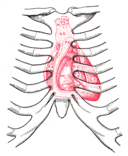
Surface anatomy
The aortic area, pulmonic area, tricuspid area and mitral area are areas on the surface of the chest where the heart is auscultated.Heart sounds result from reverberation within the blood associated with the sudden block of flow reversal by the valves closing. Because of this, auscultation to determine function of a valve is usually not performed at the position of the valve, but at the position to where the sound waves reverberate.
| Pulmonary valve Pulmonary valve The pulmonary valve is the semilunar valve of the heart that lies between the right ventricle and the pulmonary artery and has three cusps. Similar to the aortic valve, the pulmonary valve opens in ventricular systole, when the pressure in the right ventricle rises above the pressure in the... (to pulmonary trunk) |
left second intercostal space Intercostal space The intercostal space is the space between two ribs . Since there are 12 ribs on each side, there are 11 intercostal spaces, each numbered for the rib superior to it.-Structures in intercostal space:* several kinds of intercostal muscle... |
>- | right second intercostal space | >- | left fifth intercostal space | >- | left fourth intercostal space | lower left sternal border |
Recording heart sounds
Using electronic stethoscopesStethoscope
The stethoscope is an acoustic medical device for auscultation, or listening to the internal sounds of an animal body. It is often used to listen to lung and heart sounds. It is also used to listen to intestines and blood flow in arteries and veins...
, it is possible to record heart sounds via direct output to an external recording device, such as a laptop or MP3
MP3
MPEG-1 or MPEG-2 Audio Layer III, more commonly referred to as MP3, is a patented digital audio encoding format using a form of lossy data compression...
recorder. The same connection can be used to listen to the previously-recorded auscultation
Auscultation
Auscultation is the term for listening to the internal sounds of the body, usually using a stethoscope...
through the stethoscope headphones, allowing for more detailed study of murmur
Heart murmur
Murmurs are extra heart sounds that are produced as a result of turbulent blood flow that is sufficient to produce audible noise. Most murmurs can only be heard with the assistance of a stethoscope ....
s and other heart sounds, for general research as well as evaluation of a particular patient's condition.
See also
- PulsePulseIn medicine, one's pulse represents the tactile arterial palpation of the heartbeat by trained fingertips. The pulse may be palpated in any place that allows an artery to be compressed against a bone, such as at the neck , at the wrist , behind the knee , on the inside of the elbow , and near the...
- Physical examinationPhysical examinationPhysical examination or clinical examination is the process by which a doctor investigates the body of a patient for signs of disease. It generally follows the taking of the medical history — an account of the symptoms as experienced by the patient...
- Precordial examinationPrecordial examinationIn medicine, the precordial exam, also cardiac exam, is performed as part of a physical examination, or when a patient presents with chest pain suggestive of a cardiovascular pathology. In reality it's unlikely that this examination would be performed in isolation or in its entirety outside of a...
- Heart murmurHeart murmurMurmurs are extra heart sounds that are produced as a result of turbulent blood flow that is sufficient to produce audible noise. Most murmurs can only be heard with the assistance of a stethoscope ....
- Benign pediatric heart murmur
- Pulsatile TinnitusTinnitusTinnitus |ringing]]") is the perception of sound within the human ear in the absence of corresponding external sound.Tinnitus is not a disease, but a symptom that can result from a wide range of underlying causes: abnormally loud sounds in the ear canal for even the briefest period , ear...
- hearing a heartbeat sound in one or both ears
External links
- Diagram, with associated sounds at University of MichiganUniversity of MichiganThe University of Michigan is a public research university located in Ann Arbor, Michigan in the United States. It is the state's oldest university and the flagship campus of the University of Michigan...
- Flash tutorials at blaufuss.org
- Overview at University of DundeeUniversity of DundeeThe University of Dundee is a university based in the city and Royal burgh of Dundee on eastern coast of the central Lowlands of Scotland and with a small number of institutions elsewhere....
- Overview at University of WashingtonUniversity of WashingtonUniversity of Washington is a public research university, founded in 1861 in Seattle, Washington, United States. The UW is the largest university in the Northwest and the oldest public university on the West Coast. The university has three campuses, with its largest campus in the University...
- Recorded heart sounds at Auscultation Assistant at UCLA
- Heart sounds descriptions
- Heart sounds and Murmurs from the US Medical Videos Journal
- Lehrer, Steven. Understanding Pediatric Heart SoundsUnderstanding Pediatric Heart SoundsUnderstanding Pediatric Heart Sounds 2nd edition by Steven Lehrer is a book and audio CD that guides the student through the skills of pediatric heart auscultation. It provides a complete overview of pediatric heart examination, anatomy, physiology, and pathology. The audio CD presents and...
. Elsevier 2002. - Hanifin, Christopher. Heart Sounds: A Cardiac Auscultation Primer. CreateSpace, 2010.