
Arnold-Chiari malformation
Encyclopedia
Arnold–Chiari malformation, or often simply Chiari malformation, is a malformation of the brain
. It consists of a downward displacement of the cerebellar tonsil
s through the foramen magnum (the opening at the base of the skull), sometimes causing non-communicating hydrocephalus
as a result of obstruction of cerebrospinal fluid
(CSF) outflow. The cerebrospinal fluid outflow is caused by phase difference in outflow and influx of blood in the vasculature of the brain. It can cause headaches, fatigue, muscle weakness in the head and face, difficulty swallowing, dizziness, nausea, impaired coordination, and, in severe cases, paralysis.
Other conditions sometimes associated with Chiari Malformation include hydrocephalus
, syringomyelia
, spinal curvature
, tethered spinal cord syndrome
, and connective tissue disorders such as Ehlers-Danlos syndrome
and Marfan Syndrome
.
Chiari malformation is the most frequently used term for these types of malformations. The use of the term Arnold–Chiari malformation has fallen somewhat out of favor over time, although it is used to refer to the type II malformation. Current sources use "Chiari malformation" to describe four specific types of the condition, reserving the term "Arnold-Chiari" for type II only. Some sources still use "Arnold-Chiari" for all four types. This article uses the latter convention.
Chiari malformation or Arnold–Chiari malformation should not be confused with Budd-Chiari syndrome
, a hepatic condition also named for Hans Chiari
.
Brain Sagging and Pseudo-Chiari Malformation. The displacement of the cerebellar tonsils into the spinal canal may be mistaken for a Chiari I malformation, and some patients with spontaneous intracranial hypotension have undergone decompressive posterior fossa surgery.
The blockage of Cerebro-Spinal Fluid
(CSF) flow may also cause a syrinx
to form, eventually leading to syringomyelia
. Central cord symptoms such as hand weakness, dissociated sensory loss, and, in severe cases, paralysis may occur.
(MRI). The radiographic criteria for diagnosing a congenital Chiari I Malformation is a downward herniation of the cerebellar tonsil
s greater than 5 mm below the foramen magnum. Other imaging techniques involve the use of 3-D CT imaging of the brain and cine imaging (a movie of the brain) can be used to determine if the brainstem is being compressed by the pulsating arteries that surround it.
In the Syndrome of Occipitoatlantoaxial Hypermobility, cerebellar tonsillar herniation is typically only evident on an up-right MRI, due to the fact that the Chiari Malformation is gravitationally acquired by means of connective tissue weakness. 3-D CT imaging may aid in the diagnosis of related disorders such as retroflexed odontoid. Invasive cranial traction (lifting of the head off the spine) is often used as a confirmation of the diagnosis.
The diagnosis of a Chiari II Malformation can be made prenatally through Ultrasound
.
of the first and sometimes the second or even third cervical vertebrae
and part of the occipital bone
of the skull
to relieve pressure. The flow of spinal fluid may be accompanied by a shunt. Since this surgery usually involves the opening of the dura mater
and the expansion of the space beneath, a dural graft is usually applied to cover the expanded posterior fossa
.
A small number of neurological surgeons believe that detethering the spinal cord as an alternate approach relieves the compression of the brain against the skull opening (foramen magnum), obviating the need for decompression surgery and associated trauma. However, this approach is significantly less documented in the medical literature, with reports on only a handful of patients. It should be noted that the alternative spinal surgery is also not without risk.
On April 24, 2009, a young patient with Type 1 Chiari malformation was successfully treated with a minimally invasive endoscopic transnasal procedure by Dr. Richard Anderson at the Columbia University Medical Center Department of Neurosurgery.
related) is more difficult to treat than the congenital form of the disease. Individuals with this type do not respond well to the decompression surgery and often require an occipitoatlantoaxial fusion for stability. These patients are at risk of experiencing serious heart complications. Types I and II sufferers may also develop syringomyelia
. Type II is typically diagnosed at birth or prenatally. Approximately 33% of individuals with Chiari II malformation develop symptoms of brainstem damage within five years; a 1996 study found a mortality rate of 33% or more among symptomatic patients, with death frequently occurring due to respiratory failure
. 15% of individuals with Chiari II malformation die within two years of birth. Among children under two who also have myelomeningocele, it is the leading cause of death. Prognosis among children with Chiari II malformation who do not have spina bifida is linked to specific symptoms; the condition may be fatal among symptomatic children when it leads to neurological deterioration, but surgical intervention has shown promise. Types III and IV are extremely rare and patients generally do not survive past the age of two or three.
of Chiari I malformation, defined as tonsilar herniations of 3 to 5 mm or greater, is estimated to be in the range of one per 1000 to one per 5000 individuals. The incidence of symptomatic Chiari is less but unknown.
n pathologist, Hans Chiari
, first described these hindbrain malformations in the 1890s. A colleague of Professor Chiari, Dr. Julius Arnold, later contributed to the definition of the condition, and students of Dr. Arnold (Schwalbe and Gredig) suggested the term "Arnold-Chiari malformation" to henceforth refer to the condition.
Some sources credit the characterization of the condition to Cleland or Cruveilhier
.
in the tenth season episode "Internal Combustion" on February 4, 2010. Chiari was briefly mentioned in House, MD on the fifth season episode "House Divided"
and was the focus of the sixth season episode "The Choice."
Bobby Jones
- Legendary American golfer
Marissa Irwin
-model with Chiari secondary to Ehlers-Danlos syndrome
Brain
The brain is the center of the nervous system in all vertebrate and most invertebrate animals—only a few primitive invertebrates such as sponges, jellyfish, sea squirts and starfishes do not have one. It is located in the head, usually close to primary sensory apparatus such as vision, hearing,...
. It consists of a downward displacement of the cerebellar tonsil
Cerebellar tonsil
The cerebellar tonsil is analogous to a rounded lobule on the undersurface of each cerebellar hemisphere, continuous medially with the uvula of the Cerebellar vermis and superiorly by the flocculonodular lobe Synonyms include: tonsilla cerebelli, amygdala cerebelli, the latter of which is not to...
s through the foramen magnum (the opening at the base of the skull), sometimes causing non-communicating hydrocephalus
Hydrocephalus
Hydrocephalus , also known as "water in the brain," is a medical condition in which there is an abnormal accumulation of cerebrospinal fluid in the ventricles, or cavities, of the brain. This may cause increased intracranial pressure inside the skull and progressive enlargement of the head,...
as a result of obstruction of cerebrospinal fluid
Cerebrospinal fluid
Cerebrospinal fluid , Liquor cerebrospinalis, is a clear, colorless, bodily fluid, that occupies the subarachnoid space and the ventricular system around and inside the brain and spinal cord...
(CSF) outflow. The cerebrospinal fluid outflow is caused by phase difference in outflow and influx of blood in the vasculature of the brain. It can cause headaches, fatigue, muscle weakness in the head and face, difficulty swallowing, dizziness, nausea, impaired coordination, and, in severe cases, paralysis.
Classification
The Austrian pathologist Hans Chiari in the late 19th century described seemingly related anomalies of the hindbrain, the so called Chiari malformations I, II and III. Later, other investigators added a fourth (Chiari IV) malformation. The scale of severity is rated I - IV, with IV being the most severe. Types III and IV are very rare.| Type | Presentation | Other notes |
|---|---|---|
| I | A congenital malformation. Is generally asymptomatic during childhood, but often manifests with headaches and cerebellar symptoms. Herniation of cerebellar tonsil Cerebellar tonsil The cerebellar tonsil is analogous to a rounded lobule on the undersurface of each cerebellar hemisphere, continuous medially with the uvula of the Cerebellar vermis and superiorly by the flocculonodular lobe Synonyms include: tonsilla cerebelli, amygdala cerebelli, the latter of which is not to... s. |
The most common form. |
| Syndrome of occipitoatlantoaxial hypermobility | An acquired Chiari I Malformation in patients with hereditary disorders of connective tissue. Patients who exhibit extreme joint hypermobility Hypermobility Hypermobility describes joints that stretch farther than is normal. For example, some hypermobile people can bend their thumbs backwards to their wrists, bend their knee joints backwards, put their leg behind the head or other contortionist performances... and connective tissue weakness as a result of Ehlers-Danlos syndrome Ehlers-Danlos syndrome Ehlers–Danlos syndrome is a group of inherited connective tissue disorders, caused by a defect in the synthesis of collagen . The collagen in connective tissue helps tissues to resist deformation... or Marfan Syndrome Marfan syndrome Marfan syndrome is a genetic disorder of the connective tissue. People with Marfan's tend to be unusually tall, with long limbs and long, thin fingers.... are susceptible to instabilities of the craniocervical junction and thus acquiring a Chiari Malformation. This type is difficult to diagnose and treat. |
|
| II | Usually accompanied by a lumbar Lumbar In tetrapod anatomy, lumbar is an adjective that means of or pertaining to the abdominal segment of the torso, between the diaphragm and the sacrum ... myelomeningocele leading to partial or complete paralysis below the spinal defect. As opposed to the less pronounced tonsillar herniation seen with Chiari I, there is a larger cerebellar vermian displacement. Low lying torcular herophili, tectal beaking, and hydrocephalus with consequent clival hypoplasia are classic anatomic associations. The position of the torcular herophili is important for distinction from Dandy-Walker syndrome Dandy-Walker syndrome Dandy–Walker syndrome , or Dandy–Walker complex, is a congenital brain malformation involving the cerebellum and the fluid filled spaces around it. A key feature of this syndrome is the partial or even complete absence of the part of the brain located between the two cerebellar hemispheres... in which it is classically upturned. This is important because the hypoplastic cerebellum of Dandy-Walker may be difficult to distinguish from a Chiari malformation that has herniated or is ectopic on imaging. Colpocephaly Colpocephaly Colpocephaly refers to an abnormal appearance of the brain in which there is asymmetric dilatation of its lateral ventricle occipital horns, but with normal caliber frontal horns. It is typically associated with microcephaly and developmental delay... may be seen due to the associated neural tube defect. |
|
| III | Causes severe neurological defects. It is associated with an occipital Occiput The occiput is the anatomical term for the posterior portion of the head, in insects the posterior part of those head capsule.-Clinical significance:Trauma to the occiput can cause a basilar skull fracture.... encephalocele Encephalocele Encephalocele, sometimes known by the Latin name cranium bifidum, is a neural tube defect characterized by sac-like protrusions of the brain and the membranes that cover it through openings in the skull. These defects are caused by failure of the neural tube to close completely during fetal... . |
|
| IV | Characterized by a lack of cerebellar development. |
Other conditions sometimes associated with Chiari Malformation include hydrocephalus
Hydrocephalus
Hydrocephalus , also known as "water in the brain," is a medical condition in which there is an abnormal accumulation of cerebrospinal fluid in the ventricles, or cavities, of the brain. This may cause increased intracranial pressure inside the skull and progressive enlargement of the head,...
, syringomyelia
Syringomyelia
Syringomyelia is a generic term referring to a disorder in which a cyst or cavity forms within the spinal cord. This cyst, called a syrinx, can expand and elongate over time, destroying the spinal cord. The damage may result in pain, paralysis, weakness, and stiffness in the back, shoulders, and...
, spinal curvature
Spinal curvature
Although spinal curvature can refer to the normal concave and convex curvature of the spine, in clinical contexts, the phrase usually refers to deviations from the expected curvature, even when that difference is a reduction in curvature.Types include kyphosis, lordosis, and scoliosis.The thoracic...
, tethered spinal cord syndrome
Tethered spinal cord syndrome
Tethered spinal cord syndrome or occult spinal dysraphism sequence refers to a group of neurological disorders related to malformations of the spinal cord. The various forms include: tight filum terminale, lipomeningomyelocele, split cord malformations, dermal sinus tracts, dermoids, and cystoceles...
, and connective tissue disorders such as Ehlers-Danlos syndrome
Ehlers-Danlos syndrome
Ehlers–Danlos syndrome is a group of inherited connective tissue disorders, caused by a defect in the synthesis of collagen . The collagen in connective tissue helps tissues to resist deformation...
and Marfan Syndrome
Marfan syndrome
Marfan syndrome is a genetic disorder of the connective tissue. People with Marfan's tend to be unusually tall, with long limbs and long, thin fingers....
.
Chiari malformation is the most frequently used term for these types of malformations. The use of the term Arnold–Chiari malformation has fallen somewhat out of favor over time, although it is used to refer to the type II malformation. Current sources use "Chiari malformation" to describe four specific types of the condition, reserving the term "Arnold-Chiari" for type II only. Some sources still use "Arnold-Chiari" for all four types. This article uses the latter convention.
Chiari malformation or Arnold–Chiari malformation should not be confused with Budd-Chiari syndrome
Budd-Chiari syndrome
In medicine , Budd–Chiari syndrome is the clinical picture caused by occlusion of the hepatic veins. It presents with the classical triad of abdominal pain, ascites and hepatomegaly. Examples of occlusion include thrombosis of hepatic veins. The syndrome can be fulminant, acute, chronic, or...
, a hepatic condition also named for Hans Chiari
Hans Chiari
Hans Chiari was an Austrian pathologist who was a native of Vienna. He was the son of gynecologist Johann Baptist Chiari , and brother to rhinolaryngologist Ottokar Chiari ....
.
Brain Sagging and Pseudo-Chiari Malformation. The displacement of the cerebellar tonsils into the spinal canal may be mistaken for a Chiari I malformation, and some patients with spontaneous intracranial hypotension have undergone decompressive posterior fossa surgery.
Symptoms
- HeadacheHeadacheA headache or cephalalgia is pain anywhere in the region of the head or neck. It can be a symptom of a number of different conditions of the head and neck. The brain tissue itself is not sensitive to pain because it lacks pain receptors. Rather, the pain is caused by disturbance of the...
s aggravated by Valsalva maneuverValsalva maneuverThe Valsalva maneuver or Valsalva manoeuvre is performed by moderately forceful attempted exhalation against a closed airway, usually done by closing one's mouth and pinching one's nose shut...
s, such as yawning, laughing, crying, coughing, sneezing or straining - TinnitusTinnitusTinnitus |ringing]]") is the perception of sound within the human ear in the absence of corresponding external sound.Tinnitus is not a disease, but a symptom that can result from a wide range of underlying causes: abnormally loud sounds in the ear canal for even the briefest period , ear...
(ringing in the ears) - Dizziness and vertigo
- Nausea
- NystagmusNystagmusNystagmus is a condition of involuntary eye movement, acquired in infancy or later in life, that may result in reduced or limited vision.There are two key forms of Nystagmus: pathological and physiological, with variations within each type. Nystagmus may be caused by congenital disorders,...
(irregular eye movements) - Facial pain
- Muscle weakness
- Impaired gag reflex
- Restless Leg Syndrome
- Sleep ApneaSleep apneaSleep apnea is a sleep disorder characterized by abnormal pauses in breathing or instances of abnormally low breathing, during sleep. Each pause in breathing, called an apnea, can last from a few seconds to minutes, and may occur 5 to 30 times or more an hour. Similarly, each abnormally low...
- DysphagiaDysphagiaDysphagia is the medical term for the symptom of difficulty in swallowing. Although classified under "symptoms and signs" in ICD-10, the term is sometimes used as a condition in its own right. Sufferers are sometimes unaware of their dysphagia....
(difficulty swallowing) - Impaired coordination
- intercranial pressure
- pupillary dilatation
- DysautonomiaDysautonomiaDysautonomia is a broad term that describes any disease or malfunction of the autonomic nervous system. This includes postural orthostatic tachycardia syndrome , inappropriate sinus tachycardia , vasovagal syncope, mitral valve prolapse dysautonomia, pure autonomic failure, neurocardiogenic...
: tachycardiaTachycardiaTachycardia comes from the Greek words tachys and kardia . Tachycardia typically refers to a heart rate that exceeds the normal range for a resting heart rate...
(rapid heart), syncopeSyncope (medicine)Syncope , the medical term for fainting, is precisely defined as a transient loss of consciousness and postural tone characterized by rapid onset, short duration, and spontaneous recovery due to global cerebral hypoperfusion that most often results from hypotension.Many forms of syncope are...
(fainting), polydipsiaPolydipsiaPolydipsia is a medical symptom in which the patient displays excessive thirst. The word derives from the Greek πολυδιψία, which is derived from πολύς + δίψα...
(extreme thirst), chronic fatigue
The blockage of Cerebro-Spinal Fluid
Cerebrospinal fluid
Cerebrospinal fluid , Liquor cerebrospinalis, is a clear, colorless, bodily fluid, that occupies the subarachnoid space and the ventricular system around and inside the brain and spinal cord...
(CSF) flow may also cause a syrinx
Syrinx (medicine)
In medicine, a syrinx is a rare, fluid-filled neuroglial cavity within the spinal cord , in the brain stem , or in the nerves of the elbow, usually in a young age.-Etymology:...
to form, eventually leading to syringomyelia
Syringomyelia
Syringomyelia is a generic term referring to a disorder in which a cyst or cavity forms within the spinal cord. This cyst, called a syrinx, can expand and elongate over time, destroying the spinal cord. The damage may result in pain, paralysis, weakness, and stiffness in the back, shoulders, and...
. Central cord symptoms such as hand weakness, dissociated sensory loss, and, in severe cases, paralysis may occur.
Diagnosis
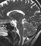
Diagnosis is made through a combination of patient history, neurological examination, and Magnetic Resonance ImagingMagnetic resonance imaging
Magnetic resonance imaging , nuclear magnetic resonance imaging , or magnetic resonance tomography is a medical imaging technique used in radiology to visualize detailed internal structures...
(MRI). The radiographic criteria for diagnosing a congenital Chiari I Malformation is a downward herniation of the cerebellar tonsil
Cerebellar tonsil
The cerebellar tonsil is analogous to a rounded lobule on the undersurface of each cerebellar hemisphere, continuous medially with the uvula of the Cerebellar vermis and superiorly by the flocculonodular lobe Synonyms include: tonsilla cerebelli, amygdala cerebelli, the latter of which is not to...
s greater than 5 mm below the foramen magnum. Other imaging techniques involve the use of 3-D CT imaging of the brain and cine imaging (a movie of the brain) can be used to determine if the brainstem is being compressed by the pulsating arteries that surround it.
In the Syndrome of Occipitoatlantoaxial Hypermobility, cerebellar tonsillar herniation is typically only evident on an up-right MRI, due to the fact that the Chiari Malformation is gravitationally acquired by means of connective tissue weakness. 3-D CT imaging may aid in the diagnosis of related disorders such as retroflexed odontoid. Invasive cranial traction (lifting of the head off the spine) is often used as a confirmation of the diagnosis.
The diagnosis of a Chiari II Malformation can be made prenatally through Ultrasound
Ultrasound
Ultrasound is cyclic sound pressure with a frequency greater than the upper limit of human hearing. Ultrasound is thus not separated from "normal" sound based on differences in physical properties, only the fact that humans cannot hear it. Although this limit varies from person to person, it is...
.
Treatment
Once symptomatic onset occurs, a common treatment is decompression surgery, in which a neurosurgeon usually removes the laminaLamina of the vertebral arch
The laminæ are two broad plates, extending dorsally and medially from the pedicles, fusing to complete the roof of the vertebral arch.Their upper borders and the lower parts of their anterior surfaces are rough for the attachment of the ligamenta flava....
of the first and sometimes the second or even third cervical vertebrae
Cervical vertebrae
In vertebrates, cervical vertebrae are those vertebrae immediately inferior to the skull.Thoracic vertebrae in all mammalian species are defined as those vertebrae that also carry a pair of ribs, and lie caudal to the cervical vertebrae. Further caudally follow the lumbar vertebrae, which also...
and part of the occipital bone
Occipital bone
The occipital bone, a saucer-shaped membrane bone situated at the back and lower part of the cranium, is trapezoidal in shape and curved on itself...
of the skull
Human skull
The human skull is a bony structure, skeleton, that is in the human head and which supports the structures of the face and forms a cavity for the brain.In humans, the adult skull is normally made up of 22 bones...
to relieve pressure. The flow of spinal fluid may be accompanied by a shunt. Since this surgery usually involves the opening of the dura mater
Dura mater
The dura mater , or dura, is the outermost of the three layers of the meninges surrounding the brain and spinal cord. It is derived from Mesoderm. The other two meningeal layers are the pia mater and the arachnoid mater. The dura surrounds the brain and the spinal cord and is responsible for...
and the expansion of the space beneath, a dural graft is usually applied to cover the expanded posterior fossa
Posterior cranial fossa
The posterior cranial fossa is part of the intracranial cavity, located between the foramen magnum and tentorium cerebelli. It contains the brainstem and cerebellum.This is the most inferior of the fossae. It houses the cerebellum, medulla and pons....
.
A small number of neurological surgeons believe that detethering the spinal cord as an alternate approach relieves the compression of the brain against the skull opening (foramen magnum), obviating the need for decompression surgery and associated trauma. However, this approach is significantly less documented in the medical literature, with reports on only a handful of patients. It should be noted that the alternative spinal surgery is also not without risk.
On April 24, 2009, a young patient with Type 1 Chiari malformation was successfully treated with a minimally invasive endoscopic transnasal procedure by Dr. Richard Anderson at the Columbia University Medical Center Department of Neurosurgery.
Prognosis
The prognosis differs dependent on the type of malformation (i.e., type I, II, III, or IV). Type I is generally adult-onset and, while not curable, is treatable and rarely fatal. Syndrome of Occipitoatlantoaxial Hypermobility (Ehlers-Danlos syndromeEhlers-Danlos syndrome
Ehlers–Danlos syndrome is a group of inherited connective tissue disorders, caused by a defect in the synthesis of collagen . The collagen in connective tissue helps tissues to resist deformation...
related) is more difficult to treat than the congenital form of the disease. Individuals with this type do not respond well to the decompression surgery and often require an occipitoatlantoaxial fusion for stability. These patients are at risk of experiencing serious heart complications. Types I and II sufferers may also develop syringomyelia
Syringomyelia
Syringomyelia is a generic term referring to a disorder in which a cyst or cavity forms within the spinal cord. This cyst, called a syrinx, can expand and elongate over time, destroying the spinal cord. The damage may result in pain, paralysis, weakness, and stiffness in the back, shoulders, and...
. Type II is typically diagnosed at birth or prenatally. Approximately 33% of individuals with Chiari II malformation develop symptoms of brainstem damage within five years; a 1996 study found a mortality rate of 33% or more among symptomatic patients, with death frequently occurring due to respiratory failure
Respiratory failure
The term respiratory failure, in medicine, is used to describe inadequate gas exchange by the respiratory system, with the result that arterial oxygen and/or carbon dioxide levels cannot be maintained within their normal ranges. A drop in blood oxygenation is known as hypoxemia; a rise in arterial...
. 15% of individuals with Chiari II malformation die within two years of birth. Among children under two who also have myelomeningocele, it is the leading cause of death. Prognosis among children with Chiari II malformation who do not have spina bifida is linked to specific symptoms; the condition may be fatal among symptomatic children when it leads to neurological deterioration, but surgical intervention has shown promise. Types III and IV are extremely rare and patients generally do not survive past the age of two or three.
Epidemiology
The prevalencePrevalence
In epidemiology, the prevalence of a health-related state in a statistical population is defined as the total number of cases of the risk factor in the population at a given time, or the total number of cases in the population, divided by the number of individuals in the population...
of Chiari I malformation, defined as tonsilar herniations of 3 to 5 mm or greater, is estimated to be in the range of one per 1000 to one per 5000 individuals. The incidence of symptomatic Chiari is less but unknown.
History
An AustriaAustria
Austria , officially the Republic of Austria , is a landlocked country of roughly 8.4 million people in Central Europe. It is bordered by the Czech Republic and Germany to the north, Slovakia and Hungary to the east, Slovenia and Italy to the south, and Switzerland and Liechtenstein to the...
n pathologist, Hans Chiari
Hans Chiari
Hans Chiari was an Austrian pathologist who was a native of Vienna. He was the son of gynecologist Johann Baptist Chiari , and brother to rhinolaryngologist Ottokar Chiari ....
, first described these hindbrain malformations in the 1890s. A colleague of Professor Chiari, Dr. Julius Arnold, later contributed to the definition of the condition, and students of Dr. Arnold (Schwalbe and Gredig) suggested the term "Arnold-Chiari malformation" to henceforth refer to the condition.
Some sources credit the characterization of the condition to Cleland or Cruveilhier
Jean Cruveilhier
Jean Cruveilhier was a French anatomist and pathologist.In 1816 he earned his doctorate in Paris, where in 1825 he succeeded Pierre Augustin Béclard as professor of anatomy...
.
Society and culture
The condition was brought to the mainstream on the series CSICSI: Crime Scene Investigation
CSI: Crime Scene Investigation is an American crime drama television series, which premiered on CBS on October 6, 2000. The show was created by Anthony E. Zuiker and produced by Jerry Bruckheimer...
in the tenth season episode "Internal Combustion" on February 4, 2010. Chiari was briefly mentioned in House, MD on the fifth season episode "House Divided"
and was the focus of the sixth season episode "The Choice."
Notable cases
Rosanne CashRosanne Cash
Rosanne Cash is an American singer-songwriter and author. She is the eldest daughter of the late country music singer Johnny Cash and his first wife, Vivian Liberto Cash Distin....
Bobby Jones
Bobby Jones (golfer)
Robert Tyre "Bobby" Jones Jr. was an American amateur golfer, and a lawyer by profession. Jones was the most successful amateur golfer ever to compete on a national and international level...
- Legendary American golfer
Marissa Irwin
Marissa Irwin
Marissa Irwin is an American model best known for her appearances on the cover of Bridal Guide and in Seventeen Magazine.A native of Canton, Ohio, she received training from the Barbizon modeling school of Akron. She now lives and works in New York City.Marissa lives with a rare and painful...
-model with Chiari secondary to Ehlers-Danlos syndrome
Ehlers-Danlos syndrome
Ehlers–Danlos syndrome is a group of inherited connective tissue disorders, caused by a defect in the synthesis of collagen . The collagen in connective tissue helps tissues to resist deformation...
External links
- Duke Chiari Research
- Chari malformation patient information from The Mount Sinai Medical Center
- Chiari Connection International—Chiari and Related Disorders Information
- Chiari & Syringomyelia Foundation
- Conquer Chiari
- World Arnold Chiari Malformation Association
- American Syringomyelia Alliance Project
- The Ann Conroy Trust
- Chiari One