
Photoacoustic Imaging
Encyclopedia

Photoacoustic spectroscopy
Photoacoustic spectroscopy is the measurement of the effect of absorbed electromagnetic energy on matter by means of acoustic detection. The discovery of the photoacoustic effect dates to 1880 when Alexander Graham Bell showed that thin discs emitted sound when exposed to a beam of sunlight that...
. In photoacoustic imaging, non-ionizing laser
Laser
A laser is a device that emits light through a process of optical amplification based on the stimulated emission of photons. The term "laser" originated as an acronym for Light Amplification by Stimulated Emission of Radiation...
pulses are delivered into biological tissues (when radio frequency
Radio frequency
Radio frequency is a rate of oscillation in the range of about 3 kHz to 300 GHz, which corresponds to the frequency of radio waves, and the alternating currents which carry radio signals...
pulses are used, the technology is referred to as thermoacoustic imaging). Some of the delivered energy will be absorbed and converted into heat, leading to transient thermoelastic expansion and thus wideband (e.g. MHz) ultrasonic
Ultrasound
Ultrasound is cyclic sound pressure with a frequency greater than the upper limit of human hearing. Ultrasound is thus not separated from "normal" sound based on differences in physical properties, only the fact that humans cannot hear it. Although this limit varies from person to person, it is...
emission. The generated ultrasonic waves are then detected by ultrasonic transducers to form images. It is known that optical absorption is closely associated with physiological properties, such as hemoglobin
Hemoglobin
Hemoglobin is the iron-containing oxygen-transport metalloprotein in the red blood cells of all vertebrates, with the exception of the fish family Channichthyidae, as well as the tissues of some invertebrates...
concentration and oxygen saturation
Oxygen saturation
Oxygen saturation or dissolved oxygen is a relative measure of the amount of oxygen that is dissolved or carried in a given medium. It can be measured with a dissolved oxygen probe such as an oxygen sensor or an optode in liquid media, usually water.It has particular significance in medicine and...
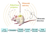
. As a result, the magnitude of the ultrasonic emission (i.e. photoacoustic signal), which is proportional to the local energy deposition, reveals physiologically specific optical absorption contrast. 2D or 3D images of the targeted areas can then be formed. Fig. 1 is a schematic illustration showing the basic principles of photoacoustic imaging.

Endogenous
Endogenous substances are those that originate from within an organism, tissue, or cell. Endogenous retroviruses are caused by ancient infections of germ cells in humans, mammals and other vertebrates...
molecules such as hemoglobin or melanin
Melanin
Melanin is a pigment that is ubiquitous in nature, being found in most organisms . In animals melanin pigments are derivatives of the amino acid tyrosine. The most common form of biological melanin is eumelanin, a brown-black polymer of dihydroxyindole carboxylic acids, and their reduced forms...
, or exogenously delivered contrast agents. As an example, Fig. 2 shows the optical absorption spectra of oxygenated hemoglobin
Hemoglobin
Hemoglobin is the iron-containing oxygen-transport metalloprotein in the red blood cells of all vertebrates, with the exception of the fish family Channichthyidae, as well as the tissues of some invertebrates...
(HbO2) and deoxygenated hemoglobin
Hemoglobin
Hemoglobin is the iron-containing oxygen-transport metalloprotein in the red blood cells of all vertebrates, with the exception of the fish family Channichthyidae, as well as the tissues of some invertebrates...
(Hb) in the visible and near infrared region. Since blood usually has orders of magnitude larger absorption than surrounding tissues, there is sufficient endogenous contrast for photoacoustic imaging to visualize blood vessels. Recent studies have shown that photoacoustic imaging can be used in vivo
In vivo
In vivo is experimentation using a whole, living organism as opposed to a partial or dead organism, or an in vitro controlled environment. Animal testing and clinical trials are two forms of in vivo research...
for tumor angiogenesis
Angiogenesis
Angiogenesis is the physiological process involving the growth of new blood vessels from pre-existing vessels. Though there has been some debate over terminology, vasculogenesis is the term used for spontaneous blood-vessel formation, and intussusception is the term for the formation of new blood...
monitoring, blood oxygenation
Oxygenation (medical)
Oxygenation occurs when oxygen molecules enter the tissues of the body. For example, blood is oxygenated in the lungs, where oxygen molecules travel from the air and into the blood...
mapping, functional brain imaging, and skin melanoma
Melanoma
Melanoma is a malignant tumor of melanocytes. Melanocytes are cells that produce the dark pigment, melanin, which is responsible for the color of skin. They predominantly occur in skin, but are also found in other parts of the body, including the bowel and the eye...
detection etc.
Advantages
| Δf | Primary contrast | Δz | δz | δx | Speed | |
|---|---|---|---|---|---|---|
| Hz | mm | µm | µm | Mvx/s | ||
| Photoacoustic microscopy | 50 M | Optical absorption | 3 | 15 | 45 | 0.5 |
| Photoacoustic microscopy | 5 M | Optical absorption | 50 | 700 | 700 | 0.5 |
| Confocal microscopy Confocal microscopy Confocal microscopy is an optical imaging technique used to increase optical resolution and contrast of a micrograph by using point illumination and a spatial pinhole to eliminate out-of-focus light in specimens that are thicker than the focal plane. It enables the reconstruction of... |
Fluorescence, scattering | 0.2 | 3-20 | 0.3-3 | 10-100 | |
| Two-photon microscopy | Fluorescence | 0.5-1.0 | 1-10 | 0.3-3 | 10-100 | |
| Optical coherence tomography Optical coherence tomography Optical coherence tomography is an optical signal acquisition and processing method. It captures micrometer-resolution, three-dimensional images from within optical scattering media . Optical coherence tomography is an interferometric technique, typically employing near-infrared light... |
50 T | Optical scattering | 1-2 | 0.5-10 | 1-10 | 20-4.000 |
| Scanning Laser Acoustic Microscopy | 300 M | Ultrasonic scattering | 1-2 | 20 | 20 | 10 |
| Acoustic microscopy Acoustic microscopy Acoustic microscopy is microscopy that employs very high or ultra high frequency ultrasound. Acoustic microscopes operate nondestructively and penetrate most solid materials to make visible images of internal features, including defects such as cracks, delaminations and voids.-Types of acoustic... |
50 M | Ultrasonic scattering | 20 | 20-100 | 80-160 | 0.1 |
| Ultrasonography | 5 M | Ultrasonic scattering | 60 | 300 | 300 | 1 |
| Table 1. Comparison of contrast mechanisms, penetration depth (Δz), axial resolution (δz), lateral resolution (δx=δy) and imaging speed of confocal microscopy, two-photon microscopy, optical coherence tomography (50 THz), ultrasound microscopy (50 MHz), ultrasound imaging (5 MHz), photoacoustic microscopy (50 MHz), and photoacoustic tomography (3.5 MHz). Speeds in Megavoxel per second of non-parallel techniques. | ||||||
Imaging systems
Two types of photoacoustic imaging systems, photoacoustic/thermoacoustic computed tomography (also known as photoacoustic/thermoacoustic tomography, i.e., PAT/TAT) and photoacoustic microscopy (PAM), have been developed. A typical PAT system uses an unfocused ultrasound detector to acquire the photoacoustic signals, and the image is reconstructed by inversely solving the photoacoustic equations. A PAM system, on the other hand, uses a spherically focused ultrasound detector with 2D point-by-point scanning and requires no reconstruction algorithm.General equation
Given the heating function , the generation and propagation of photoacoustic wave pressure
  in an acoustically homogeneous inviscid medium is governed by
where
 is the speed of sound in medium,
  is the thermal expansion coefficient, and
  is the specific heat capacity at constant pressure. Eq. (1) holds under thermal confinement to ensure that heat conduction is negligible during the laser pulse excitation. The thermal confinement occurs when the laser pulsewidth is much shorter than the thermal relaxation time.
The forward solution of Eq. (1) is given by

In stress confinement, which occurs when the laser pulsewidth is much shorter than the stress relaxation time, Eq. (2) can be further derived as

where
 is the initial photoacoustic pressure.
Universal reconstruction algorithm
In PAT system, the acoustic pressure is detected by scanning an ultrasound transducer over a surface that encloses the photoacoustic source. To reconstruct the internal source distribution, we need to solve the inverse problem of equation (3) (i.e. to obtain ).
).A representative method applied for PAT reconstruction is known as the universal back projection algorithm. This method is suitable for three imaging geometries: planar, spherical, and cylindrical surfaces.
The universal backprojection formula is

where
 is the solid angle subtended by the entire surface
  with respect to the reconstruction point
  inside
  , and
, and
A typical PAT/TAT system
A typical PAT system is shown as the left part in Fig. 3. The laser beam is expanded and diffused to cover the whole region of interest. Photoacoustic waves are generated proportional to the distribution of optical absorption in the target, and are detected by an ultrasonic transducer. TAT system is the same as PAT system except using a microwave excitation source instead of laser. A typical TAT system is shown as the right part in Fig. 3. Although single-element transducers have been employed in these two systems, the detection scheme can be extended to using ultrasound arrays as well.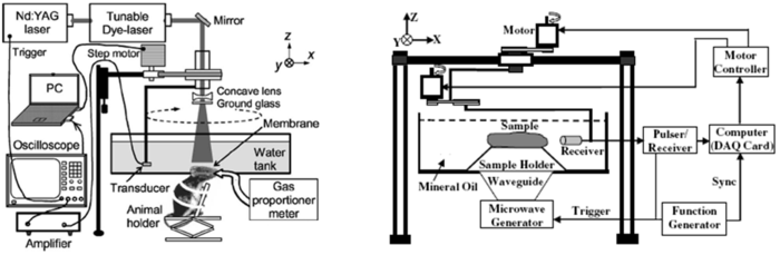
Biomedical applications of PAT/TAT
Intrinsic optical/microwave absorption contrast and diffraction-limited high spatial resolution of ultrasound make PAT and TAT promising imaging modalities for wide biomedical applications:Brain lesion detection

HemodynamicsHemodynamicsHemodynamics, meaning literally "blood movement" is the study of blood flow or the circulation.All animal cells require oxygen for the conversion of carbohydrates, fats and proteins into carbon dioxide , water and energy in a process known as aerobic respiration...
monitoring
Since HbO2 and Hb are the dominant absorbing compounds in biological tissues in the visible spectral range, multiple wavelength photoacoustic measurements can be used to reveal the relative concentration of these two chromophores. Thus, the relative total concentration of hemoglobin (HbT) and the hemoglobin oxygen saturation
Oxygen saturation
Oxygen saturation or dissolved oxygen is a relative measure of the amount of oxygen that is dissolved or carried in a given medium. It can be measured with a dissolved oxygen probe such as an oxygen sensor or an optode in liquid media, usually water.It has particular significance in medicine and...
(sO2) can be derived. Therefore, cerebral hemodynamic changes associated with brain function can be successfully detected with PAT.
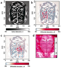

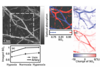
Parietal bone
The parietal bones are bones in the human skull which, when joined together, form the sides and roof of the cranium. Each bone is roughly quadrilateral in form, and has two surfaces, four borders, and four angles. It is named from the Latin pariet-, wall....
; L, lambda; M, midline; A, activated regions corresponding to whisker stimulation).
Fig. 6 shows functional changes of sO2 and HbT in the rat cerebral cortex as a result of the physiological modulations. Under the hyperoxia status, the averaged sO2 level,
Breast cancerBreast cancerBreast cancer is cancer originating from breast tissue, most commonly from the inner lining of milk ducts or the lobules that supply the ducts with milk. Cancers originating from ducts are known as ductal carcinomas; those originating from lobules are known as lobular carcinomas...
diagnosis

Photoacoustic microscopy (PAM)
Fig. 8 shows a representative PAM set-up. A tunable dye laser is pumped by a Q-switched pulsed Nd:YAG (neodymium: yttrium aluminum garnet) laser. A short laser pulse at a certain wavelength between 532-770 nm is generated to irradiate the target tissue to induce acoustic pressure waves. Laser pulses of ~6 mJ/cm2 at the focus will be delivered at 10 Hz repetition rate. An optical fiberOptical fiber
An optical fiber is a flexible, transparent fiber made of a pure glass not much wider than a human hair. It functions as a waveguide, or "light pipe", to transmit light between the two ends of the fiber. The field of applied science and engineering concerned with the design and application of...
of 0.6 mm core diameter is coaxially positioned on a three-dimensional mechanical stage with changeable ultrasound
Ultrasound
Ultrasound is cyclic sound pressure with a frequency greater than the upper limit of human hearing. Ultrasound is thus not separated from "normal" sound based on differences in physical properties, only the fact that humans cannot hear it. Although this limit varies from person to person, it is...
transducers between 25-75 MHz.

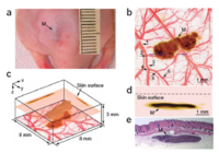
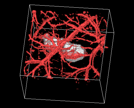
PAM has multiple important applications in functional imaging. Two examples are shown in Figs. 9 and 10. PAM can detect changes in oxygenated/deoxygenated hemoglobin in small vessels. As shown in Fig. 9, arterioles (red) and venules (blue) are clearly delineated with high spatial resolution. Fig. 10 shows the capability of PAM to image skin melanoma by using dual wavelength to obtain the morphological relationship between the melanoma and its surrounding blood vessels structures in vivo.
External links
- http://labs.seas.wustl.edu/bme/Wang/index.html Optical Imaging Laboratory, Department of Biomedical Engineering, Washington University in St. Louis
- http://www.medphys.ucl.ac.uk/research/mle/index.htm Photoacoustic Imaging Group, Department of Medical Physics and Bioengineering, University College London
- http://fluor.unibe.ch/optoacoustics/index.htm Optoacoustic Imaging Lab, Department of Biomedical Photonics, Institute of Applied Physics, University of Bern
- http://bmo.tnw.utwente.nl/bmo/techniques.htm Biomedical Optics, Biophysical Engineering group, Faculty of Science and Technology, University of Twente, Enschede, The Netherlands
- http://foil.northwestern.edu Functional Optical Imaging Laboratory (FOIL), Department of Biomedical Engineering, Northwestern University
- http://www.bme.ogi.edu/biomedicaloptics/wang/ Biophotonics and Imaging Laboratory (BAIL), Department of Biomedical Engineering, Oregon Health & Science University
- http://www.optosonics.com/ OptoSonics, Inc.
- http://www.fairwaymed.com/ Fairway Medical Technologies, Inc.
- http://www.engr.uconn.edu/ece/labs/optlab/ Biomedical Optical Imaging Laboratory, Department of Electrical & Computer Engineering, University of Connecticut
- http://csc.mat.univie.ac.at/pai/ An interdisciplinary consortium of experts in photoacoustic imaging in Austria where research in physics and mathematics is heavily driven by input from biology and medicine
- http://www3.interscience.wiley.com/journal/116325199/abstract Recent advances in application of acoustic, acousto-optic and photoacoustic methods in biology and medicine
- http://ultrasound.bme.utexas.edu/ Ultrasound Imaging and Therapeutics Research Laboratory, Department of Biomedical Engineering, University of Texas at Austin