

Visual extinction
Encyclopedia
Neurological disorder
A neurological disorder is a disorder of the body's nervous system. Structural, biochemical or electrical abnormalities in the brain, spinal cord, or in the nerves leading to or from them, can result in symptoms such as paralysis, muscle weakness, poor coordination, loss of sensation, seizures,...
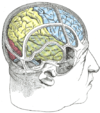
which occurs following damage to the parietal lobe
Parietal lobe
The parietal lobe is a part of the Brain positioned above the occipital lobe and behind the frontal lobe.The parietal lobe integrates sensory information from different modalities, particularly determining spatial sense and navigation. For example, it comprises somatosensory cortex and the...
of the brain. It is similar to, but distinct from, hemispatial neglect
Hemispatial neglect
Hemispatial neglect, also called hemiagnosia, hemineglect, unilateral neglect, spatial neglect, unilateral visual inattention, hemi-inattention or neglect syndrome is a neuropsychological condition in which, after damage to one hemisphere of the brain, a deficit in attention to and awareness of...
. Visual extinction has the characteristic symptom of difficulty to perceive contralesional stimuli when presented simultaneously with an ipsilesional stimulus, but the ability to correctly identify them when not presented simultaneously. Under simultaneous presentation, the contralesional stimulus is apparently ignored by the patient, or extinguished. This deficiency may lead to difficulty on behalf of the patient with processing the stimuli’s 3D position.
History
Visual Extinction is the result of unilateral cerebral damage and has always been poorly understood. Researchers have been studying visual extinction in great depth since the 1990s. It has since been commonly associated with damage to the right hemisphereCerebral hemisphere
A cerebral hemisphere is one of the two regions of the eutherian brain that are delineated by the median plane, . The brain can thus be described as being divided into left and right cerebral hemispheres. Each of these hemispheres has an outer layer of grey matter called the cerebral cortex that is...
of the brain
Human brain
The human brain has the same general structure as the brains of other mammals, but is over three times larger than the brain of a typical mammal with an equivalent body size. Estimates for the number of neurons in the human brain range from 80 to 120 billion...
. Studies have suggested that visual extinction may be a result of sensory imbalance. This imbalance is due to weak or delayed afferent inputs in the hemisphere affected by the extinction.
Research done by Pavlovskaya, Sagi, Soroker and Ring show that visual extinction is dependent on simple stimuli properties. These properties are thought to reflect connectivity constraints during the early steps of visual processing.
Physiological Basis
Visual extinction arises from damage to the parietal lobe of the brain. This damage most frequently arises following a stroke or other infarction in that area – however, any traumatic event sufficient to cause widespread tissue damage in the area may cause sufficient harm. Although both sides of the brain are vulnerable to such damage, it is more unusual for extinction to occur when the left hemisphere is damaged than the right.When presented with simultaneous stimuli, the patient will ignore the contralesional stimuli, and only report the ipsilesional. Their ability to report the stimuli correctly when presented singly indicates that this is not a problem with vision per se. This bias against contralesional stimuli is evident even when patients are presented with two signals within the ipsilesional visual field, whose processing remains intact following the damage, since it is done in the opposite brain hemisphere. This implies that extinction can be somewhat controlled by biasing the point of visual fixation
Fixation (visual)
Fixation or visual fixation is the maintaining of the visual gaze on a single location. Humans typically alternate saccades and visual fixations, the notable exception being in smooth pursuit, controlled by a different neural substrate that appear to have developed for hunting prey...
contralesionally, such that all relevant stimuli are contained within the functioning side of the visual field, findings supported in experiment
Fatigue and habituation effects have previously been connected to visual extinction; experiments such as those done by Vuilleumier and Rafal eliminate the connection of these effects with visual extinction.
Historically, it was believed that the parietal damage weakened afferent neuron input to the visual cortex
Visual cortex
The visual cortex of the brain is the part of the cerebral cortex responsible for processing visual information. It is located in the occipital lobe, in the back of the brain....
, and so the extinction event was caused by the signals originating in the contralesional field being lost during transmission. However, this does not account for the ability of extinction patients being able to correctly identify contralesional stimuli in isolation. A more current theory is called the "race theory."
Race theory holds that stimuli are competing for processing power within the brain. Following parietal damage, all contralesional messages are lowered in priority for the brain’s processing in some way – potentially by signal transmission delay – such that when presented with alternative stimuli, the contralesional stimulus doesn’t arrive at the decision center simultaneously with the ipsilesional stimulus, and thus is denied conscious processing.
The race theory sees some support in findings by Marzi et al., where patients who show extinction also display aggravated delays in their reaction time to stimuli, both when compared to patients who have other forms of right-brain damage and undamaged patients. In the study, patients were shown to be able to focus their attention and improve reaction time, but still have subnormal performance. This theory is given further support by Gorea and Sagi’s inducing of extinction-like events in undamaged patients when presented with simultaneous stimuli of different intensities
Light intensity
Several measures of light are commonly known as intensity. These are obtained by dividing either a power or a luminous flux by a solid angle, a planar area, or a combination of the two...
, with the lower intensity stimulus being extinguished by the mere presence of the high intensity stimulus.
Extinction frequency may also be affected by reporting method, due to how visual processing interacts with the rest of the brain. Reported extinctions drop when the patient is told to relay the extinction news by a method other than speaking – particularly, the use of the eyes in a feedback loop (looking to the side if there is a single stimuli, and up if there are two). This may work by eliminating the need for hemispheric transfer of information
Corpus callosum
The corpus callosum , also known as the colossal commissure, is a wide, flat bundle of neural fibers beneath the cortex in the eutherian brain at the longitudinal fissure. It connects the left and right cerebral hemispheres and facilitates interhemispheric communication...
– routing the response to the motor cortex
Motor cortex
Motor cortex is a term that describes regions of the cerebral cortex involved in the planning, control, and execution of voluntary motor functions.-Anatomy of the motor cortex :The motor cortex can be divided into four main parts:...
instead of the visual cortex – and so shortening the path and response time, a conclusion supported by increased detection of isolated contralesional stimuli under such circumstances.
Although extinction is compared with hemispatial neglect, the two are different disorders. It is possible to exhibit one without the other, and the neural basis for each appears to be different, hemispatial neglect arising from tempo-parietal junction damage. Although patients may exhibit these disorders simultaneously, this occurs in less than half of the afflicted population.
Characteristics
- When measuring the ERPEvent-related potentialAn event-related potential is any measured brain response that is directly the result of a thought or perception. More formally, it is any stereotyped electrophysiological response to an internal or external stimulus....
of the right parietal lobe during extinction events, EEGs do not display the P1 or N1 waves otherwise evidenced in healthy bilateral response. - When presented with stimuli, eye saccadeSaccadeA saccade is a fast movement of an eye, head or other part of an animal's body or device. It can also be a fast shift in frequency of an emitted signal or other quick change. Saccades are quick, simultaneous movements of both eyes in the same direction...
s from fixation point are more common in extinction patients, are longer in duration, and show ipsilesional bias in movement. - Some evidence for a slight increase in detection of eccentric stimuli in the ipsilesional visual fieldVisual fieldThe term visual field is sometimes used as a synonym to field of view, though they do not designate the same thing. The visual field is the "spatial array of visual sensations available to observation in introspectionist psychological experiments", while 'field of view' "refers to the physical...
– i.e. slightly improved peripheral vision. - Patients show increased difference in reaction time (crossed-uncrossed difference) for processing in contralesional and ipsilesional visual hemispheres.
Diagnosis
Diagnosis is achieved through several related methods. One approach involves having patients sit in front of a computer screen, on which either stars or triangles are projected simultaneously in different areas of the screen. Patients are then asked to identify the locations and identities of the shapes. Another similar approach is to project colored letters on the screen, again asking patients to identify the letter and color in different parts of the screen. From this information, a diagnosis of visual extinction can be attained. A positive diagnosis results from a patient's inability to correctly identify shapes or letters projected simultaneously in different areas of the screen. However, the most common test for visual extinction is the finger confrontation model. In this test, the doctor asks the patient to note which of his hands have moving fingers. This can be used immediately following a stroke, for the doctor’s quick diagnosis.Treatment
Without any sort of treatment, visual extinction usually worsens in severity of symptoms or remains completely stagnant. Simple exercises, such as readingReading (process)
Reading is a complex cognitive process of decoding symbols for the intention of constructing or deriving meaning . It is a means of language acquisition, of communication, and of sharing information and ideas...
and copying tasks, can be useful in treating the symptoms and increasing brain activity, though the damaged area can never be completely healed since the dead brain cells do not regenerate. An individual should keep up with exercises designed to maintain or improve function in order to create the best chance of improvement and/or maintenance at his or her current state.
Treatment methods for patients who suffer from visual extinction generally involve use and training of an individual's vision
Visual perception
Visual perception is the ability to interpret information and surroundings from the effects of visible light reaching the eye. The resulting perception is also known as eyesight, sight, or vision...
. A doctor may instruct a patient to scan rows of lights in different ways in an attempt to regenerate function. Some light boards do exist that are used specifically to aid this task. Other methods exist that are designed to force the patient to focus on multiple stimuli at the same time. These cueing and scanning methods do yield results, but they are not consistent across all patients. In successful cases, generally forty hours of retraining the patients' vision were necessary as well as some additional work done at home with similar exercises.
Other computer scanning methods of treatment were shown to be effective with specialized equipment designed specifically for scanning and cueing exercises to force patients to focus on multiple stimuli simultaneously. Again, the results of these methods were not consistent, but in laboratory settings rehabilitation was successful. Of the individuals who recovered some function, not all maintained the function they recovered, losing it after about five months.
In another, single-stimulus rehabilitatory approach, the patient is shown a computer screen with a box in each corner and a fixation cross at the center. When a stimulus appears in any of the boxes, the patient is instructed to indicate seeing it as quickly as possible. This task is eased somewhat by an indicating icon before the stimulus – either an arrow that points to the box the stimulus is most likely (but not guaranteed) to be in, or a cross to indicate that the stimulus could appear in any box. Following thirty hour-long sessions, patients demonstrated a significant decrease in both extinction and hemispatial neglect.
In patients whose brain damage was in the right hemisphere, left limb activation (LLA) was another treatment that proved effective. The theory behind this method of treatment is that any use of the damaged side of the brain will enhance all functions related to the damaged hemisphere and that use of the opposite (healthy) side will only cause further impairment. LLA therefore is merely the concentrated use of the left side of the body in patients whose damage is in the right hemisphere (since the right hemisphere controls the left side of the body). This forces the patient to exercise spatial awareness using the damaged side of the brain, increasing activity around the lesion. While generally a treatment used for visual neglect (a different but related neurological disorder), LLA still increases brain activity around damaged area in patients with visual extinction and generates improvement in some patients. The reason this treatment does not work in some cases of visual extinction is that damage can be on either side of the brain as opposed to in visual neglect where the damage is always on the right.
Society & Culture
This condition does not inhibit patients from social interaction. In fact, most people would not be able to distinguish a visual extinction patient from a non-visual extinction patient in passing. Patients have selective spatial interactions, typically within the range of six degrees of the angle of vision. When two visual stimuli are presented to a patient, they can be processed as a single object due to the corresponding neuronal functions which are linked through long-range lateral interactions. Visual Extinction is often mistaken for attentional deficit. Some researchers believe visual extinction may be connected to a restriction in attention capacity. Attention allows a person to identify and react to pertinent objects in space, while ignoring other irrelevant objects. Patients with visual extinction, especially those with unilateral damage to the right parietal lobe, may be unable to attend and orient to objects in collateral space, therefore presenting neglect to visual stimuli.A delay in reaction time is observed in many patients, but it is unknown whether this is a primary result of the stroke
Stroke
A stroke, previously known medically as a cerebrovascular accident , is the rapidly developing loss of brain function due to disturbance in the blood supply to the brain. This can be due to ischemia caused by blockage , or a hemorrhage...
or resulting from the visual extinction. Detrimental social aspects may be repercussions of the stroke, which caused the visual extinction, but not from the visual extinction condition alone.
Visual Extinction and Image Preprocessing
Visual extinction has been involved in several studies regarding preprocessing of images in the brain. A study performed by Dr Vuilleumier examined the effect of using faces as the stimulus in studying an extinction event. Subjects were presented with two simultaneous stimuli – some combination of a schematic face, a shape, a word, and a scrambled face. Faces in the contralesional field were less likely to be extinguished than other stimuli, but faces in the ipsilesional field appeared to induce more extinction events. The appearance of a face was prioritized in attention, despite the handicap presented of extinction damage.Visual extinction has also been used to demonstrate brain bias towards gestalt
Gestalt
Die Gestalt is a German word for form or shape. It is used in English to refer to a concept of 'wholeness'. Gestalt may also refer to:* Gestalt psychology , a theory of mind and brain, describing the Gestalt effect....
processing. When presented with a figure containing illusory contours
Illusory contours
Illusory contours or subjective contours are a form of visual illusion where contours are perceived without a luminance or color change across the contour. Friedrich Schumann discovered illusory contours.-Examples:...
, patients were able to correctly report the presence of stimuli in both contralesional and ipsilesional hemispheres, due to their unconscious processing of the whole field to produce the illusion. This experiment implied that the attention center prioritizes the visualization of surfaces over other stimuli – therefore, although under race model the ipsilesional stimuli should extinguish the contralesional, the creation of the gestalt takes priority over detection of both. Further, a study using Gabor signals (alternating blurred and noisy black and white bars, commonly used by opticians in diagnostic tests) investigated how the orientation of these signals affected their extinction rate. Bilateral stimuli were least extinguished when both stimuli were oriented horizontally, although both stimuli being oriented vertically also showed a reduction in extinguishing rate when compared to one stimulus vertical and one horizontal – in what could be assumed by the brain to represent two different surfaces.
Combining the brain bias towards facial processing and surface discovery, a followup study by Dr. Vuilleumier exposed subjects to paired visual stimuli in three phases. In the initial phase, stimuli were simply bilateral pairs, and extinction frequency was measured. In the next stage, the stimuli were surrounded with an oval, so that they appeared to represent eyes in a face. The extinction rate for this appearance was greatly reduced. Finally, the patients were shown the stimuli minus the contextual oval – however, the reduced extinction rate persisted, as though the patients had learned to group these stimuli. The effect did not occur when the stimuli were surrounded by a nonoval shape, pointing towards facial processing as being the key to this phenomenon.
Visual feedback has also been studied in relation to visual extinction. Patients were asked to touch a known target in a darkened room. A light attached to the patient’s hand was sometimes briefly illuminated, to provide information
Feedback
Feedback describes the situation when output from an event or phenomenon in the past will influence an occurrence or occurrences of the same Feedback describes the situation when output from (or information about the result of) an event or phenomenon in the past will influence an occurrence or...
about where the hand was in relation to the target. In some of these trials, a distracting light was also lit, which induced an extinction event in the patient. Although the patient reported in such cases that he had not seen the indicator light on his hand, their performance was correspondingly better, similar to the results when visual feedback had been available. Although the patient was not cognizant of having received the information, they were able to correctly act upon it, in a manner similar to blindsight
Blindsight
Blindsight is a phenomenon in which people who are perceptually blind in a certain area of their visual field demonstrate some response to visual stimuli...
.
Visual extinction has also been researched with regard to the effect of repetition on visual detection rate. Patients were shown a colored (red or green) letter (O or E), one to each visual field, and then asked variably to report the color or shape of one letter or the other. Extinction was found to be increased in the contralesional field when the patient was asked to report on a repeated characteristic – if both stimuli had been the same shape, or same color – regardless of whether the other characteristic had also been changed. This is another example of repetition blindness
Repetition blindness
Repetition Blindness is a phenomenon observed in Rapid Serial Visual Presentation. Subjects are less likely to detect the repetition of a target stimulus than they are to detect a second, different target....
.
Future & Research
Researchers conduct studies involving first-episode stroke patients. These patients typically have unilateral cerebral damage and/or contralateral extinction. Current research is focusing on the more detailed role of the parietal lobe in visual extinction.The ability to understand which factors in visual processing determine whether a contralesional event, occurring on the half of the patient's brain or body opposing the site of a lesion, is observed or eliminated can provide crucial insights connecting to the mechanisms of attention and operation.
Routine eye examination
Eye examination
An eye examination is a battery of tests performed by an ophthalmologist, optometrist, or orthoptist assessing vision and ability to focus on and discern objects, as well as other tests and examinations pertaining to the eyes....
s have begun to incorporate the knowledge of visual extinction. Such examinations screen for simple, unsuspected visual field defects or abnormalities, while screening for the more severe extinction, by testing all quadrants of the visual field.