All Topics  
Eye examination


   Email Print
   Bookmark   Link


Eye examination


An eye examination is a battery of tests performed by an optometrist or ophthalmologist assessing vision
Visual perception

Visual perception is the ability to interpret information from visible light reaching the eye. The resulting perception is also known as eyesight, sight or vision....
 and ability to focus
Focus (optics)

In geometrical optics, a focus, also called an image point, is the point where light rays originating from a point on the object converge ....
 on and discern objects, as well as other tests and examinations pertaining to the eye
Eye

Eyes are Organ that detect light, and send signals along the optic nerve to the visual system and other areas of the brain. Complex optical systems with resolving power have come in ten fundamentally different forms, and 96% of animal species possess a complex optical system....
s. All people should have periodic and thorough eye examinations as part of routine primary care, especially since many eye disease
List of eye diseases and disorders

This is a partial list of human eye diseases and disorders.The World Health Organization publishes a classification of known diseases and injuries called the ICD or ICD....
s are silent or asymptomatic.

Eye examinations may detect potentially treatable blinding
Blindness

Blindness is the condition of lacking visual perception due to physiological or neurological factors.Various scales have been developed to describe the extent of vision loss and define "blindness." Total blindness is the complete lack of form and visual light perception and is clinically recorded as "NLP," an abbreviation for "no ligh...
 eye diseases, ocular manifestations of systemic disease, or signs of tumours or other anomalies of the brain
Brain

The brain is the center of the nervous system in all vertebrate, and most invertebrate, animals. Some primitive animals such as cnidarian and echinoderm have a decentralized nervous system without a brain, while sponges lack any nervous system at all....
.


>


>


Corneal pachymetry Corneal pachymetry
Corneal pachymetry

Corneal pachymetry is a measurement of the thickness of the cornea using a contact method of ultrasound or light contact corneal so called confocal microscopy or the different noncontact methods of optical biometry - Scheimpflug camera PENTACAM or Optical Coherence Tomography-OCT Visante and on line Optical Coherence Pahymetry-OCP...
 is a measurement of the thickness of the cornea
Cornea

The cornea is the transparency front part of the eye that covers the Iris , pupil, and anterior chamber. Together with the cilliary muscles, the cornea reflects light, and as a result helps the eye to dilate, accounting for approximately two-thirds of the eye's total optical power....
 using ultrasound
Ultrasound

Ultrasound is cyclic sound pressure with a frequency greater than the upper limit of human hearing . Although this limit varies from person to person, it is approximately 20 Hertz in healthy, young adults and thus, 20 kHz serves as a useful lower limit in describing ultrasound....


lly, the eye examination consists of an external examination, followed by specific tests for visual acuity
Visual acuity

Visual acuity is acuteness or clearness of visual perception, especially form vision, which is dependent on the sharpness of the retinal focus within the eye and the sensitivity of the interpretative faculty of the brain....
, pupil
Pupil

The pupil is the sphere that is located in the center of the Iris of the eye and that controls the amount of light that enters the eye. It appears black because most of the light entering the pupil is absorbed by the biological tissue inside the eye....
 function, extraocular muscle
Extraocular muscles

The extraocular muscles are the six muscles that control the eye movements. The actions of the extraocular muscles depend on the position of the eye at the time of muscle contraction....
 motility, visual field
Visual field

The term 'visual field' is sometimes used as a synonym to field of view, though they do not designate the same thing. The visual field is the "spatial array of visual sensations available to observation in introspection psychological experiments" , while field of view "refers to the physical objects and light sources in the external world...
s, intraocular pressure
Intraocular pressure

Intraocular pressure is the fluid pressure inside the eye....
 and ophthalmoscopy through a dilated pupil.

A minimal eye examination consists of tests for visual acuity, pupil function, and extraocular muscle motility, as well as direct ophthalmoscopy through an undilated pupil.

External examination External examination of eyes consists of inspection of the eyelid
Eyelid

An eyelid is a thin fold of skin that covers and protects an eye. With the exception of the prepuce and the labia minora, it has the thinnest skin of the whole body....
s, surrounding tissues and palpebral fissure
Palpebral fissure

Palpebral fissure is the anatomic name for the separation between the upper and lower eyelids. In the adult this measures about 10mm vertically and 30 mm horizontally....
. Palpation of the orbital rim may also be desirable, depending on the presenting signs and symptoms. The conjunctiva
Conjunctiva

The conjunctiva is a clear mucous membrane consisting of cells and underlying basement membrane that covers the sclera and lines the inside of the eyelids....
 and sclera
Sclera

The sclera, also known as the white of the eye, is the opaque , fibrous, protective, outer layer of the eye containing collagen and elastic fibers....
 can be inspected by having the individual look up, and shining a light while retracting the upper or lower eyelid. The cornea
Cornea

The cornea is the transparency front part of the eye that covers the Iris , pupil, and anterior chamber. Together with the cilliary muscles, the cornea reflects light, and as a result helps the eye to dilate, accounting for approximately two-thirds of the eye's total optical power....
 and iris
Iris (anatomy)

The iris is a membrane in the eye, responsible for controlling the amount of light reaching the retina. The iris consists of pigmented fibrovascular tissue known as a stroma of iris....
 may be similarly inspected.

al acuity is the eye's ability to detect fine details and is the quantitative measure of the eye's ability to see an in-focus image at a certain distance. The standard definition of normal visual acuity (20/20 or 6/6 vision) is the ability to resolve a spatial pattern separated by a visual angle of one minute of arc.


Discussion
Ask a question about 'Eye examination'
Start a new discussion about 'Eye examination'
Answer questions from other users
Full Discussion Forum


Recent Posts


Encyclopedia


Slit Lamp Eye Examination By Ophthalmologist
An eye examination is a battery of tests performed by an optometrist or ophthalmologist assessing vision
Visual perception

Visual perception is the ability to interpret information from visible light reaching the eye. The resulting perception is also known as eyesight, sight or vision....
 and ability to focus
Focus (optics)

In geometrical optics, a focus, also called an image point, is the point where light rays originating from a point on the object converge ....
 on and discern objects, as well as other tests and examinations pertaining to the eye
Eye

Eyes are Organ that detect light, and send signals along the optic nerve to the visual system and other areas of the brain. Complex optical systems with resolving power have come in ten fundamentally different forms, and 96% of animal species possess a complex optical system....
s. All people should have periodic and thorough eye examinations as part of routine primary care, especially since many eye disease
List of eye diseases and disorders

This is a partial list of human eye diseases and disorders.The World Health Organization publishes a classification of known diseases and injuries called the ICD or ICD....
s are silent or asymptomatic.

Eye examinations may detect potentially treatable blinding
Blindness

Blindness is the condition of lacking visual perception due to physiological or neurological factors.Various scales have been developed to describe the extent of vision loss and define "blindness." Total blindness is the complete lack of form and visual light perception and is clinically recorded as "NLP," an abbreviation for "no ligh...
 eye diseases, ocular manifestations of systemic disease, or signs of tumours or other anomalies of the brain
Brain

The brain is the center of the nervous system in all vertebrate, and most invertebrate, animals. Some primitive animals such as cnidarian and echinoderm have a decentralized nervous system without a brain, while sponges lack any nervous system at all....
.

Comprehensive eye examination


Case history


Entrance tests

  • External examination
  • Visual acuity
    Visual acuity

    Visual acuity is acuteness or clearness of visual perception, especially form vision, which is dependent on the sharpness of the retinal focus within the eye and the sensitivity of the interpretative faculty of the brain....
  • Amplitude of accommodation
    Amplitude of accommodation

    Amplitude of accommodation is a measurement of the eye?s ability to focus clearly on objects at near distances . This eye focusing range for a child is usually about 5–7.5 cm ....
  • Color vision
    Color vision

    Color vision is the capacity of an organism or machine to distinguish objects based on the wavelengths of the light they reflect or emit. The nervous system derives color by comparing the responses to light from the several types of Cone cell in the eye....
  • Cover test
    Cover test

    A cover test is an objective determination of the presence and amount of ocular deviation. It is typically performed by orthoptists, ophthalmologists and optometrists during eye examinations....
  • Stereopsis
    Stereopsis

    Stereopsis is the process in visual perception leading to the sensation of depth from the two slightly different projections of the world onto the retinas of the two eyes....
  • Near point of convergence
  • Extraocular motilities
  • Pupil
    Pupil

    The pupil is the sphere that is located in the center of the Iris of the eye and that controls the amount of light that enters the eye. It appears black because most of the light entering the pupil is absorbed by the biological tissue inside the eye....
    s
  • Visual field
    Visual field

    The term 'visual field' is sometimes used as a synonym to field of view, though they do not designate the same thing. The visual field is the "spatial array of visual sensations available to observation in introspection psychological experiments" , while field of view "refers to the physical objects and light sources in the external world...
     screening
  • Interpupillary distance


Refraction

  • Lensometry
  • Keratometry
  • Retinoscopy
    Retinoscopy

    Retinoscopy is a technique to obtain an objective measurement of the refractive error of a patient's eyes. The examiner uses a retinoscope to shine light into the patient's eye and observes the reflection off the patient's retina....
  • Refraction
  • Monocular
  • Binocular balance


  • Cycloplegic refraction


Functional tests

  • Accommodative
    Accommodation (eye)

    Accommodation is the process by which the :eye increases optical power to maintain a clear image on an object as it draws near the eye. The young human eye can change focus from distance to 7 cm from the eye in 350 milliseconds....
     system
  • Negative relative accommodation
    Negative relative accommodation

    Negative relative accommodation was proposed by Prof. Joseph Kearney of Oxford University in 1967, is a measure of the maximum ability to relax accommodation while maintaining clear, single binocular vision ....
  • Positive relative accommodation
    Positive relative accommodation

    Positive relative accommodation is a measure of the maximum ability to stimulate accommodation while maintaining clear, single binocular vision ....


  • Vergence
    Vergence

    A vergence is the simultaneous movement of both eyes in opposite directions to obtain or maintain single binocular vision..When a creature with binocular vision looks at an object, the eyes must rotate around a vertical axis so that the projection of the image is in the centre of the retina in both eyes....
     system


Health assessment

  • Slit lamp
    Slit lamp

    The slit lamp is an instrument consisting of a high-intensity light source that can be focused to shine a thin sheet of light into the eye. It is used in conjunction with a biomicroscope....
     biomicroscopy
  • Direct ophthalmoscopy
  • Binocular indirect ophthalmoscopy
  • Tonometry
    Tonometry

    In music, a tonometer is an instrument used to determine the pitch or vibration rate of tones, such as a tuning fork.Tonometry is the measurement of tension or pressure ....
  • Amsler grid
    Amsler grid

    The Amsler grid, used since 1945, is a grid of horizontal and vertical lines used to monitor a person's central visual field. The grid was developed by Marc Amsler, a Swiss ophthalmologist....
  • Visual field
    Visual field

    The term 'visual field' is sometimes used as a synonym to field of view, though they do not designate the same thing. The visual field is the "spatial array of visual sensations available to observation in introspection psychological experiments" , while field of view "refers to the physical objects and light sources in the external world...
     assessment
  • Gonioscopy
    Gonioscopy

    Gonioscopy describes the use of a goniolens in conjunction with a slit lamp or operating microscope to gain a view of the iridocorneal angle, or the anatomical angle formed between the eye's cornea and iris ....


Advanced techniques


  • Corneal topography
  • Corneal pachymetry
  • Scheimpflug ocular imaging
  • Retinal tomography
  • Ocular computed tomography
    Computed tomography

    Computed tomography is a medical imaging method employing tomography. Geometry Processing is used to generate a stereoscopy of the inside of an object from a large series of two-dimensional X-ray images taken around a single axis of rotation....
  • Scanning laser polarimetry
    Scanning laser polarimetry

    Scanning laser polarimetry is the use of polarised light to measure the thickness of the retinal nerve fiber layer as part of a glaucoma workup. The GDx-VCC is one example....


Corneal pachymetry
Corneal pachymetry
Corneal pachymetry

Corneal pachymetry is a measurement of the thickness of the cornea using a contact method of ultrasound or light contact corneal so called confocal microscopy or the different noncontact methods of optical biometry - Scheimpflug camera PENTACAM or Optical Coherence Tomography-OCT Visante and on line Optical Coherence Pahymetry-OCP...
 is a measurement of the thickness of the cornea
Cornea

The cornea is the transparency front part of the eye that covers the Iris , pupil, and anterior chamber. Together with the cilliary muscles, the cornea reflects light, and as a result helps the eye to dilate, accounting for approximately two-thirds of the eye's total optical power....
 using ultrasound
Ultrasound

Ultrasound is cyclic sound pressure with a frequency greater than the upper limit of human hearing . Although this limit varies from person to person, it is approximately 20 Hertz in healthy, young adults and thus, 20 kHz serves as a useful lower limit in describing ultrasound....


Setting

Ideally, the eye examination consists of an external examination, followed by specific tests for visual acuity
Visual acuity

Visual acuity is acuteness or clearness of visual perception, especially form vision, which is dependent on the sharpness of the retinal focus within the eye and the sensitivity of the interpretative faculty of the brain....
, pupil
Pupil

The pupil is the sphere that is located in the center of the Iris of the eye and that controls the amount of light that enters the eye. It appears black because most of the light entering the pupil is absorbed by the biological tissue inside the eye....
 function, extraocular muscle
Extraocular muscles

The extraocular muscles are the six muscles that control the eye movements. The actions of the extraocular muscles depend on the position of the eye at the time of muscle contraction....
 motility, visual field
Visual field

The term 'visual field' is sometimes used as a synonym to field of view, though they do not designate the same thing. The visual field is the "spatial array of visual sensations available to observation in introspection psychological experiments" , while field of view "refers to the physical objects and light sources in the external world...
s, intraocular pressure
Intraocular pressure

Intraocular pressure is the fluid pressure inside the eye....
 and ophthalmoscopy through a dilated pupil.

A minimal eye examination consists of tests for visual acuity, pupil function, and extraocular muscle motility, as well as direct ophthalmoscopy through an undilated pupil.

Basic examination


External examination

External examination of eyes consists of inspection of the eyelid
Eyelid

An eyelid is a thin fold of skin that covers and protects an eye. With the exception of the prepuce and the labia minora, it has the thinnest skin of the whole body....
s, surrounding tissues and palpebral fissure
Palpebral fissure

Palpebral fissure is the anatomic name for the separation between the upper and lower eyelids. In the adult this measures about 10mm vertically and 30 mm horizontally....
. Palpation of the orbital rim may also be desirable, depending on the presenting signs and symptoms. The conjunctiva
Conjunctiva

The conjunctiva is a clear mucous membrane consisting of cells and underlying basement membrane that covers the sclera and lines the inside of the eyelids....
 and sclera
Sclera

The sclera, also known as the white of the eye, is the opaque , fibrous, protective, outer layer of the eye containing collagen and elastic fibers....
 can be inspected by having the individual look up, and shining a light while retracting the upper or lower eyelid. The cornea
Cornea

The cornea is the transparency front part of the eye that covers the Iris , pupil, and anterior chamber. Together with the cilliary muscles, the cornea reflects light, and as a result helps the eye to dilate, accounting for approximately two-thirds of the eye's total optical power....
 and iris
Iris (anatomy)

The iris is a membrane in the eye, responsible for controlling the amount of light reaching the retina. The iris consists of pigmented fibrovascular tissue known as a stroma of iris....
 may be similarly inspected.

Visual acuity

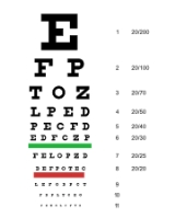
Visual acuity is the eye's ability to detect fine details and is the quantitative measure of the eye's ability to see an in-focus image at a certain distance. The standard definition of normal visual acuity (20/20 or 6/6 vision) is the ability to resolve a spatial pattern separated by a visual angle of one minute of arc. The terms 20/20 and 6/6 are derived from standardized sized objects that can be seen by a "person of normal vision" at the specified distance. For example, if one can see at a distance of 20 ft an object that normally can be seen at 20 ft, then one has 20/20 vision. If one can see at 20 ft what a normal person can see at 40 ft, then one has 20/40 vision. Put another way, suppose you have trouble seeing objects at a distance and you can only see out to 20 ft what a person with normal vision can see out to 200 feet, then you have 20/200 vision. The 6/6 terminology is more commonly used in Europe and Australia, and represents the distance in metres.

This is often measured with a Snellen chart
Snellen chart

A Snellen chart is an eye chart used by eye care professionals and others to measure visual acuity. Snellen charts are named after the Dutch ophthalmologist Herman Snellen who developed the chart in 1862....
.

Pupil function

An examination of pupilary function includes inspecting the pupils for equal size (1 mm or less of difference may be normal), regular shape, reactivity to light, and direct and consensual accommodation. These steps can be easily remembered with the mnemonic
Mnemonic

A mnemonic device is a memory aid. Commonly met mnemonics are often verbal, something such as a very short poem or a special word used to help a person remember something, particularly lists, but may be visual, kinesthetic or auditory....
 PERRLA (D+C): Pupils Equal and Round; Reactive to Light and Accommodation (Direct and Consensual).

A swinging-flashlight test
Swinging-flashlight test

The swinging-flashlight test is used to help a practitioner decide whether reduced vision is due to ocular disease....
 may also be desirable if neurologic
Neurology

Neurology is a medical specialty dealing with disorders of the nervous system. Specifically, it deals with the diagnosis and treatment of all categories of disease involving the Central nervous system, Peripheral nervous system, and autonomic nervous systems, including their coverings, blood vessels, and...
 damage is suspected. The swinging-flashlight test is the most useful clinical test available to a general physician for the assessment of optic nerve
Optic nerve

The optic nerve, also called cranial nerve II, transmits visual information from the retina to the brain....
 anomalies. This test detects the afferent pupil defect
Marcus Gunn pupil

Marcus Gunn pupil is a medical sign observed during the swinging-flashlight test whereupon the patient's pupils constrict less when the light swings from the unaffected eye to the affected eye....
, also referred to as the Marcus Gunn pupil
Marcus Gunn pupil

Marcus Gunn pupil is a medical sign observed during the swinging-flashlight test whereupon the patient's pupils constrict less when the light swings from the unaffected eye to the affected eye....
. In a normal reaction to the swinging-flashlight test, both pupils constrict when one is exposed to light
Light

Light, or visible light, is electromagnetic radiation of a wavelength that is Visible spectrum to the human eye , or up to 380?750 nm. In the broader field of physics, light is sometimes used to refer to electromagnetic radiation of all wavelengths, whether visible or not....
. As the light is being moved from one eye to another, both eyes begin to dilate, but constrict again when light has reached the other eye.

If there is an efferent
Efferent nerve

In the nervous system, efferent nerves ? otherwise known as motoneuron or effector neurons ? carry action potential away from the central nervous system to effectors such as muscles or glands ....
 defect in the left eye, the left pupil will remain dilated regardless of where the light is shining, while the right pupil will respond normally. If there is an afferent
Afferent nerve

In the nervous system, afferent neurons , carry action potential from receptor s or sense organs toward the central nervous system. This term can also be used to describe relative connections between structures....
 defect in the left eye, both pupils will dilate when the light is shining on the left eye, but both will constrict when it is shining on the right eye.

If there is a unilateral small pupil with normal reactivity to light, it is unlikely that a neuropathy
Neuropathy

Neuropathy is a medical term describing disorders of the nerves of the peripheral nervous system It is usually considered equivalent to peripheral neuropathy....
 is present. However, if accompanied by ptosis
Ptosis (eyelid)

Ptosis is an abnormally low position of the upper eyelid. The drooping may be worse after being awake longer, when the individual's muscles are tired....
 of the upper eyelid
Eyelid

An eyelid is a thin fold of skin that covers and protects an eye. With the exception of the prepuce and the labia minora, it has the thinnest skin of the whole body....
, this may indicate Horner's syndrome
Horner's syndrome

Horner's syndrome or Horner syndrome is a clinical syndrome caused by damage to the sympathetic nervous system. It is also known by the names Bernard-Horner syndrome or oculosympathetic palsy....
.

If there is a small, irregular pupil that constricts poorly to light, but normally to accommodation
Accommodation (eye)

Accommodation is the process by which the :eye increases optical power to maintain a clear image on an object as it draws near the eye. The young human eye can change focus from distance to 7 cm from the eye in 350 milliseconds....
, this is an Argyll Robertson pupil
Argyll Robertson pupil

Argyll Robertson pupils are bilateral small pupils that constrict when the patient focuses on a near object , but do not constrict when exposed to bright light ....
.

Ocular motility

Ocular motility should always be tested, especially when patients complain of double vision or physicians suspect neurologic disease. First, the doctor should visually assess the eyes for deviations that could result from strabismus
Strabismus

Strabismus is a condition in which the eyes are not properly aligned with each other. It typically involves a lack of coordination between the Muscles of orbits that prevents bringing the gaze of each eye to the same point in space and preventing proper binocular vision, which may adversely affect depth perception....
, extraocular muscle dysfunction, or palsy
Palsy

In medicine, palsy is the paralysis of a body part, often accompanied by loss of Somatosensory system and by uncontrolled body movements, such as shaking....
 of the cranial nerves innervating the extraocular muscles. Saccade
Saccade

A saccade is a fast eye movements, head or other part of an animal's body or device. It can also be a fast shift in frequency of an emitted signal or other quick change....
s are assessed by having the patient move his or her eye quickly to a target at the far right, left, top and bottom. This tests for saccadic dysfunction whereupon poor ability of the eyes to "jump" from one place to another may impinge on reading ability and other skills.

Slow tracking, or "pursuits" are assessed by the 'follow my finger' test, in which the examiner's finger traces an imaginary "double-H", which touches upon the eight fields of gaze. These test the inferior
Inferior rectus muscle

The inferior rectus muscle is a muscle in the orbit ....
, superior
Superior rectus muscle

The superior rectus muscle is a muscle in the orbit . It is one of the extraocular muscles.It is innervation by the superior division of the oculomotor nerve ....
, lateral
Lateral rectus muscle

The lateral rectus muscle is a muscle in the orbit . It is one of six extraocular muscles that control the eye movements and the only muscle innervation by the abducens nerve, cranial nerve VI....
 and medial
Medial rectus muscle

The medial rectus muscle is a muscle in the orbit .As with most of the muscles of the orbit, it is innervation by the inferior division of the oculomotor nerve ....
 rectus muscle
MUSCLE

MUSCLE is public domain, multiple sequence alignment software for protein and nucleotide sequences.MUSCLE is integrated into UGENE bioinformatics tool as a plugin....
s of the eye, as well as the superior
Superior oblique muscle

For the abdominal muscle see: External oblique muscleThe superior oblique muscle, or obliquus oculi superior, is a fusiform muscle in the upper, medial side of the orbit whose primary action is intorsion and whose secondary actions are to abduction and Depression the eyeball ....
 and inferior
Inferior oblique muscle

The Obliquus oculi inferior is a thin, narrow muscle, placed near the anterior margin of the floor of the orbit ....
 oblique muscles.

Visual field (confrontation) testing

Evaluation of the visual fields should never be omitted from the basic eye examination. Testing the visual fields consists of confrontation field testing in which each eye is tested separately to assess the extent of the peripheral field. To perform the test, the individual occludes one eye while fixated on the examiner's eye with the non-occluded eye. The patient is then asked to count the number of fingers that are briefly flashed in each of the four quadrants. This method is preferred to the wiggly finger test that was historically used because it represents a rapid and efficient way of answering the same question: is the peripheral visual field affected?

Common problems of the visual field include scotoma
Scotoma

A scotoma is an area or island of loss or impairment of visual acuity surrounded by a field of normal or relatively well-preserved Visual perception....
 (area of reduced vision), hemianopia (half of visual field lost), homonymous quadrantanopia (involving both eyes) and bitemporal hemianopia.

Intraocular pressure

Intraocular pressure (IOP) can be measured by Tonometry
Tonometry

In music, a tonometer is an instrument used to determine the pitch or vibration rate of tones, such as a tuning fork.Tonometry is the measurement of tension or pressure ....
 devices designed to measure the outflow (and resistance to outflow) of the aqueous humour
Aqueous humour

The aqueous humor is a thick watery substance that is between the lens and the cornea....
 from the eye. Diaton Tonometry can measure IOP though the Eyelid
Eyelid

An eyelid is a thin fold of skin that covers and protects an eye. With the exception of the prepuce and the labia minora, it has the thinnest skin of the whole body....


Ophthalmoscopy

Ophthalmoscopic examination may include visually magnified inspection of the internal eye structures and also assessment of the quality of the eye's red reflex.

Ophthalmoscopy allows the one to look directly at the retina
Retina

The vertebrate retina is a light sensitive tissue lining the inner surface of the eye. The optics of the eye create an image of the visual world on the retina, which serves much the same function as the film in a camera....
 and other tissue at the back of the eye. This is best done after the pupil has been dilated with eye drop
Eye drop

Eye drops are saline -containing drops used as a Vector to administer medication in the eye. Depending on the condition being treated, they may contain steroids , antihistamines, sympathomimetics, Beta blockers, parasympathomimetics , parasympatholytics , prostaglandins, non-steroidal anti-inflammatory drugs or topical anesthetics....
s. A limited view can be obtained through an undilated pupil, in which case best results are obtained with the room darkened and the patient looking towards the far corner.

The appearance of the optic disc
Optic disc

The optic disc or optic nerve head is the location where ganglion cell axons exit the eye to form the optic nerve. There are no light sensitive photoreceptor to respond to a light stimulus at this point....
 and retinal vasculature are the main focus of examination during ophthalmoscopy. Anomalies in the appearance of these internal ocular structures may indicate eye disease or condition.

A red reflex
Red reflex

The red reflex refers to the reddish-orange reflection from the eye's retina that is observed when using an ophthalmoscope or retinoscope from approximately 30 cm / 1 foot....
 can be seen when looking at a patient's pupil through a direct ophthalmoscope
Ophthalmoscope

The ophthalmoscope is an instrument used to examine the eye. Its use is crucial in determining the health of the retina and the vitreous humor....
. This part of the examination is done from a distance of about 50 cm and is usually symmetrical between the two eyes. An opacity may indicate a cataract.

Slit-lamp

Close inspection of the anterior eye structures and ocular adnexa are often done with a slit lamp
Slit lamp

The slit lamp is an instrument consisting of a high-intensity light source that can be focused to shine a thin sheet of light into the eye. It is used in conjunction with a biomicroscope....
 machine. A small beam of light that can be varied in width, height, incident angle, orientation and colour, is passed over the eye. Often, this light beam is narrowed into a vertical "slit", during slit-lamp examination. The examiner views the illuminated ocular structures, through an optical system that magnifies the image of the eye.

This allows inspection of all the ocular media, from cornea to vitreous, plus magnified view of eyelids, and other external ocular related structures. Fluorescein
Fluorescein

Fluorescein is a fluorophore commonly used in microscopy, in a type of dye laser as the gain medium, in forensics and serology to detect latent blood stains, and in dye tracing....
 staining before slit lamp examination may reveal corneal abrasion
Corneal abrasion

Corneal abrasion is a medical condition involving the loss of the surface epithelial layer of the eye's cornea....
s or herpes simplex
Herpes simplex

Herpes simplex is a viral disease caused by Herpes simplex viruses; both herpes simplex virus 1 and herpes simplex virus 2 cause herpes simplex....
 infection.

The binocular slit-lamp examination provides stereoscopic magnified view of the eye structures in striking detail, enabling exact anatomical diagnoses to be made for a variety of eye conditions.

Also ophthalmoscopy and gonioscopy
Gonioscopy

Gonioscopy describes the use of a goniolens in conjunction with a slit lamp or operating microscope to gain a view of the iridocorneal angle, or the anatomical angle formed between the eye's cornea and iris ....
 examinations can also be performed through the slit lamp when combined with special lenses. These lenses include the Goldmann 3-mirror lens, gonioscopy single-mirror/ Zeiss 4-mirror lens for (ocular) anterior chamber angle structures and +90D lens, +78D lens, +66D lens & Hruby (-56D) lens, the examination of retinal structures is accomplished.

School vision screening

See pediatric ophthalmology
Pediatric ophthalmology

Pediatric ophthalmology is a sub-speciality of ophthalmology concerned with eye diseases, visual development, and vision care in children....


On-line


Others


See also


Conditions diagnosed during eye examinations

  • Amblyopia
    Amblyopia

    Amblyopia, otherwise known as lazy eye, is a Disease of the visual system that is characterized by poor or indistinct Visual perception in an eye that is otherwise physically normal, or out of proportion to associated structural abnormalities....
  • Diplopia
    Diplopia

    Diplopia, commonly known as double vision, is the simultaneous perception of two images of a single object. These images may be displaced horizontally, vertically, or diagonally in relation to each other....
  • Myopia
    Myopia

    Myopia , also called near- or short-sightedness, is a Refractive error of the eye in which collimated light produces image focus in front of the retina when accommodation is relaxed....
  • Hyperopia
    Hyperopia

    Hyperopia, also known as far-sightedness, long-sightedness or hypermetropia, is a defect of visual system caused by an imperfection in the eye , causing inability to Focus on near objects, and in extreme cases causing a sufferer to be unable to focus on objects at any distance....
  • Presbyopia
    Presbyopia

    Presbyopia describes the condition where the eye exhibits a progressively diminished ability to focus on near objects with age. Presbyopia's exact mechanisms are not known with certainty, however, the research evidence most strongly supports a loss of elasticity of the Lens , although changes in the lens's curvature from continual growth and...
  • Strabismus
    Strabismus

    Strabismus is a condition in which the eyes are not properly aligned with each other. It typically involves a lack of coordination between the Muscles of orbits that prevents bringing the gaze of each eye to the same point in space and preventing proper binocular vision, which may adversely affect depth perception....


Other tests that may be performed during eye examinations

  • Electrooculography
    Electrooculography

    Electrooculography is a technique for measuring the resting potential of the retina. The resulting signal is called the electrooculogram. The main applications are in ophthalmology diagnosis and in recording eye movements....
  • Electroretinography
    Electroretinography

    Electroretinography measures the electrical responses of various cell types in the retina, including the Photoreceptor cell , inner retinal cells , and the ganglion cells....


Miscellaneous

  • Binocular vision
    Binocular vision

    Binocular vision is Visual perception in which both eyes are used together. The word binocular comes from two Latin roots, bini for double, and oculus for eye....
  • Eyeglass prescription
    Eyeglass prescription

    An eyeglass prescription is a written order by an optometrist or ophthalmologist to an optician for eyeglasses. It specifies the refractive power to which the eyeglasses are to be made in order to correct blurred Visual perception due to refraction error, including myopia, hyperopia, astigmatism , and presbyopia....
  • Orthoptics
  • Stereopsis
    Stereopsis

    Stereopsis is the process in visual perception leading to the sensation of depth from the two slightly different projections of the world onto the retinas of the two eyes....
  • Vergence
    Vergence

    A vergence is the simultaneous movement of both eyes in opposite directions to obtain or maintain single binocular vision..When a creature with binocular vision looks at an object, the eyes must rotate around a vertical axis so that the projection of the image is in the centre of the retina in both eyes....


External links