

RESOLFT
Encyclopedia
RESOLFT, an acronym for REversible Saturable OpticaL Fluorescence Transitions, denotes a group of optical microscopy techniques with very high resolution. Using standard far field visible light optics a resolution far below the diffraction limit down to molecular scales can be obtained.
With conventional microscopy
techniques, it is not possible to distinguish features that are located at distances less than about half the wavelength
used (i.e. about 200 nm for visible light
). This diffraction limit is based on the wave nature of light
. In conventional microscopes the limit is determined by the used wavelength and the numerical aperture
of the optical system. The RESOLFT concept surmounts this limit by temporarily switching the molecules to a state in which they cannot send a (fluorescence-) signal upon illumination. This concept is different from for example electron microscopy where instead the used wavelength is much smaller.
. Within RESOLFT the principle of STED microscopy
and GSD microscopy
are generalized. Structures that are normally too close to each other to be distinguished are read out sequentially.
Within this concept all methods to operate on molecules that have at least two distinguishable states A and B, can be switched reversibly between these two and the switching of at least one transition can be optically induced are explained in one common framework. In most cases fluorescent markers are used. Transitions between states of these marker molecules are optically driven. One of the state is bright (A), i.e. can generate a signal and the another one is dark (B) and cannot give a signal. One transition between them can be induced by light (e.g. A-B, bright to dark).
The sample is illuminated inhomogeneously with the illumination intensity being very small (zero under ideal conditions) at one position. Only at this place the molecules are never in the dark state B (if A is the pre-existing state) and remain fully in A. The area where molecules are mostly in A (being able to give a signal) can be made very small (smaller than the conventional diffraction limit) by increasing the transition light intensity (see below). Upon detection of the signal (mostly fluorescence) its now known that only molecules in the small area around the illumination intensity minimum can contribute to the signal. Scanning the whole sample, i.e. shifting the illumination profile accordingly allows to reconstruct a high resolution image.
The transition back from B to A can be spontaneous or also driven by light of another wavelength. The molecules have to be switchable several times in order to be present in state A or B at different times during scanning the sample. The method also works if the bright and the dark state are reversed, one then obtains a negative image and has to apply image deconvolution
to obtain a positive.
Upon weak illumination we see that the area is quite large where the illumination is so low that most molecules reside in state A. The shape of the illumination profile does not need to be altered. Increasing the illumination brightness already results in a smaller area where the intensity is below the amount for efficient switching to the dark state. Consequently also the area where molecules can reside in state A is diminished. The (fluorescence) signal during a following readout originates from a very small spot and one can obtain very sharp images.
In the RESOLFT concept the resolution can be approximated by

, whereby is the characteristic intensity required for saturating the transition (half of the molecules in state A and half in state B), and
  denotes the intensity applied. If the minima are produced by focusing optics with a numerical aperture
  , the minimal distance at which two identical objects can be discerned is
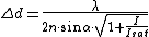 which can be regarded as an extension of Abbe
’s equation. The diffraction-unlimited nature of the RESOLFT family of concepts is reflected by the fact that the minimal resolvable distance can be continuously decreased by increasing
  . Hence the quest for nanoscale resolution comes down to maximizing this quantity. This is possible by increasing
  or by lowering
  .
.
)
Within the STED microscopy (STimulated Emission Depletion microscopy) a fluorescent dye molecule is driven between its electronic ground state and its excited state while sending out fluorescence photons. This is the standard operation mode in fluorescence microscopy and depicts state A. In state B the dye is permanently kept in its electronic ground state through stimulated emission
. The dye can fluoresce in state A and not in state B, the RESOLFT concept applies.
)
GSD microscopy (Ground State Depletion microscopy) also uses fluorescent markers. In state A, the molecule can freely be driven between the ground and the first excited state and fluorescence can be sent out. In the dark state B the ground state of the molecule is depopulated, a transition to a long lived excited state takes place from which fluorescence is not emitted. As long as the molecule is in the dark state, it's not available for cycling between ground and excited state, fluorescence is hence turned off.
can be switched on and off by light of appropriate wavelength. They can be used in a RESOLFT type microscope.
During illumination with light these proteins change their conformation. In the process they gain or loose their ability to emit fluorescence. The fluorescing state corresponds to state A, the non-fluorescing to state B and the RESOLFT concept applies again. The reversible transition (e.g. from B back to A) takes place either spontaneously or again driven by light. Inducing conformational changes in proteins can be achieved already at much lower switching light intensities as compared to stimulated emission or ground state depletion (some W/cm²).
With conventional microscopy
Microscopy
Microscopy is the technical field of using microscopes to view samples and objects that cannot be seen with the unaided eye...
techniques, it is not possible to distinguish features that are located at distances less than about half the wavelength
Wavelength
In physics, the wavelength of a sinusoidal wave is the spatial period of the wave—the distance over which the wave's shape repeats.It is usually determined by considering the distance between consecutive corresponding points of the same phase, such as crests, troughs, or zero crossings, and is a...
used (i.e. about 200 nm for visible light
Visible spectrum
The visible spectrum is the portion of the electromagnetic spectrum that is visible to the human eye. Electromagnetic radiation in this range of wavelengths is called visible light or simply light. A typical human eye will respond to wavelengths from about 390 to 750 nm. In terms of...
). This diffraction limit is based on the wave nature of light
Light
Light or visible light is electromagnetic radiation that is visible to the human eye, and is responsible for the sense of sight. Visible light has wavelength in a range from about 380 nanometres to about 740 nm, with a frequency range of about 405 THz to 790 THz...
. In conventional microscopes the limit is determined by the used wavelength and the numerical aperture
Numerical aperture
In optics, the numerical aperture of an optical system is a dimensionless number that characterizes the range of angles over which the system can accept or emit light. By incorporating index of refraction in its definition, NA has the property that it is constant for a beam as it goes from one...
of the optical system. The RESOLFT concept surmounts this limit by temporarily switching the molecules to a state in which they cannot send a (fluorescence-) signal upon illumination. This concept is different from for example electron microscopy where instead the used wavelength is much smaller.
Working principle
RESOLFT microscopy is an optical microscopy with very high resolution that can image details in samples that cannot be imaged with conventional or confocal microscopyConfocal microscopy
Confocal microscopy is an optical imaging technique used to increase optical resolution and contrast of a micrograph by using point illumination and a spatial pinhole to eliminate out-of-focus light in specimens that are thicker than the focal plane. It enables the reconstruction of...
. Within RESOLFT the principle of STED microscopy
STED microscopy
Stimulated Emission Depletion microscopy, or STED microscopy, is a fluorescence microscopy technique that uses the non-linear de-excitation of fluorescent dyes to overcome the resolution limit imposed by diffraction with standard confocal laser scanning microscopes and conventional far-field...
and GSD microscopy
GSD microscopy
Ground State Depletion Microscopy, or GSD Microscopy, is an implementation of the RESOLFT concept. The method was proposed in 1995 and experimentally demonstrated in 2007. It is the second concept to overcome the diffraction barrier in far-field optical microscopy published by Stefan Hell...
are generalized. Structures that are normally too close to each other to be distinguished are read out sequentially.
Within this concept all methods to operate on molecules that have at least two distinguishable states A and B, can be switched reversibly between these two and the switching of at least one transition can be optically induced are explained in one common framework. In most cases fluorescent markers are used. Transitions between states of these marker molecules are optically driven. One of the state is bright (A), i.e. can generate a signal and the another one is dark (B) and cannot give a signal. One transition between them can be induced by light (e.g. A-B, bright to dark).
The sample is illuminated inhomogeneously with the illumination intensity being very small (zero under ideal conditions) at one position. Only at this place the molecules are never in the dark state B (if A is the pre-existing state) and remain fully in A. The area where molecules are mostly in A (being able to give a signal) can be made very small (smaller than the conventional diffraction limit) by increasing the transition light intensity (see below). Upon detection of the signal (mostly fluorescence) its now known that only molecules in the small area around the illumination intensity minimum can contribute to the signal. Scanning the whole sample, i.e. shifting the illumination profile accordingly allows to reconstruct a high resolution image.
The transition back from B to A can be spontaneous or also driven by light of another wavelength. The molecules have to be switchable several times in order to be present in state A or B at different times during scanning the sample. The method also works if the bright and the dark state are reversed, one then obtains a negative image and has to apply image deconvolution
Deconvolution
In mathematics, deconvolution is an algorithm-based process used to reverse the effects of convolution on recorded data. The concept of deconvolution is widely used in the techniques of signal processing and image processing...
to obtain a positive.
Resolution below the diffraction-limit
Despite the diffraction-limit the area where molecules reside in state A (bright state) can be made arbitrarily small.- One has to illuminate the sample inhomogeneously so that an isolated zero intensity point is created. This can be achieved e.g. by interference.
- At low intensities (lower than the blue line in the image) most marker molecules are in the bright state, if the intensity is above, most markers are in the dark state.
Upon weak illumination we see that the area is quite large where the illumination is so low that most molecules reside in state A. The shape of the illumination profile does not need to be altered. Increasing the illumination brightness already results in a smaller area where the intensity is below the amount for efficient switching to the dark state. Consequently also the area where molecules can reside in state A is diminished. The (fluorescence) signal during a following readout originates from a very small spot and one can obtain very sharp images.
In the RESOLFT concept the resolution can be approximated by
, whereby
is the characteristic intensity required for saturating the transition (half of the molecules in state A and half in state B), and denotes the intensity applied. If the minima are produced by focusing optics with a numerical aperture , the minimal distance at which two identical objects can be discerned is which can be regarded as an extension of AbbeErnst Karl Abbe
- See also :*Abbe prism*Abbe refractometer*Abbe error*Aberration in optical systems*Calculation of glass properties* German inventors and discoverers-External links:*...
’s equation. The diffraction-unlimited nature of the RESOLFT family of concepts is reflected by the fact that the minimal resolvable distance
can be continuously decreased by increasing . Hence the quest for nanoscale resolution comes down to maximizing this quantity. This is possible by increasing or by lowering .Variants
For switching of the molecules different processes are used. However, all have in common that at least two distinguishable state are used. Typically the fluorescence property is the distinction, however this is not essential also absorption or scattering properties could be exploited.STED Microscopy
(Main article STED microscopySTED microscopy
Stimulated Emission Depletion microscopy, or STED microscopy, is a fluorescence microscopy technique that uses the non-linear de-excitation of fluorescent dyes to overcome the resolution limit imposed by diffraction with standard confocal laser scanning microscopes and conventional far-field...
)
Within the STED microscopy (STimulated Emission Depletion microscopy) a fluorescent dye molecule is driven between its electronic ground state and its excited state while sending out fluorescence photons. This is the standard operation mode in fluorescence microscopy and depicts state A. In state B the dye is permanently kept in its electronic ground state through stimulated emission
Stimulated emission
In optics, stimulated emission is the process by which an atomic electron interacting with an electromagnetic wave of a certain frequency may drop to a lower energy level, transferring its energy to that field. A photon created in this manner has the same phase, frequency, polarization, and...
. The dye can fluoresce in state A and not in state B, the RESOLFT concept applies.
GSD microscopy
(Main article GSD microscopyGSD microscopy
Ground State Depletion Microscopy, or GSD Microscopy, is an implementation of the RESOLFT concept. The method was proposed in 1995 and experimentally demonstrated in 2007. It is the second concept to overcome the diffraction barrier in far-field optical microscopy published by Stefan Hell...
)
GSD microscopy (Ground State Depletion microscopy) also uses fluorescent markers. In state A, the molecule can freely be driven between the ground and the first excited state and fluorescence can be sent out. In the dark state B the ground state of the molecule is depopulated, a transition to a long lived excited state takes place from which fluorescence is not emitted. As long as the molecule is in the dark state, it's not available for cycling between ground and excited state, fluorescence is hence turned off.
SPEM and SSIM
SPEM (Saturated Pattern Excitation Microscopy) and SSIM (Saturated Structured Illumination Microscopy) are exploiting the RESOLFT concept using saturated excitation to produce negative images, i.e. fluorescence occurs from everywhere except at a very small region around the geometrical focus of the microscope. Also non point-like patterns are used for illumination. Mathematical image reconstruction is necessary to obtain positive images again.RESOLFT with switchable proteins
Some fluorescent proteinsGreen fluorescent protein
The green fluorescent protein is a protein composed of 238 amino acid residues that exhibits bright green fluorescence when exposed to blue light. Although many other marine organisms have similar green fluorescent proteins, GFP traditionally refers to the protein first isolated from the...
can be switched on and off by light of appropriate wavelength. They can be used in a RESOLFT type microscope.
During illumination with light these proteins change their conformation. In the process they gain or loose their ability to emit fluorescence. The fluorescing state corresponds to state A, the non-fluorescing to state B and the RESOLFT concept applies again. The reversible transition (e.g. from B back to A) takes place either spontaneously or again driven by light. Inducing conformational changes in proteins can be achieved already at much lower switching light intensities as compared to stimulated emission or ground state depletion (some W/cm²).
RESOLFT with switchable organic dyes
Just as with proteins, also some organic dyes can change their structure upon illumination. The ability to fluoresce of such organic dyes can be turned on and off through visible light. Again the applied light intensities can be quite low (some 100 W/cm²).See also
- Department of NanoBiophotonics at the Max-Planck-Institute for Biophysical Chemistry (Göttingen, Germany)
- Gutafsson Lab, University of California, San Francisco