
Parasympathetic nervous system
Overview
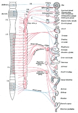
The parasympathetic nervous system
(PSNS) is one of the two main divisions of the autonomic nervous system
(ANS). The ANS is responsible for regulation of internal organs and glands, which occurs unconsciously. To be specific, the parasympathetic system is responsible for stimulation of "rest-and-digest" activities that occur when the body is at rest, including sexual arousal, salivation, lacrimation
(tears), urination
, digestion
, and defecation
. Its action is described as being complementary to that of one of the other main branches of the ANS, the sympathetic nervous system
, which is responsible for stimulating activities associated with the fight-or-flight response
.
Sympathetic
and parasympathetic divisions typically function in opposition to each other.
Nervous system
The nervous system is an organ system containing a network of specialized cells called neurons that coordinate the actions of an animal and transmit signals between different parts of its body. In most animals the nervous system consists of two parts, central and peripheral. The central nervous...
(PSNS) is one of the two main divisions of the autonomic nervous system
Autonomic nervous system
The autonomic nervous system is the part of the peripheral nervous system that acts as a control system functioning largely below the level of consciousness, and controls visceral functions. The ANS affects heart rate, digestion, respiration rate, salivation, perspiration, diameter of the pupils,...
(ANS). The ANS is responsible for regulation of internal organs and glands, which occurs unconsciously. To be specific, the parasympathetic system is responsible for stimulation of "rest-and-digest" activities that occur when the body is at rest, including sexual arousal, salivation, lacrimation
Tears
Tears are secretions that clean and lubricate the eyes. Lacrimation or lachrymation is the production or shedding of tears....
(tears), urination
Urination
Urination, also known as micturition, voiding, peeing, weeing, pissing, and more rarely, emiction, is the ejection of urine from the urinary bladder through the urethra to the outside of the body. In healthy humans the process of urination is under voluntary control...
, digestion
Digestion
Digestion is the mechanical and chemical breakdown of food into smaller components that are more easily absorbed into a blood stream, for instance. Digestion is a form of catabolism: a breakdown of large food molecules to smaller ones....
, and defecation
Defecation
Defecation is the final act of digestion by which organisms eliminate solid, semisolid or liquid waste material from the digestive tract via the anus. Waves of muscular contraction known as peristalsis in the walls of the colon move fecal matter through the digestive tract towards the rectum...
. Its action is described as being complementary to that of one of the other main branches of the ANS, the sympathetic nervous system
Sympathetic nervous system
The sympathetic nervous system is one of the three parts of the autonomic nervous system, along with the enteric and parasympathetic systems. Its general action is to mobilize the body's nervous system fight-or-flight response...
, which is responsible for stimulating activities associated with the fight-or-flight response
Fight-or-flight response
The fight-or-flight response was first described by Walter Bradford Cannon....
.
Sympathetic
Sympathetic nervous system
The sympathetic nervous system is one of the three parts of the autonomic nervous system, along with the enteric and parasympathetic systems. Its general action is to mobilize the body's nervous system fight-or-flight response...
and parasympathetic divisions typically function in opposition to each other.