

Neonatal jaundice
Encyclopedia
Neonatal jaundice or Neonatal hyperbilirubinemia is a yellowing of the skin
and other tissues of a newborn infant
. A bilirubin
level of more than 85 umol/l (5 mg/dL) manifests clinical jaundice
in neonates whereas in adults a level of 34 umol/l (2 mg/dL) would look icteric. In newborns jaundice is detected by blanching the skin with digital pressure so that it reveals underlying skin and subcutaneous tissue. Jaundice newborns have an apparent icteric sclera
, and yellowing of the face, extending down onto the chest.
In neonates the dermal icterus is first noted in the face and as the bilirubin level rises proceeds caudal to the trunk and then to the extremities.
This condition is common in newborns affecting over half (50 -60%) of all babies in the first week of life.
Notoriously inaccurate rules of thumb have been applied to the physical exam of the jaundiced infant. Some include estimation of serum bilirubin
based on appearance. One such rule of thumb includes infants whose jaundice is restricted to the face and part of the trunk above the umbilicus
, have the bilirubin
less than 204 umol/l (12 mg/dL) (less dangerous level). Infants whose palms and soles are yellow, have serum bilirubin level over 255 umol/l (15 mg/dL) (more serious level).
Studies have shown that trained examiners assessment of levels of jaundice show moderate agreement with icterometer bilirubin measurements.
In infants jaundice can be measured using invasive
or non-invasive methods. In non-invasive method Ingram icterometers and Transcutaneous bilirubinometers are used.
s develop visible jaundice due to elevation of unconjugated bilirubin concentration during their first week. This common condition is called physiological jaundice. This pattern of hyperbilirubinemia has been classified into two functionally distinct periods.
Phase one
Phase two - bilirubin levels decline to about 34 umol/l (2 mg/dL) for two weeks, eventually mimicking adult values.
1. Increase bilirubin load on liver cells
:
2. Defective hepatic uptake of bilirubin from blood plasma
:
3. Defective billirubin conjugation:
4. Defective bilirubin excretion
The sign which helps to differentiate pathological jaundice of neonates from physiological jaundice of neonates are presence of intrauterine retardation, stigma of intrauterine infections (e.g. cataracts, microcephaly
, hepatosplenomegaly
etc.), cephalhematoma
, bruising, signs of intra ventricular hemorrhage etc.
History of illness is noteworthy. Family history of jaundice and anemia, family history of neonatal or early infant death due to liver disease, maternal illness suggestive of viral infection (fever, rash or lymphadenopathy
), Maternal drugs (e.g. Sulphonamides, anti-malarials causing hemolysis in G-6-PD deficiency) are suggestive of pathological jaundice in neonates.
as it is replaced with adult hemoglobin
and the relatively immature hepatic metabolic pathways which are unable to conjugate and so excrete bilirubin as quickly as an adult. This causes an accumulation of bilirubin in the blood (hyperbilirubinemia), leading to the symptoms of jaundice.
If the neonatal jaundice does not clear up with simple phototherapy, other causes such as biliary atresia
, PFIC, bile duct paucity, Alagille's syndrome, alpha 1 and other pediatric liver diseases should be considered. The evaluation for these will include blood work and a variety of diagnostic tests. Prolonged neonatal jaundice is serious and should be followed up promptly.
Severe neonatal jaundice may indicate the presence of other conditions contributing to the elevated bilirubin levels, of which there are a large variety of possibilities (see below). These should be detected or excluded as part of the differential diagnosis
to prevent the development of complications. They can be grouped into the following categories:
during birth helps stimulate milk production in the mother's body, so infants born by cesarean section are at higher risk for this condition.
Despite the advantages of breast feeding, there is a strong association of breast feeding with neonatal hyperbilirubinemia and thus risk of kernicterus
, though this is uncommon. Serum bilirubin levels may reach as high as 30 mg/dL. Jaundice should be managed either with phototherapy or with exchange blood transfusion as is needed. Breast feeds however need not be discontinued. The child should be kept well hydrated and extra feeds given.
This method is more accurate and less subjective in estimating jaundice.
Ingram icterometer: In this method a piece of transparent plastic known as Ingram icterometer is used. Ingram icterometer is painted in five transverse strips of graded yellow lines. The instrument is pressed against the nose and the yellow colour of the blanched skin is matched with the graded yellow lines and biluribin level is assigned.
Transcutaneous bilirubinometer: This is hand held, portable and rechargeable but expensive and sophisticated. When pressure is applied to the photoprobe, a xenon tube generates a strobe light, and this light passes through the subcutaneous tissue. The reflected light returns through the second fiber optic bundle to the spectrophotometric module. The intensity of the yellow color in this light, after correcting for the hemoglobin, is measured and instantly displayed in arbitrary units.
 Infants with neonatal jaundice are treated with colored light called phototherapy. Physicians randomly assigned 66 infants 35 weeks of gestation to receive phototherapy. After 15±5 the levels of bilirubin, a yellowish bile pigment that in excessive amounts causes jaundice, were decreased down to 0.27±0.25 mg/dl/h in the blue light. This suggests that blue light therapy helps reduce high bilirubin levels that cause neonatal jaundice.
Exposing infants to high levels of colored light changes trans-bilirubin to the more water soluble cis-form which is excreted in the bile. Scientists studied 616 capillary blood samples from jaundiced newborn infants. These samples were randomly divided into three groups. One group contained 133 samples and would receive phototherapy with blue light. Another group contained 202 samples would receive room light, or white light. The final group contained 215 samples, and were left in a dark room. The total bilirubin levels were checked at 0, 2, 4, 6, 24, and 48 hours. There was a significant decrease in bilirubin in the first group exposed to phototherapy after two hours, but no change occurred in the white light and dark room group. After 6 hours, there was a significant change in bilirubin level in the white light group but not the dark room group. It took 48 hours to record a change in the dark room group’s bilirubin level. Phototherapy is the most effective way of breaking down a neonate’s bilirubin.
Phototherapy works through a process of isomerization that changes trans-bilirubin into the water-soluble cis-bilirubin isomer.
In phototherapy, blue light is typically used because it is more effective at breaking down bilirubin (Amato, Inaebnit, 1991). Two matched groups of newborn infants with jaundice were exposed to intensive green or blue light phototherapy. The efficiency of the treatment was measured by the rate of decline of serum bilirubin, which in excessive amounts causes jaundice, concentration after 6, 12 and 24 hours of light exposure. A more rapid response was obtained using the blue lamps than the green lamps. However, a shorter phototherapy recovery period was noticed in babies exposed to the green lamps(1). Green light is not commonly used because exposure time must be longer to see dramatic results(1).
Ultraviolet light therapy may increase the risk of or skin moles, in childhood. While an increased number of moles is related to an increased risk of skin cancer, it is not ultraviolet light that is used for treating neonatal jaundice. Rather, it is simply a specific frequency of blue light that does not carry these risks.
Increased feedings help move bilirubin through the neonate’s metabolic system.
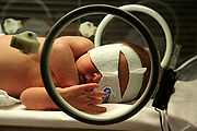
The light can be applied with overhead lamps, which means that the baby's eyes need to be covered, or with a device called a Biliblanket
, which sits under the baby's clothing close to its skin.
). Quick and accurate treatment of neonatal jaundice helps to reduce the risk of neonates developing kernicterus.
An effect of kernicterus is a fever. A male full term neonate had hyperbilirubinemia (kernicterus) and jaundice at the age of 4 days old. He displayed symptoms of increased lethargy, refusal to eat, and had a fever. The neonate who was diagnosed with kernicterus displayed symptoms of a fever.
Another effect of kernicterus is seizures. The Neonatal Unit at Allied Hospital Faisalabad studied 200 neonates of either gender who presented seizures during their hospital stay from April 2003 to June 2004. The seizures were evaluated and one cause of the seizures was kernicterus. 4.5%, or 9 neonates, displayed seizures caused by kernicterus.
High pitched crying is an effect of kernicterus. Scientists used a computer to record and measure cranial nerves 8, 9 and 12 in 50 infants who were divided into two groups equally depending upon bilirubin concentrations. Of the 50 infants, 43 had tracings of high pitched crying.
Exchange transfusions performed to lower high bilirubin levels are an aggressive treatment.
has issued guidelines for managing this disease, which can be obtained for free.
Skin
-Dermis:The dermis is the layer of skin beneath the epidermis that consists of connective tissue and cushions the body from stress and strain. The dermis is tightly connected to the epidermis by a basement membrane. It also harbors many Mechanoreceptors that provide the sense of touch and heat...
and other tissues of a newborn infant
Infant
A newborn or baby is the very young offspring of a human or other mammal. A newborn is an infant who is within hours, days, or up to a few weeks from birth. In medical contexts, newborn or neonate refers to an infant in the first 28 days after birth...
. A bilirubin
Bilirubin
Bilirubin is the yellow breakdown product of normal heme catabolism. Heme is found in hemoglobin, a principal component of red blood cells. Bilirubin is excreted in bile and urine, and elevated levels may indicate certain diseases...
level of more than 85 umol/l (5 mg/dL) manifests clinical jaundice
Jaundice
Jaundice is a yellowish pigmentation of the skin, the conjunctival membranes over the sclerae , and other mucous membranes caused by hyperbilirubinemia . This hyperbilirubinemia subsequently causes increased levels of bilirubin in the extracellular fluid...
in neonates whereas in adults a level of 34 umol/l (2 mg/dL) would look icteric. In newborns jaundice is detected by blanching the skin with digital pressure so that it reveals underlying skin and subcutaneous tissue. Jaundice newborns have an apparent icteric sclera
Sclera
The sclera , also known as the white or white of the eye, is the opaque , fibrous, protective, outer layer of the eye containing collagen and elastic fiber. In the development of the embryo, the sclera is derived from the neural crest...
, and yellowing of the face, extending down onto the chest.
In neonates the dermal icterus is first noted in the face and as the bilirubin level rises proceeds caudal to the trunk and then to the extremities.
This condition is common in newborns affecting over half (50 -60%) of all babies in the first week of life.
Notoriously inaccurate rules of thumb have been applied to the physical exam of the jaundiced infant. Some include estimation of serum bilirubin
Bilirubin
Bilirubin is the yellow breakdown product of normal heme catabolism. Heme is found in hemoglobin, a principal component of red blood cells. Bilirubin is excreted in bile and urine, and elevated levels may indicate certain diseases...
based on appearance. One such rule of thumb includes infants whose jaundice is restricted to the face and part of the trunk above the umbilicus
Navel
The navel is a scar on the abdomen caused when the umbilical cord is removed from a newborn baby...
, have the bilirubin
Bilirubin
Bilirubin is the yellow breakdown product of normal heme catabolism. Heme is found in hemoglobin, a principal component of red blood cells. Bilirubin is excreted in bile and urine, and elevated levels may indicate certain diseases...
less than 204 umol/l (12 mg/dL) (less dangerous level). Infants whose palms and soles are yellow, have serum bilirubin level over 255 umol/l (15 mg/dL) (more serious level).
Studies have shown that trained examiners assessment of levels of jaundice show moderate agreement with icterometer bilirubin measurements.
In infants jaundice can be measured using invasive
Invasive
Invasive may refer to:*A military invasion*An invasive species*An invasive medical procedure*The invasively progressive spread of disease from one organ in the body to another, especially in reference to cancer...
or non-invasive methods. In non-invasive method Ingram icterometers and Transcutaneous bilirubinometers are used.
Physiological jaundice
Most infantInfant
A newborn or baby is the very young offspring of a human or other mammal. A newborn is an infant who is within hours, days, or up to a few weeks from birth. In medical contexts, newborn or neonate refers to an infant in the first 28 days after birth...
s develop visible jaundice due to elevation of unconjugated bilirubin concentration during their first week. This common condition is called physiological jaundice. This pattern of hyperbilirubinemia has been classified into two functionally distinct periods.
Phase one
- Term infants - jaundice lasts for about 10 days with a rapid rise of serum bilirubin up to 204 umol/l (12 mg/dL).
- Preterm infants - jaundice lasts for about two weeks, with a rapid rise of serum bilirubin up to 255 umol/l (15 mg/dL).
Phase two - bilirubin levels decline to about 34 umol/l (2 mg/dL) for two weeks, eventually mimicking adult values.
- Preterm infants - phase two can last more than one month.
- Exclusively breastfed infants - phase two can last more than one month.
Causes
Possible mechanisms involved in physiological jaundice1. Increase bilirubin load on liver cells
Hepatocyte
A hepatocyte is a cell of the main tissue of the liver. Hepatocytes make up 70-80% of the liver's cytoplasmic mass.These cells are involved in:* Protein synthesis* Protein storage* Transformation of carbohydrates...
:
- Increased red blood cell (RBC) volume
- Increased labeled bilirubin
- Increased circulation of bilirubin in the liver
- Decreased RBC survival
2. Defective hepatic uptake of bilirubin from blood plasma
Blood plasma
Blood plasma is the straw-colored liquid component of blood in which the blood cells in whole blood are normally suspended. It makes up about 55% of the total blood volume. It is the intravascular fluid part of extracellular fluid...
:
- Decreased ligadin (Y protein)
- Increased binding of Y proteins by other anionsIonAn ion is an atom or molecule in which the total number of electrons is not equal to the total number of protons, giving it a net positive or negative electrical charge. The name was given by physicist Michael Faraday for the substances that allow a current to pass between electrodes in a...
- Decreased liver uptake especially in phase two
3. Defective billirubin conjugation:
- Decreased UDPG activity
4. Defective bilirubin excretion
Pathological Jaundice of Neonates (Unconjugated Pathological Hyperbilirubinemia)
Any of the following features characterizes pathological jaundice:- Clinical jaundice appearing in the first 24 hours.
- Increases in the level of total bilirubin by more than 8.5 umol/l (0.5 mg/dL) per hour or (85 umol/l) 5 mg/dL per 24 hours.
- Total bilirubin more than 331.5 umol/l (19.5 mg/dL) (hyperbilirubinemia).
- Direct bilirubin more than 34 umol/l (2.0 mg/dL).
Differentiating Physiological and Pathological Jaundice
The aim of clinical assessment is to distinguish physiological from pathological jaundice.The sign which helps to differentiate pathological jaundice of neonates from physiological jaundice of neonates are presence of intrauterine retardation, stigma of intrauterine infections (e.g. cataracts, microcephaly
Microcephaly
Microcephaly is a neurodevelopmental disorder in which the circumference of the head is more than two standard deviations smaller than average for the person's age and sex. Microcephaly may be congenital or it may develop in the first few years of life...
, hepatosplenomegaly
Hepatosplenomegaly
Hepatosplenomegaly is the simultaneous enlargement of both the liver and the spleen . Hepatosplenomegaly can occur as the result of acute viral hepatitis or infectious mononucleosis, or it can be the sign of a serious and life threatening lysosomal storage disease...
etc.), cephalhematoma
Cephalhematoma
A cephalohematoma or cephalohaematoma is a hemorrhage of blood between the skull and the periosteum of a newborn baby secondary to rupture of blood vessels crossing the periosteum...
, bruising, signs of intra ventricular hemorrhage etc.
History of illness is noteworthy. Family history of jaundice and anemia, family history of neonatal or early infant death due to liver disease, maternal illness suggestive of viral infection (fever, rash or lymphadenopathy
Lymphadenopathy
Lymphadenopathy is a term meaning "disease of the lymph nodes." It is, however, almost synonymously used with "swollen/enlarged lymph nodes". It could be due to infection, auto-immune disease, or malignancy....
), Maternal drugs (e.g. Sulphonamides, anti-malarials causing hemolysis in G-6-PD deficiency) are suggestive of pathological jaundice in neonates.
Causes of jaundice
In neonates, jaundice tends to develop because of two factors - the breakdown of fetal hemoglobinFetal hemoglobin
Fetal hemoglobin, or foetal haemoglobin, is the main oxygen transport protein in the fetus during the last seven months of development in the uterus and in the newborn until roughly 6 months old...
as it is replaced with adult hemoglobin
Hemoglobin
Hemoglobin is the iron-containing oxygen-transport metalloprotein in the red blood cells of all vertebrates, with the exception of the fish family Channichthyidae, as well as the tissues of some invertebrates...
and the relatively immature hepatic metabolic pathways which are unable to conjugate and so excrete bilirubin as quickly as an adult. This causes an accumulation of bilirubin in the blood (hyperbilirubinemia), leading to the symptoms of jaundice.
If the neonatal jaundice does not clear up with simple phototherapy, other causes such as biliary atresia
Biliary atresia
Biliary atresia, also known as "extrahepatic ductopenia" and "progressive obliterative cholangiopathy" is a congenital or acquired disease of the liver and one of the principle forms of chronic rejection of a transplanted liver allograft. As a birth defect in newborn infants, it has an occurrence...
, PFIC, bile duct paucity, Alagille's syndrome, alpha 1 and other pediatric liver diseases should be considered. The evaluation for these will include blood work and a variety of diagnostic tests. Prolonged neonatal jaundice is serious and should be followed up promptly.
Severe neonatal jaundice may indicate the presence of other conditions contributing to the elevated bilirubin levels, of which there are a large variety of possibilities (see below). These should be detected or excluded as part of the differential diagnosis
Differential diagnosis
A differential diagnosis is a systematic diagnostic method used to identify the presence of an entity where multiple alternatives are possible , and may also refer to any of the included candidate alternatives A differential diagnosis (sometimes abbreviated DDx, ddx, DD, D/Dx, or ΔΔ) is a...
to prevent the development of complications. They can be grouped into the following categories:
Intrinsic causes of hemolysis
- Membrane conditions
- SpherocytosisSpherocytosisSpherocytosis is an auto-hemolytic anemia characterized by the production of red blood cells , or erythrocytes, that are sphere-shaped, rather than bi-concave disk shaped. Spherocytes are found in hereditary spherocytosis and autoimmune hemolytic anemia.It almost always refers to hereditary...
- Hereditary ellipsoidosis
- Spherocytosis
- Systemic conditions
- SepsisSepsisSepsis is a potentially deadly medical condition that is characterized by a whole-body inflammatory state and the presence of a known or suspected infection. The body may develop this inflammatory response by the immune system to microbes in the blood, urine, lungs, skin, or other tissues...
- Arteriovenous malformationArteriovenous malformationArteriovenous malformation or AVM is an abnormal connection between veins and arteries, usually congenital. This pathology is widely known because of its occurrence in the central nervous system, but can appear in any location. An arteriovenous malformation is a vascular anomaly. It is a...
- Sepsis
- Enzyme conditions
- Glucose-6-phosphate dehydrogenase deficiencyGlucose-6-phosphate dehydrogenase deficiencyGlucose-6-phosphate dehydrogenase deficiency is an X-linked recessive hereditary disease characterised by abnormally low levels of glucose-6-phosphate dehydrogenase , a metabolic enzyme involved in the pentose phosphate pathway, especially important in red blood cell metabolism. G6PD deficiency is...
(also called G6PD deficiency) - Pyruvate kinase deficiencyPyruvate kinase deficiencyPyruvate kinase deficiency, also called erythrocyte pyruvate kinase deficiency, is an inherited metabolic disorder of the enzyme pyruvate kinase which affects the survival of red blood cells and causes them to deform into echinocytes on peripheral blood smears.Both autosomal dominant and recessive...
- Glucose-6-phosphate dehydrogenase deficiency
- Globin synthesis defect
- sickle cell disease
- Alpha-thalassemiaAlpha-thalassemiaAlpha-thalassemia is a form of thalassemia involving the genes HBA1 and HBA2. Alpha-thalassemia is due to impaired production of 1,2,3, or 4 alpha globin chains, leading to a relative excess of beta globin chains...
Extrinsic causes of hemolysis
- AlloimmunityAlloimmunityAlloimmunity is a condition in which the body gains immunity against antigens of another individual of the same species, which are perceived as foreign....
(The neonatal or cord bloodCord bloodUmbilical cord blood is blood that remains in the placenta and in the attached umbilical cord after childbirth. Cord blood is collected because it contains stem cells which can be used to treat hematopoietic and genetic disorders.-Collection:...
gives a positive direct Coombs test and the maternal blood gives a positive indirect Coombs test)- Hemolytic disease of the newborn (ABO)Hemolytic disease of the newborn (ABO)In ABO hemolytic disease of the newborn maternal IgG antibodies with specificity for the ABO blood group system pass through the placenta to the fetal circulation where they can cause hemolysis of fetal red blood cells which can lead to fetal anemia and HDN...
- Hemolytic disease of the newborn (ABO)
-
- Rh diseaseRh diseaseRh disease is one of the causes of hemolytic disease of the newborn...
- Hemolytic disease of the newborn (anti-Kell)Hemolytic disease of the newborn (anti-Kell)Hemolytic disease of the newborn is the second most common cause of severe hemolytic diseases of newborns after Rh disease. Anti-Kell1 is becoming relatively more important as prevention of Rh disease is also becoming more effective....
- Hemolytic disease of the newborn (anti-Rhc)Hemolytic disease of the newborn (anti-Rhc)Hemolytic disease of the newborn can range from a mild to a severe disease. It is the third most common cause of severe HDN. Rh disease is the most common and hemolytic disease of the newborn is the second most common cause of severe HDN.It occurs more commonly in women who are Rh D...
- Other blood type mismatches causing hemolytic disease of the newbornHemolytic disease of the newbornHemolytic disease of the newborn, also known as hemolytic disease of the fetus and newborn, HDN, HDFN, or erythroblastosis fetalis, is an alloimmune condition that develops in a fetus, when the IgG molecules produced by the mother pass through the placenta...
- Breast milk feeding.
- Rh disease
Non-hemolytic causes
- Cephalohematoma
- PolycythemiaPolycythemiaPolycythemia is a disease state in which the proportion of blood volume that is occupied by red blood cells increases...
- SepsisSepsisSepsis is a potentially deadly medical condition that is characterized by a whole-body inflammatory state and the presence of a known or suspected infection. The body may develop this inflammatory response by the immune system to microbes in the blood, urine, lungs, skin, or other tissues...
- HypothyroidismHypothyroidismHypothyroidism is a condition in which the thyroid gland does not make enough thyroid hormone.Iodine deficiency is the most common cause of hypothyroidism worldwide but it can be caused by other causes such as several conditions of the thyroid gland or, less commonly, the pituitary gland or...
- Gilbert's syndromeGilbert's syndromeGilbert's syndrome , often shortened to GS, also called Gilbert-Meulengracht syndrome, is the most common hereditary cause of increased bilirubin and is found in up to 5% of the population...
- Crigler-Najjar syndromeCrigler-Najjar syndromeCrigler-Najjar Syndrome or CNS is a rare disorder affecting the metabolism of bilirubin, a chemical formed from the breakdown of blood. The disorder results in an inherited form of non-hemolytic jaundice, which results in high levels of unconjugated bilirubin and often leads to brain damage in...
Hepatic causes
- Infections
- SepsisSepsisSepsis is a potentially deadly medical condition that is characterized by a whole-body inflammatory state and the presence of a known or suspected infection. The body may develop this inflammatory response by the immune system to microbes in the blood, urine, lungs, skin, or other tissues...
- Hepatitis B
- TORCH infections
- Sepsis
- Metabolic
- GalactosemiaGalactosemiaGalactosemia is a rare genetic metabolic disorder that affects an individual's ability to metabolize the sugar galactose properly. Although the sugar lactose can metabolize to galactose, galactosemia is not related to and should not be confused with lactose intolerance...
- Alpha-1-antitrypsin deficiency
- Cystic fibrosisCystic fibrosisCystic fibrosis is a recessive genetic disease affecting most critically the lungs, and also the pancreas, liver, and intestine...
- Galactosemia
- Drugs
- Total parenteral nutritionTotal parenteral nutritionParenteral nutrition is feeding a person intravenously, bypassing the usual process of eating and digestion. The person receives nutritional formulae that contain nutrients such as glucose, amino acids, lipids and added vitamins and dietary minerals...
- Idiopathic
Breast feeding jaundice
"Breastfeeding jaundice" or "lack of breastfeeding jaundice," is caused by insufficient breast milk intake, resulting in inadequate quantities of bowel movements to remove bilirubin from the body. This can usually be ameliorated by frequent breastfeeding sessions of sufficient duration to stimulate adequate milk production. Passage of the baby through the vaginaVagina
The vagina is a fibromuscular tubular tract leading from the uterus to the exterior of the body in female placental mammals and marsupials, or to the cloaca in female birds, monotremes, and some reptiles. Female insects and other invertebrates also have a vagina, which is the terminal part of the...
during birth helps stimulate milk production in the mother's body, so infants born by cesarean section are at higher risk for this condition.
Breast milk jaundice
Whereas breast feeding jaundice is a mechanical problem, breast milk jaundice is more of a biochemical problem. The term applies to jaundice in a newborn baby.- First, at birth, the gut is sterile, and normal gut flora takes time to establish. The bacteria in the adult gut convert conjugated bilirubinBilirubinBilirubin is the yellow breakdown product of normal heme catabolism. Heme is found in hemoglobin, a principal component of red blood cells. Bilirubin is excreted in bile and urine, and elevated levels may indicate certain diseases...
to stercobilinogenStercobilinogenStercobilinogen is a chemical created by bacteria in the gut. It is made of broken-down hemoglobin. It is further processed to become the chemical that gives feces its brown color....
which is then oxidized to stercobilinStercobilinStercobilin is a tetrapyrrolic bile pigment and is one end-product of heme catabolism. It is the chemical responsible for the brown color of human fecal material and was originally isolated from feces in 1932...
and excreted in the stool. In the absence of sufficient bacteria, the bilirubin is de-conjugated by brush border β-glucuronidase and reabsorbed. This process of re-absorption is called enterohepatic circulationEnterohepatic circulationEnterohepatic circulation refers to the circulation of biliary acids from the liver, where they are produced and secreted in the bile, to the small intestine, where it aids in digestion of fats and other substances, back to the liver....
. It has been suggested that bilirubin uptake in the gut (enterohepatic circulation) is increased in breast fed babies, possibly as the result of increased levels of epidermal growth factor (EGF) in breast milk .
- Second, the breast-milk of some women contains a metabolite of progesterone called 3-alpha-20-beta pregnanediol. This substance inhibits the action of the enzyme uridine diphosphoglucuronic acid (UDPGA) glucuronyl transferase responsible for conjugation and subsequent excretion of bilirubin. In the newborn liver, activity of glucuronyl transferase is only at 0.1-1% of adult levels, so conjugation of bilirubin is already reduced. Further inhibition of bilirubin conjugation leads to increased levels of bilirubin in the blood . However, these results have not been supported by subsequent studies.
- Third, an enzyme in breast milk called lipoprotein lipaseLipoprotein lipaseLipoprotein lipase is a member of the lipase gene family, which includes pancreatic lipase, hepatic lipase, and endothelial lipase. It is a water soluble enzyme that hydrolyzes triglycerides in lipoproteins, such as those found in chylomicrons and very low-density lipoproteins , into two free...
produces increased concentration of nonesterified free fatty acids that inhibit hepatic glucuronyl transferase, which again leads to decreased conjugation and subsequent excretion of bilirubin .
Despite the advantages of breast feeding, there is a strong association of breast feeding with neonatal hyperbilirubinemia and thus risk of kernicterus
Kernicterus
Kernicterus is damage to the brain centers of infants caused by increased levels of unconjugated bilirubin. This may be due to several underlying pathologic processes. Newborn babies are often polycythemic. When they break down the erythrocytes, one of the byproducts is bilirubin, which circulates...
, though this is uncommon. Serum bilirubin levels may reach as high as 30 mg/dL. Jaundice should be managed either with phototherapy or with exchange blood transfusion as is needed. Breast feeds however need not be discontinued. The child should be kept well hydrated and extra feeds given.
Non-invasive measurement of jaundice
Clinical Assessment Kramer's ruleThis method is more accurate and less subjective in estimating jaundice.
Ingram icterometer: In this method a piece of transparent plastic known as Ingram icterometer is used. Ingram icterometer is painted in five transverse strips of graded yellow lines. The instrument is pressed against the nose and the yellow colour of the blanched skin is matched with the graded yellow lines and biluribin level is assigned.
Transcutaneous bilirubinometer: This is hand held, portable and rechargeable but expensive and sophisticated. When pressure is applied to the photoprobe, a xenon tube generates a strobe light, and this light passes through the subcutaneous tissue. The reflected light returns through the second fiber optic bundle to the spectrophotometric module. The intensity of the yellow color in this light, after correcting for the hemoglobin, is measured and instantly displayed in arbitrary units.
Treatment
The bilirubin levels for initiative of phototherapy varies depends on the age and health status of the newborn. However any newborn with a total serum bilirubin greater than 359 umol/l ( 21 mg/dL ) should receive phototherapy.Phototherapy
The use of phototherapy was first discovered, accidentally, at Rochford Hospital in Essex, England. The ward sister (Charge Nurse) of the premature baby unit, firmly believed that the infants under her care benefited from fresh air and sunlight in the courtyard. Although this led to the first noticing of jaundice being improved with sunlight, further studies only progressed when a vial of blood sent for bilirubin measurement sat on a windowsill in the lab for several hours. The results indicated a much lower level of bilirubin than expected based on the patient's visible jaundice. Further investigation lead to the determination that blue light, wavelength of 420-448 nm, oxidized the bilirubin to biliverdin, a soluble product that does not contribute to kernicterus. Although some pediatricians began using phototherapy in the United Kingdom following Dr. Cremer's publishing the above facts in the Lancet in 1958, most hospitals only began to regularly use phototherapy ten years later when an American group independently made the same discovery.Exposing infants to high levels of colored light changes trans-bilirubin to the more water soluble cis-form which is excreted in the bile. Scientists studied 616 capillary blood samples from jaundiced newborn infants. These samples were randomly divided into three groups. One group contained 133 samples and would receive phototherapy with blue light. Another group contained 202 samples would receive room light, or white light. The final group contained 215 samples, and were left in a dark room. The total bilirubin levels were checked at 0, 2, 4, 6, 24, and 48 hours. There was a significant decrease in bilirubin in the first group exposed to phototherapy after two hours, but no change occurred in the white light and dark room group. After 6 hours, there was a significant change in bilirubin level in the white light group but not the dark room group. It took 48 hours to record a change in the dark room group’s bilirubin level. Phototherapy is the most effective way of breaking down a neonate’s bilirubin.
Phototherapy works through a process of isomerization that changes trans-bilirubin into the water-soluble cis-bilirubin isomer.
In phototherapy, blue light is typically used because it is more effective at breaking down bilirubin (Amato, Inaebnit, 1991). Two matched groups of newborn infants with jaundice were exposed to intensive green or blue light phototherapy. The efficiency of the treatment was measured by the rate of decline of serum bilirubin, which in excessive amounts causes jaundice, concentration after 6, 12 and 24 hours of light exposure. A more rapid response was obtained using the blue lamps than the green lamps. However, a shorter phototherapy recovery period was noticed in babies exposed to the green lamps(1). Green light is not commonly used because exposure time must be longer to see dramatic results(1).
Ultraviolet light therapy may increase the risk of or skin moles, in childhood. While an increased number of moles is related to an increased risk of skin cancer, it is not ultraviolet light that is used for treating neonatal jaundice. Rather, it is simply a specific frequency of blue light that does not carry these risks.
Increased feedings help move bilirubin through the neonate’s metabolic system.
The light can be applied with overhead lamps, which means that the baby's eyes need to be covered, or with a device called a Biliblanket
Biliblanket
A biliblanket is a portable phototherapy device for the treatment of neonatal jaundice . BiliBlanket is a trademark of General Electric's Datex-Ohmeda subsidiary, but its name has become the generic, colloquial term for a range of similar products and that's the term used in the medical...
, which sits under the baby's clothing close to its skin.
Exchange transfusions
Much like with phototherapy the level at which exchange transfusions should occur depends on the health status and age of the newborn. It should however be used for any newborn with a total serum bilirubin of greater than 428 umol/l ( 25 mg/dL ).Complications
Prolonged hyperbilirubinemia (severe jaundice) can result into chronic bilirubin encephalopathy (kernicterusKernicterus
Kernicterus is damage to the brain centers of infants caused by increased levels of unconjugated bilirubin. This may be due to several underlying pathologic processes. Newborn babies are often polycythemic. When they break down the erythrocytes, one of the byproducts is bilirubin, which circulates...
). Quick and accurate treatment of neonatal jaundice helps to reduce the risk of neonates developing kernicterus.
An effect of kernicterus is a fever. A male full term neonate had hyperbilirubinemia (kernicterus) and jaundice at the age of 4 days old. He displayed symptoms of increased lethargy, refusal to eat, and had a fever. The neonate who was diagnosed with kernicterus displayed symptoms of a fever.
Another effect of kernicterus is seizures. The Neonatal Unit at Allied Hospital Faisalabad studied 200 neonates of either gender who presented seizures during their hospital stay from April 2003 to June 2004. The seizures were evaluated and one cause of the seizures was kernicterus. 4.5%, or 9 neonates, displayed seizures caused by kernicterus.
High pitched crying is an effect of kernicterus. Scientists used a computer to record and measure cranial nerves 8, 9 and 12 in 50 infants who were divided into two groups equally depending upon bilirubin concentrations. Of the 50 infants, 43 had tracings of high pitched crying.
Exchange transfusions performed to lower high bilirubin levels are an aggressive treatment.
Guidelines
American Academy of PediatricsAmerican Academy of Pediatrics
The American Academy of Pediatrics is the major professional association of pediatricians in the United States. The AAP was founded in 1930 by 35 pediatricians to address pediatric healthcare standards. It currently has 60,000 members in primary care and sub-specialist areas...
has issued guidelines for managing this disease, which can be obtained for free.
External links
- BiliTool - Hyperbilirubinemia Risk Assessment for Newborns
- Children's Liver Disease Foundation - information on jaundice in babies
- Neonatal jaundice - Southern Illinois University School of MedicineSouthern Illinois University School of MedicineSouthern Illinois University School of Medicine is an allopathic medical school located in Springfield, Illinois, the state capital. It is part of the Southern Illinois University system, which includes a campus in Edwardsville as well as the flagship in Carbondale. The medical school was founded...
- Neonatal Jaundice at www.neonataljaundice.net
- Using LED to cure Neonatal Jaundice at Medicaid Phototherapy Unit