
Intravenous pyelogram
Encyclopedia
An intravenous pyelogram (also known as IVP, pyelography, intravenous urogram or IVU) is a radiological
procedure used to visualize abnormalities of the urinary system
, including the kidney
s, ureter
s, and bladder
.
is given to a patient via a needle or cannula
into the vein
, typically in the arm. The contrast is excreted or removed from the bloodstream via the kidneys, and the contrast media becomes visible on x-rays almost immediately after injection. X-rays are taken at specific time intervals to capture the contrast as it travels through the different parts of the urinary system. This gives a comprehensive view of the patient's anatomy and some information on the functioning of the renal system.
are now visible. At 9 – 13 minutes the contrast begins to empty into the ureters and travel to the bladder
which has now begun to fill. To visualize the bladder correctly, a post micturition x-ray is taken, so that the bulk of the contrast (which can mask a pathology) is emptied.
An IVP can be performed in either emergency or routine circumstances.
and a positive hematuria
test. In this circumstance the attending physician requires to know whether a patient has a kidney stone and if it is causing any obstruction in the urinary system.
Patients with a positive find for kidney stones but with no obstruction are usually discharged with a follow-up appointment with a urologist.
Patients with a kidney stone and obstruction are usually required to stay in hospital for monitoring or further treatment.
An Emergency IVP is carried out roughly as follows:
If no obstruction is evident on this film a post-micturition film is taken and the patient is sent back to the Emergency department. If an obstruction is visible, a post-micturition film is still taken, but is followed up with a series of radiographs taken at a "double time" interval. For example, at 30 minutes post-injection, 1 hour, 2 hours, 4 hours, and so forth, until the obstruction is seen to resolve. This time delay can give important information to the urologist on where and how severe the obstruction is.
At this point, compression may or may not be applied (this is contraindicated in cases of obstruction).
In pyelography, compression involves pressing on the lower abdominal area, which results in distension of the upper urinary tract.
The ureters are assessed and compared for:
The bladder is assessed for:
has been required to stop 48 hours pre and post procedure, as it known to cause a reaction with the contrast agent. However the newest guidelines published by the Royal College of Radiologists suggests this is not as important for patients having <100mls of contrast, who have a normal renal function
. If renal impairment is found before administration of the contrast, metformin should be stopped 48 hours before and after the procedure.
, lithotripsy
, ureteric stent
insertion and radiofrequency ablation
. Sometimes no treatment is necessary as stones <5mm can be passed without any intervention.
(CT), which gives greater detail on anatomy and function.
Radiology
Radiology is a medical specialty that employs the use of imaging to both diagnose and treat disease visualized within the human body. Radiologists use an array of imaging technologies to diagnose or treat diseases...
procedure used to visualize abnormalities of the urinary system
Urinary system
The urinary system is the organ system that produces, stores, and eliminates urine. In humans it includes two kidneys, two ureters, the bladder and the urethra.-Kidney:...
, including the kidney
Kidney
The kidneys, organs with several functions, serve essential regulatory roles in most animals, including vertebrates and some invertebrates. They are essential in the urinary system and also serve homeostatic functions such as the regulation of electrolytes, maintenance of acid–base balance, and...
s, ureter
Ureter
In human anatomy, the ureters are muscular tubes that propel urine from the kidneys to the urinary bladder. In the adult, the ureters are usually long and ~3-4 mm in diameter....
s, and bladder
Urinary bladder
The urinary bladder is the organ that collects urine excreted by the kidneys before disposal by urination. A hollow muscular, and distensible organ, the bladder sits on the pelvic floor...
.
Procedure
An injection of x-ray contrast mediumContrast medium
A medical contrast medium is a substance used to enhance the contrast of structures or fluids within the body in medical imaging...
is given to a patient via a needle or cannula
Cannula
A cannula or canula is a tube that can be inserted into the body, often for the delivery or removal of fluid or for the gathering of data...
into the vein
Vein
In the circulatory system, veins are blood vessels that carry blood towards the heart. Most veins carry deoxygenated blood from the tissues back to the heart; exceptions are the pulmonary and umbilical veins, both of which carry oxygenated blood to the heart...
, typically in the arm. The contrast is excreted or removed from the bloodstream via the kidneys, and the contrast media becomes visible on x-rays almost immediately after injection. X-rays are taken at specific time intervals to capture the contrast as it travels through the different parts of the urinary system. This gives a comprehensive view of the patient's anatomy and some information on the functioning of the renal system.
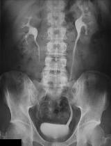
Normal Appearances
Immediately after the contrast is administered, it appears on an x-ray as a 'renal blush'. This is the contrast being filtered through the cortex. At an interval of 3 minutes, the renal blush is still evident (to a lesser extent) but the calyces and renal pelvisRenal pelvis
The renal pelvis or pyelum is the funnel-like dilated proximal part of the ureter in the kidney.In humans, the renal pelvis is the point of convergence of two or three major calyces...
are now visible. At 9 – 13 minutes the contrast begins to empty into the ureters and travel to the bladder
Urinary bladder
The urinary bladder is the organ that collects urine excreted by the kidneys before disposal by urination. A hollow muscular, and distensible organ, the bladder sits on the pelvic floor...
which has now begun to fill. To visualize the bladder correctly, a post micturition x-ray is taken, so that the bulk of the contrast (which can mask a pathology) is emptied.
An IVP can be performed in either emergency or routine circumstances.
Emergency IVP
This procedure is carried out on patients who present to an Emergency department, usually with severe renal colicRenal colic
Renal colic is a type of abdominal pain commonly caused by kidney stones.-Presentation:The pain typically begins in the abdomen and often radiates to the hypochondrium or the groin. The pain is often colicky due to ureteric peristalsis, but may be constant...
and a positive hematuria
Hematuria
In medicine, hematuria, or haematuria, is the presence of red blood cells in the urine. It may be idiopathic and/or benign, or it can be a sign that there is a kidney stone or a tumor in the urinary tract , ranging from trivial to lethal...
test. In this circumstance the attending physician requires to know whether a patient has a kidney stone and if it is causing any obstruction in the urinary system.
Patients with a positive find for kidney stones but with no obstruction are usually discharged with a follow-up appointment with a urologist.
Patients with a kidney stone and obstruction are usually required to stay in hospital for monitoring or further treatment.
An Emergency IVP is carried out roughly as follows:
- plain KUB or Abdominal x-rayAbdominal x-rayAn abdominal x-ray is an x-ray of the abdomen. It is sometimes abbreviated to AXR, or KUB .-Diagnostic Tool:...
; - an injection of contrast mediaContrast mediumA medical contrast medium is a substance used to enhance the contrast of structures or fluids within the body in medical imaging...
, typically 50 ml; - delayed Abdominal x-ray, taken at roughly 15 minutes post injection.
If no obstruction is evident on this film a post-micturition film is taken and the patient is sent back to the Emergency department. If an obstruction is visible, a post-micturition film is still taken, but is followed up with a series of radiographs taken at a "double time" interval. For example, at 30 minutes post-injection, 1 hour, 2 hours, 4 hours, and so forth, until the obstruction is seen to resolve. This time delay can give important information to the urologist on where and how severe the obstruction is.
Routine IVP
This procedure is most common for patients who have unexplained microscopic or macroscopic hematuria. It is used to ascertain the presence of a tumour or similar anatomy-altering disorders. The sequence of images are roughly as follows:- plain or Control KUB image;
- immediate x-ray of just the renal area;
- 5 minute x-ray of just the renal area.
At this point, compression may or may not be applied (this is contraindicated in cases of obstruction).
In pyelography, compression involves pressing on the lower abdominal area, which results in distension of the upper urinary tract.
- If compression is applied: a 10 minutes post-injection x-ray of the renal area is taken, followed by a KUB on release of the compression.
- If compression is not given: a standard KUB is taken to show the ureters emptying. This may sometimes be done with the patient lying in a prone position.
- A post-micturition x-ray is taken afterwards. This is usually a coned bladder view.
Image Assessment
The kidneys are assessed and compared for:- Regular appearance, smooth outlines, size, position, equal filtration and flow.
The ureters are assessed and compared for:
- Size, a smooth regular and symmetrical appearance. A 'standing column' is suggestive of a partial obstruction.
The bladder is assessed for:
- Regular smooth appearance and complete voiding.
Contraindications
Historically, the drug metforminMetformin
Metformin is an oral antidiabetic drug in the biguanide class. It is the first-line drug of choice for the treatment of type 2 diabetes, in particular, in overweight and obese people and those with normal kidney function. Its use in gestational diabetes has been limited by safety concerns...
has been required to stop 48 hours pre and post procedure, as it known to cause a reaction with the contrast agent. However the newest guidelines published by the Royal College of Radiologists suggests this is not as important for patients having <100mls of contrast, who have a normal renal function
Creatinine
Creatinine is a break-down product of creatine phosphate in muscle, and is usually produced at a fairly constant rate by the body...
. If renal impairment is found before administration of the contrast, metformin should be stopped 48 hours before and after the procedure.
Diagnoses
- Chronic PyelonephritisPyelonephritisPyelonephritis is an ascending urinary tract infection that has reached the pyelum or pelvis of the kidney. It is a form of nephritis that is also referred to as pyelitis...
- Kidney stoneKidney stoneA kidney stone, also known as a renal calculus is a solid concretion or crystal aggregation formed in the kidneys from dietary minerals in the urine...
s - Renal cell carcinomaRenal cell carcinomaRenal cell carcinoma is a kidney cancer that originates in the lining of the proximal convoluted tubule, the very small tubes in the kidney that filter the blood and remove waste products. RCC is the most common type of kidney cancer in adults, responsible for approximately 80% of cases...
or RCC - Transitional cell carcinomaTransitional cell carcinomaTransitional cell carcinoma is a type of cancer that typically occurs in the urinary system: the kidney, urinary bladder, and accessory organs. It is the most common type of bladder cancer and cancer of the ureter, urethra, and urachus...
, or TCC - Polycystic kidneysPolycystic kidney diseaseAutosomal dominant polycystic kidney disease is an inherited systemic disorder that predominantly affects the kidneys, but may affect other organs including the liver, pancreas, brain, and arterial blood vessels...
- Anatomical variations, i.e. horseshoe kidneyHorseshoe kidneyHorseshoe kidney, also known as renal fusion or super kidney, is a congenital disorder affecting about 1 in 400 people. In this disorder, the patient's kidneys fuse together to form a horseshoe-shape during development in the womb...
or a duplex collecting system - Obstruction (commonly at the pelvic-ureteric junction or PUJ and the vesicoureteric junction or VUJ)
Other tests
An IVP can and should be used in conjunction with the following tests:- UltrasoundUltrasoundUltrasound is cyclic sound pressure with a frequency greater than the upper limit of human hearing. Ultrasound is thus not separated from "normal" sound based on differences in physical properties, only the fact that humans cannot hear it. Although this limit varies from person to person, it is...
- CystoscopyCystoscopyCystoscopy is endoscopy of the urinary bladder via the urethra. It is carried out with a cystoscope.Diagnostic cystoscopy is usually carried out with local anaesthesia...
- CTComputed tomographyX-ray computed tomography or Computer tomography , is a medical imaging method employing tomography created by computer processing...
- MRI
- Video cystometrography or VCMG
- Blood testBlood testA blood test is a laboratory analysis performed on a blood sample that is usually extracted from a vein in the arm using a needle, or via fingerprick....
- Urine analysis
Treatment
Depending on the outcome and diagnosis following an IVP, treatment may be required for the patient. These include surgerySurgery
Surgery is an ancient medical specialty that uses operative manual and instrumental techniques on a patient to investigate and/or treat a pathological condition such as disease or injury, or to help improve bodily function or appearance.An act of performing surgery may be called a surgical...
, lithotripsy
Lithotripsy
Lithotripsy refers to the physical destruction of gallstones or kidney stones. The term is derived from the Greek words meaning "breaking stones" .Forms include:* Extracorporeal shock wave lithotripsy...
, ureteric stent
Ureteric stent
A ureteral stent, sometimes as well called ureteric stent, is a thin tube inserted into the ureter to prevent or treat obstruction of the urine flow from the kidney. The length of the stents used in adult patients varies between 24 to 30 cm. Additionally, stents come in differing diameters or...
insertion and radiofrequency ablation
Radiofrequency ablation
Radio frequency ablation is a medical procedure where part of the electrical conduction system of the heart, tumor or other dysfunctional tissue is ablated using the heat generated from the high frequency alternating current to treat a medical disorder...
. Sometimes no treatment is necessary as stones <5mm can be passed without any intervention.
The Future of the intravenous pyelogram
The IVP is now becoming more and more obsolete. It has largely been taken over by Computed tomographyComputed tomography
X-ray computed tomography or Computer tomography , is a medical imaging method employing tomography created by computer processing...
(CT), which gives greater detail on anatomy and function.