

Muscles of the hip
Encyclopedia
Human anatomy
Human anatomy is primarily the scientific study of the morphology of the human body. Anatomy is subdivided into gross anatomy and microscopic anatomy. Gross anatomy is the study of anatomical structures that can be seen by the naked eye...
, the muscles of the hip joint are those muscle
Muscle
Muscle is a contractile tissue of animals and is derived from the mesodermal layer of embryonic germ cells. Muscle cells contain contractile filaments that move past each other and change the size of the cell. They are classified as skeletal, cardiac, or smooth muscles. Their function is to...
s that cause movement in the hip
Hip (anatomy)
In vertebrate anatomy, hip refer to either an anatomical region or a joint.The hip region is located lateral to the gluteal region , inferior to the iliac crest, and overlying the greater trochanter of the femur, or "thigh bone"...
. Most modern anatomists define 17 of these muscles, although some additional muscles may sometimes be considered. These are often divided into four groups according to their orientation around the hip joint: the gluteal group
Gluteal muscles
The gluteal muscles are the three muscles that make up the buttocks: the gluteus maximus muscle, gluteus medius muscle and gluteus minimus muscle.-The gluteal muscles:...
, the lateral rotator group
Lateral rotator group
The Lateral rotator group are a group of muscles of the hip which all externally rotate the femur in the hip joint. It consists of the following muscles: * piriformis* gemellus superior* obturator internus* gemellus inferior* obturator externus...
, the adductor group
Adductor muscles of the hip
In human anatomy, the adductor muscles of the hip is a group of muscles of the thigh.-Muscles:The adductor group is made up of:*Adductor brevis*Adductor longus*Adductor magnus*Adductor minimus This is often considered to be a part of adductor magnus....
, and the iliopsoas group.
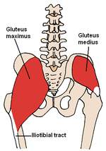
Gluteal group
The gluteal muscles include the gluteus maximus, gluteus medius, gluteus minimus, and tensor fasciae lataeTensor fasciae latae
The tensor fasciae latae or tensor fasciæ latæ is a muscle of the thigh. The English name for this muscle is the muscle that stretches the band on the side...
. They cover the lateral surface of the ilium
Ilium (bone)
The ilium is the uppermost and largest bone of the pelvis, and appears in most vertebrates including mammals and birds, but not bony fish. All reptiles have an ilium except snakes, although some snake species have a tiny bone which is considered to be an ilium.The name comes from the Latin ,...
. The gluteus maximus, which forms most of the muscle of the buttocks
Buttocks
The buttocks are two rounded portions of the anatomy, located on the posterior of the pelvic region of apes and humans, and many other bipeds or quadrupeds, and comprise a layer of fat superimposed on the gluteus maximus and gluteus medius muscles. Physiologically, the buttocks enable weight to...
, originates primarily on the ilium and sacrum
Sacrum
In vertebrate anatomy the sacrum is a large, triangular bone at the base of the spine and at the upper and back part of the pelvic cavity, where it is inserted like a wedge between the two hip bones. Its upper part connects with the last lumbar vertebra, and bottom part with the coccyx...
and inserts on the gluteal tuberosity
Gluteal tuberosity
The lateral ridge of the linea aspera is very rough, and runs almost vertically upward to the base of the greater trochanter. It is termed the gluteal tuberosity, and gives attachment to part of the Glutæus maximus: its upper part is often elongated into a roughened crest, on which a more or less...
of the femur
Femur
The femur , or thigh bone, is the most proximal bone of the leg in tetrapod vertebrates capable of walking or jumping, such as most land mammals, birds, many reptiles such as lizards, and amphibians such as frogs. In vertebrates with four legs such as dogs and horses, the femur is found only in...
as well as the iliotibial tract
Iliotibial tract
The iliotibial tract or iliotibial band is a longitudinal fibrous reinforcement of the fascia lata. It is attached to the anterolateral iliac tubercle portion of the external lip of the iliac crest and to the lateral condyle of the tibia...
, a tract of strong fibrous tissue that runs along the lateral thigh
Thigh
In humans the thigh is the area between the pelvis and the knee. Anatomically, it is part of the lower limb.The single bone in the thigh is called the femur...
to the tibia
Tibia
The tibia , shinbone, or shankbone is the larger and stronger of the two bones in the leg below the knee in vertebrates , and connects the knee with the ankle bones....
and fibula. The gluteus medius and gluteus minimus originate anterior to the gluteus maximus on the ilium and both insert on the greater trochanter
Greater trochanter
The greater trochanter of the femur is a large, irregular, quadrilateral eminence and a part of the skeletal system.It is directed a little lateralward and backward, and, in the adult, is about 1 cm lower than the head...
of the femur. The tensor fasciae latae shares its origin with the gluteus maximus at the ilium and also shares the insertion at the iliotibial tract.
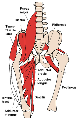
Adductor group
The adductor brevis, adductor longus, adductor magnus, pectineus, and gracilisGracilis muscle
The gracilis is the most superficial muscle on the medial side of the thigh. It is thin and flattened, broad above, narrow and tapering below.-Origin and insertion:...
make up the adductor group. The adductors all originate on the pubis
Pubis (bone)
In vertebrates, the pubic bone is the ventral and anterior of the three principal bones composing either half of the pelvis.It is covered by a layer of fat, which is covered by the mons pubis....
and insert on the medial, posterior surface of the femur, with the exception of the gracilis which inserts just below the medial condyle
Medial condyle
Medial condyle can refer to:* Medial condyle of tibia* Medial condyle of femur...
of the tibia.
Iliopsoas group
The iliacus and psoas major comprise the iliopsoas group. The iliopsoas is a large muscle that runs from the transverse processes of the T-12 to L-5 vertebrae, joins with the iliacus via its tendonTendon
A tendon is a tough band of fibrous connective tissue that usually connects muscle to bone and is capable of withstanding tension. Tendons are similar to ligaments and fasciae as they are all made of collagen except that ligaments join one bone to another bone, and fasciae connect muscles to other...
, and connects to the lesser trochanter
Lesser trochanter
The lesser trochanter of the femur is a conical eminence, which varies in size in different subjects-Anatomy:It projects from the lower and back part of the base of the femur neck.From its apex three well-marked borders extend:...
of the femur. The iliacus originates on the iliac fossa
Iliac fossa
The iliac fossa is a large, smooth, concave surface located on the internal surface of the ilium...
of the ilium. Together these muscles are commonly referred to as the "iliopsoas".
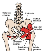
Lateral rotator group
This group consists of the externus and internus obturators, the piriformis, the superior and inferior gemelli, and the quadratus femoris. These six originate at or below the acetabulumAcetabulum
The acetabulum is a concave surface of the pelvis. The head of the femur meets with the pelvis at the acetabulum, forming the hip joint.-Structure:...
of the ilium and insert on or near the greater trochanter of the femur.
Movement of the hip joint
FlexionFlexion
In anatomy, flexion is a position that is made possible by the joint angle decreasing. The skeletal and muscular systems work together to move the joint into a "flexed" position. For example the elbow is flexed when the hand is brought closer to the shoulder...
of the hip occurs when the angle between the torso
Torso
Trunk or torso is an anatomical term for the central part of the many animal bodies from which extend the neck and limbs. The trunk includes the thorax and abdomen.-Major organs:...
and thigh
Thigh
In humans the thigh is the area between the pelvis and the knee. Anatomically, it is part of the lower limb.The single bone in the thigh is called the femur...
is decreased. When this angle is increased extension occurs; beyond normal standing posture, the movement is called hip hyperextension. Hip rotation occurs when the femur moves along its longitudinal axis. When the anterior surface of the femur turns outward, this is lateral rotation of the hip. The movement of the anterior surface of the femur inward is medial rotation of the hip. Medial rotation of the hip is carried out by gluteus medius and gluteus minimus. Hip abduction
Anatomical terms of motion
The movement of body structures is accomplished by the contraction of muscles. Muscles may move parts of the skeleton relatively to each other, or may move parts of internal organs relatively to each other. All such movements are classified by the directions in which the affected structures are moved...
occurs when the femur moves outward to the side, as in taking the thighs apart. Hip adduction
Adduction
Adduction is a movement which brings a part of the anatomy closer to the middle sagittal plane of the body. It is opposed to abduction.-Upper limb:* of arm at shoulder ** Subscapularis** Teres major** Pectoralis major** Infraspinatus...
occurs when the femur moves back to the midline.
Many of the hip muscles are responsible for more than one type of movement in the hip, as different areas of the muscle act on tendons in different ways.
The psoas is the primary hip flexor, assisted by the iliacus. The pectineus, the adductors longus, brevis, and magnus, as well as the tensor fasciae latae are also involved in flexion.
The gluteus maximus is the main hip extensor, but the inferior portion of the adductor magnus also plays a role.
The adductor group is responsible for hip adduction. Abduction primarily occurs via the gluteus medius as well as the gluteus minimus.
Medial rotation is performed by the glutaei medius and minimus, as well as the tensor fasciae latae and assisted by the adductors brevis and longus and the superior portion of the adductor magnus.
Each muscle of the lateral rotator group causes lateral rotation of the thigh. These muscles are aided by the gluteus maximus and the inferior portion of the adductor magnus.
Other hip muscles
Additional muscles, such as the rectus femoris and the sartoriusSartorius muscle
The Sartorius muscle – the longest muscle in the human body – is a long thin muscle that runs down the length of the thigh. Its upper portion forms the lateral border of the femoral triangle.-Origin and insertion:...
, can cause some movement in the hip joint. However these muscles are primarily concerned with the movement of the knee
Knee
The knee joint joins the thigh with the leg and consists of two articulations: one between the fibula and tibia, and one between the femur and patella. It is the largest joint in the human body and is very complicated. The knee is a mobile trocho-ginglymus , which permits flexion and extension as...
, and are therefore not generally classified as muscles of the hip.
The hamstring muscle group, which mostly originates from the ischial tuberosity inserting on the tibia/fibula, has a large moment assisting with hip extension.
Sources
- Calais-Germain, Blandine. "Anatomy of Movement", Eastland Press, 1993. ISBN 0-939616-17-3
- Martini, Frederic; Timmons, Michael; McKinnley, Michael. "Human Anatomy", 3rd Edition, Prentice-Hall, 2000. ISBN 0-13-010011-0
- Marieb, Elaine. "Essentials of Human Anatomy and Physiology", 6th Edition. Addison Wesley Longman, 2000. ISBN 0-8053-4940-5
- Netter, Frank H. "Atlas of Human Anatomy", 2nd Edition, Icon Learning Systems, 2001. ISBN 0-914168-81-9