
Knee
Overview
Joint
A joint is the location at which two or more bones make contact. They are constructed to allow movement and provide mechanical support, and are classified structurally and functionally.-Classification:...
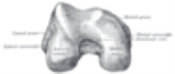
joins the thigh with the leg and consists of two articulations: one between the fibula and tibia
Tibia
The tibia , shinbone, or shankbone is the larger and stronger of the two bones in the leg below the knee in vertebrates , and connects the knee with the ankle bones....
, and one between the femur and patella. It is the largest joint in the human body and is very complicated. The knee is a mobile trocho-ginglymus (i.e. a pivotal hinge joint), which permits flexion
Flexion
In anatomy, flexion is a position that is made possible by the joint angle decreasing. The skeletal and muscular systems work together to move the joint into a "flexed" position. For example the elbow is flexed when the hand is brought closer to the shoulder...
and extension
Extension (kinesiology)
In kinesiology, extension is a movement of a joint that results in increased angle between two bones or body surfaces at a joint. Extension usually results in straightening of the bones or body surfaces involved. For example, extension is produced by extending the flexed elbow. Straightening of...
as well as a slight medial and lateral rotation.