

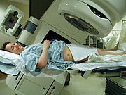
External beam radiotherapy
Encyclopedia

Radiation
In physics, radiation is a process in which energetic particles or energetic waves travel through a medium or space. There are two distinct types of radiation; ionizing and non-ionizing...
is pointed at a particular part of the body. In contrast to internal radiotherapy (brachytherapy
Brachytherapy
Brachytherapy , also known as internal radiotherapy, sealed source radiotherapy, curietherapy or endocurietherapy, is a form of radiotherapy where a radiation source is placed inside or next to the area requiring treatment...
), in which the radiation source is inside the body, external beam radiotherapy directs the radiation at the tumour from outside the body. Kilovoltage ("superficial") X-rays are used for treating skin cancer and superficial structures. Megavoltage ("deep") X-rays are used to treat deep-seated tumours (e.g. bladder, bowel, prostate, lung, or brain).
While X-ray and electron beams are by far the most widely used sources for external beam radiotherapy, a small number of centers operate experimental and pilot programs employing heavier particle beams, particularly proton sources.
Photons
Conventionally, the energy of diagnostic and therapeutic gamma- and X-rays is expressed in kilovolts or megavoltMegavolt
*A megavolt is 1 million volts in electronics and physics.*Megavolt is a villain in the Disney animated series Darkwing Duck.*Megavolt is a villain in the Teenage Mutant Ninja Turtles ...
s (kV or MV), whilst the energy of therapeutic electrons is expressed in terms of megaelectronvolts (MeV). In the first case, this voltage is the maximum electric potential used by a linear accelerator to produce the photon
Photon
In physics, a photon is an elementary particle, the quantum of the electromagnetic interaction and the basic unit of light and all other forms of electromagnetic radiation. It is also the force carrier for the electromagnetic force...
beam. The beam is made up of a spectrum of energies: the maximum energy is approximately equal to the beam's maximum electric potential times the electron charge. Thus a 1 MV beam will produce photons of no more than about 1 MeV. The mean X-ray energy is only about 1/3 of the maximum energy. Beam quality and hardness may be improved by special filters, which improve the homogeneity of the X-ray spectrum.
In the medical field, useful X-rays are produced when electrons are accelerated to a high energy. Some examples of X-ray energies used in medicine are:
- diagnostic X-rays - 20 to 150 kV
- superficial X-raysSuperficial X-raysSuperficial X-rays are low energy X-rays that do not penetrate very far before they are absorbed. They are produced by linear accelerators operating at voltages in the 50–200 kV range, and therefore have an energy in the 50–200 keV range...
- 50 to 200 kV - orthovoltage X-raysOrthovoltage X-raysOrthovoltage X-rays are produced by X-ray generators operating at voltages in the 200–500 kV range, and therefore an energy in the 200–500 keV range . When used to treat patients, radiation oncologists find that they penetrate to a useful depth of about 4–6 cm...
- 200 to 500 kV - supervoltage X-rays - 500 to 1000 kV
- megavoltage X-raysMegavoltage X-raysMegavoltage X-rays are produced by linear accelerators operating at voltages in excess of 1000 kV range, and therefore have an energy in the MeV range...
- 1 to 25 MV
Of these energy ranges, megavoltage X-rays are by far most common in radiotherapy. Orthovoltage X-rays do have limited applications, and the other energy ranges are not typically used clinically.
Medically useful photon beams can also be derived from a radioactive source such as cobalt-60
Cobalt-60
Cobalt-60, , is a synthetic radioactive isotope of cobalt. Due to its half-life of 5.27 years, is not found in nature. It is produced artificially by neutron activation of . decays by beta decay to the stable isotope nickel-60...
, iridium
Iridium
Iridium is the chemical element with atomic number 77, and is represented by the symbol Ir. A very hard, brittle, silvery-white transition metal of the platinum family, iridium is the second-densest element and is the most corrosion-resistant metal, even at temperatures as high as 2000 °C...
-192, caesium-137 or radium
Radium
Radium is a chemical element with atomic number 88, represented by the symbol Ra. Radium is an almost pure-white alkaline earth metal, but it readily oxidizes on exposure to air, becoming black in color. All isotopes of radium are highly radioactive, with the most stable isotope being radium-226,...
-226 (which is no longer used clinically). Such photon beams, derived from radioactive decay, are more or less monochromatic and are properly termed gamma ray
Gamma ray
Gamma radiation, also known as gamma rays or hyphenated as gamma-rays and denoted as γ, is electromagnetic radiation of high frequency . Gamma rays are usually naturally produced on Earth by decay of high energy states in atomic nuclei...
s. The usual energy range is between 300 keV to 1.5 MeV, and is specific to the isotope.
Therapeutic radiation is mainly generated in the radiotherapy department using the following equipment:
- Orthovoltage units. These are also known as "deep" and "superficial" machines depending on their energy range. Orthovoltage units have essentially the same design as diagnostic X-ray machines. These machines are generally limited to less than 600 kV.
- Linear accelerators ("linacs") which produce megavoltage X-rays. Commercially available medical linacs produce X-rays and electrons with an energy range from 4 MeV up to around 25 MeV. The X-rays themselves are produced by the rapid deceleration of electrons in a target material, typically a tungstenTungstenTungsten , also known as wolfram , is a chemical element with the chemical symbol W and atomic number 74.A hard, rare metal under standard conditions when uncombined, tungsten is found naturally on Earth only in chemical compounds. It was identified as a new element in 1781, and first isolated as...
alloy, which produces an X-ray spectrum via bremsstrahlungBremsstrahlungBremsstrahlung is electromagnetic radiation produced by the deceleration of a charged particle when deflected by another charged particle, typically an electron by an atomic nucleus. The moving particle loses kinetic energy, which is converted into a photon because energy is conserved. The term is...
radiation. The shape and intensity of the beam produced by a linac may be modified or collimated by a variety of means. Thus, conventional, conformal, intensity-modulated, tomographic, and stereotactic radiotherapy are all produced by specially modified linear accelerators. - Cobalt units which produce stable, dichromatic beams of 1.17 and 1.33 MeV, resulting in an average beam energy of 1.25 MeV. The role of the cobalt unit has partly been replaced by the linear accelerator, which can generate higher energy radiation. Cobalt treatment still has a useful role to play in certain applications and is still in widespread use worldwide, since the machinery is relatively reliable and simple to maintain compared to the modern linear accelerator.
Electrons
X-rays are generated by bombarding a high atomic number material with electrons. If the target is removed (and the beam current decreased) a high energy electron beam is obtained. Electron beams are useful for treating superficial lesions because the maximum of dose deposition occurs near the surface. The dose then decreases rapidly with depth, sparing underlying tissue. Electron beams usually have nominal energies in the range 4-20 MeV. Depending on the energy this translates to a treatment range of approximately 1–5 cm (in water-equivalent tissue). Energies above 18 MeV are used very rarely. Although the X-ray target is removed in electron mode, the beam must be fanned out by sets of thin scattering foils in order to achieve flat and symmetric dose profiles in the treated tissue.Hadron therapy
HadronHadron
In particle physics, a hadron is a composite particle made of quarks held together by the strong force...
therapy involves the therapeutic use of proton
Proton
The proton is a subatomic particle with the symbol or and a positive electric charge of 1 elementary charge. One or more protons are present in the nucleus of each atom, along with neutrons. The number of protons in each atom is its atomic number....
s, neutron
Neutron
The neutron is a subatomic hadron particle which has the symbol or , no net electric charge and a mass slightly larger than that of a proton. With the exception of hydrogen, nuclei of atoms consist of protons and neutrons, which are therefore collectively referred to as nucleons. The number of...
s, and heavier ion
Ion
An ion is an atom or molecule in which the total number of electrons is not equal to the total number of protons, giving it a net positive or negative electrical charge. The name was given by physicist Michael Faraday for the substances that allow a current to pass between electrodes in a...
s (fully ionized atomic nuclei). Of these, proton therapy
Proton therapy
Proton therapy is a type of particle therapy which uses a beam of protons to irradiate diseased tissue, most often in the treatment of cancer. The chief advantage of proton therapy is the ability to more precisely localize the radiation dosage when compared with other types of external beam...
is by far the most common, though still quite rare compared to other forms of external beam radiotherapy.
Multi-leaf collimator
A typical multi-leaf collimator (MLC) consists of 2 sets of 20-40 leaves, each around 5mm thick and several cm in the other two dimensions. Newer MLCs now have up to 160 leaves. Each leaf in the MLC is aligned parallel to the radiation field and can be moved independently to block part of the field. This allows the dosimetrist to match the radiation field to the shape of the tumor (by adjusting the position of the leaves), thus minimizing the amount of healthy tissue being exposed to radiation. On a machine without an MLC this must be accomplished using several hand-crafted blocks.Intensity modulated radiation therapy
Intensity modulated radiation therapy (IMRT) is an advanced radiotherapy technique used to minimize the amount of normal tissue being irradiated in the treatment field. In some systems this intensity modulation is achieved by moving the leaves in the MLC during the course of treatment, thereby delivering a radiation field with a non-uniform (i.e. modulated) intensity. With IMRT, radiation oncologists are able to break up the radiation beam into many "beamlets." This allows radiation oncologists to vary the intensity of each beamlet. With IMRT, doctors are often able to further limit the amount of radiation received by healthy tissue near the tumor. Doctors have found this sometimes allowed them to safely give a higher dose of radiation to the tumor, potentially increasing the chance of a cure.Image-guided radiation therapy
Image-guided radiation therapyImage-guided radiation therapy
Highly trained oncologists called radiation oncologists use image guided radiation therapy, or IGRT, to help better deliver radiation therapy to cancerous tumors. This is very useful since tumors can move between treatments due to differences in organ filling or movements while breathing...
(IGRT) augments radiotherapy with imaging to increase the accuracy and precision of target localization, thereby reducing the amount of healthy tissue in the treatment field.
The more advanced the treatment techniques become in terms of dose deposition accuracy, the higher become the requirements for IGRT. In order to allow patients to benefit from sophisticated treatment techniques as IMRT or Hadron Therapy, patient alignment accuracies of 0.5 mm and less become desirable. Therefore, new methods like stereoscopic digital kilovoltage imaging based patient position verification (PPVS) to alignment estimation based on in-situ Cone-Beam CT enrich the range of modern IGRT approaches.
See also
- CyberKnifeCyberknifeThe CyberKnife is a frameless robotic radiosurgery system used for treating benign tumors, malignant tumors and other medical conditions. The system was invented by John R. Adler, a Stanford University Professor of Neurosurgery and Radiation Oncology, and Peter and Russell Schonberg of Schonberg...
- Gamma knife
- TomotherapyTomoTherapyTomotherapy is a type of radiation therapy in which the radiation is delivered slice-by-slice...
- Radiation therapyRadiation therapyRadiation therapy , radiation oncology, or radiotherapy , sometimes abbreviated to XRT or DXT, is the medical use of ionizing radiation, generally as part of cancer treatment to control malignant cells.Radiation therapy is commonly applied to the cancerous tumor because of its ability to control...
- IOERT
- IORT
- BrachytherapyBrachytherapyBrachytherapy , also known as internal radiotherapy, sealed source radiotherapy, curietherapy or endocurietherapy, is a form of radiotherapy where a radiation source is placed inside or next to the area requiring treatment...
- Boron neutron capture therapyBoron Neutron Capture TherapyBoron neutron capture therapy is an experimental form of radiotherapy that uses a neutron beam that interacts with boron injected into a patient...
General references
- Radiotherapy physics in practice, edited by JR Williams and DI Thwaites, Oxford University Press UK (2nd edition 2000), ISBN 0-19-262878-X
- Linear Particle Accelerator (Linac)Animation by Ionactive
-
- http://www.myradiotherapy.com
-