

Brachytherapy
Encyclopedia
Brachytherapy also known as internal radiotherapy, sealed source radiotherapy, curietherapy or endocurietherapy, is a form of radiotherapy where a radiation source
is placed inside or next to the area requiring treatment. Brachytherapy is commonly used as an effective treatment for cervical
,
prostate
,
breast
,
and skin cancer
and can also be used to treat tumours in many other body sites.
Brachytherapy can be used alone or in combination with other therapies such as surgery, External Beam Radiotherapy
(EBRT) and chemotherapy
.
In contrast to EBRT in which high-energy x-rays are directed at the tumour from outside the body, brachytherapy involves the precise placement of radiation sources directly at the site of the cancerous tumour.
A key feature of brachytherapy is that the irradiation only affects a very localized area around the radiation sources. Exposure to radiation of healthy tissues further away from the sources is therefore reduced. In addition, if the patient moves or if there is any movement of the tumour within the body during treatment, the radiation sources retain their correct position in relation to the tumour. These characteristics of brachytherapy provide advantages over EBRT - the tumour can be treated with very high doses of localised radiation, whilst reducing the probability of unnecessary damage to surrounding healthy tissues.
A course of brachytherapy can be completed in less time than other radiotherapy techniques. This can help reduce the chance of surviving cancer cells dividing and growing in the intervals between each radiotherapy dose. Patients typically have to make fewer visits to the radiotherapy clinic compared with EBRT, and the treatment is often performed on an outpatient basis. This makes treatment accessible and convenient for many patients. These features of brachytherapy reflect that most patients are able to tolerate the brachytherapy procedure very well.
Brachytherapy represents an effective treatment option for many types of cancer. Treatment results have demonstrated that the cancer cure rates of brachytherapy are either comparable to surgery and EBRT, or are improved when used in combination with these techniques. In addition, brachytherapy is associated with a low risk of serious adverse side effects.
suggested to Henri-Alexandre Danlos
that a radioactive source could be inserted into a tumour.
It was found that the radiation caused the tumour to shrink. Independently, Alexander Graham Bell
also suggested the use of radiation in this way. In the early twentieth century, techniques for the application of brachytherapy were pioneered at the Curie institute in Paris by Danlos and at St Luke's and Memorial Hospital in New York by Robert Abbe
.
Following initial interest in brachytherapy in Europe and the US, its use declined in the middle of the twentieth century due to the problem of radiation exposure to operators from the manual application of the radioactive sources.
However, the development of remote afterloading systems, which allow the radiation to be delivered from a shielded safe, and the use of new radioactive sources in the 1950s and 1960s, reduced the risk of unnecessary radiation exposure to the operator and patients.
This, together with more recent advancements in three dimensional imaging modalities, computerised treatment planning systems and delivery equipment has made brachytherapy a safe and effective treatment for many types of cancer today.
per hour (Gy/h).
 The placement of radiation sources in the target area can be temporary or permanent.
, prostate
, breast
, and skin
.
Brachytherapy can also be used in the treatment of tumours of the brain
, eye
, head and neck region (lip, floor of mouth, tongue, nasopharynx and oropharynx), respiratory tract (trachea
and bronchi), digestive tract (oesophagus, gall bladder, bile-ducts
, rectum
, anus
), urinary tract (bladder
, urethra
, penis
), female reproductive tract (uterus
, vagina
, vulva
), and soft tissues.
As the radiation sources can be precisely positioned at the tumour treatment site, brachytherapy enables a high dose of radiation to be applied to a small area. Furthermore, because the radiation sources are placed in or next to the target tumour, the sources maintain their position in relation to the tumour when the patient moves or if there is any movement of the tumour within the body. Therefore, the radiation sources remain accurately targeted. This enables clinicians to achieve a high level of dose conformity – i.e. ensuring the whole of the tumour receives an optimal level of radiation. It also reduces the risk of damage to healthy tissue, organs or structures around the tumour, thus enhancing the chance of cure and preservation of organ function.
The use of HDR brachytherapy enables overall treatment times to be reduced compared with EBRT.
Patients receiving brachytherapy generally have to make fewer visits for radiotherapy compared with EBRT, and overall radiotherapy treatment plans can be completed in less time.
Many brachytherapy procedures are performed on an outpatient basis. This convenience may be particularly relevant for patients who have to work, older patients, or patients who live some distance from treatment centres, to ensure that they have access to radiotherapy treatment and adhere to treatment plans. Shorter treatment times and outpatient procedures can also help improve the efficiency of radiotherapy clinics.
Brachytherapy can be used with the aim of curing the cancer in cases of small or locally advanced tumours, provided the cancer has not metastasized (spread to other parts of the body). In appropriately selected cases, brachytherapy for primary tumours often represents a comparable approach to surgery, achieving the same probability of cure and with similar side effects.
However, in locally advanced tumours, surgery may not routinely provide the best chance of cure and is often not technically feasible to perform. In these cases radiotherapy, including brachytherapy, offers the only chance of cure.
In more advanced disease stages, brachytherapy can be used as palliative treatment for symptom relief from pain and bleeding.
In cases where the tumour is not easily accessible or is too large to ensure an optimal distribution of irradiation to the treatment area, brachytherapy can be combined with other treatments, such as EBRT and/or surgery. Combination therapy of brachytherapy exclusively with chemotherapy is rare.
and is a standard of care in many countries.
Cervical cancer can be treated with either LDR, PDR or HDR brachytherapy.
Used in combination with EBRT, brachytherapy can provide better outcomes than EBRT alone.
The precision of brachytherapy enables a high dose of targeted radiation to be delivered to the cervix, while minimising radiation exposure to adjacent tissues and organs.
The chances of staying free of disease (disease-free survival) and of staying alive (overall survival) are similar for LDR, PDR and HDR treatments.
However, a key advantage of HDR treatment is that each dose can be delivered on an outpatient basis with a short administration time providing greater convenience for many patients.
can be given either as permanent LDR seed implantation or as temporary HDR brachytherapy.
Permanent seed implantation is suitable for patients with a localised tumour and good prognosis
and has been shown to be a highly effective treatment to prevent the cancer from returning. The survival rate is similar to that found with EBRT or surgery (radical prostatectomy), but with fewer side effects such as impotence and incontinence
. The procedure can be completed quickly and patients are usually able to go home on the same day of treatment and return to normal activities after 1 to 2 days.
Permanent seed implantation is often a less invasive treatment option compared to the surgical removal of the prostate.
Temporary HDR brachytherapy is a newer approach to treating prostate cancer, but is currently less common than seed implantation. It is predominately used as to provide an extra dose in addition to EBRT (known as ‘”boost” therapy) as it offers an alternative method to deliver a high dose of radiation therapy that conforms to the shape of the tumour within the prostate, while sparing radiation exposure to surrounding tissues.
HDR brachytherapy as a boost for prostate cancer also means that the EBRT course can be shorter than when EBRT is used alone.
or mastectomy
surgery, and is an integral component of breast-conserving therapy.
Brachytherapy can be used after surgery, before chemotherapy or palliatively in the case of advanced disease. Brachytherapy to treat breast cancer
is usually performed with HDR temporary brachytherapy. Post surgery, breast brachytherapy can be used as a “boost” following irradiation of the whole breast using EBRT.
More recently, brachytherapy alone is applied in a technique called APBI (accelerated partial breast irradiation), involving delivery of radiation to only the immediate region surrounding the original tumour.
The main benefit of breast brachytherapy compared to EBRT is that a high dose of radiation can be precisely applied to the tumour while sparing radiation to healthy breast tissues and underlying structures such as the ribs and lungs. APBI can typically be completed over the course of a week. The option of brachytherapy may be particularly important in ensuring that working women, the elderly or women without easy access to a treatment centre, are able to benefit from breast-conserving therapy due to the short treatment course compared with EBRT (which often requires more visits over the course of 1–2 months). Brachytherapy has demonstrated excellent local control of breast cancer at follow-up of up to 6 years post treatment. A study is underway to compare patient outcomes of APBI in comparison to EBRT at up to 10 years after treatment.
There are two methods that can be used to deliver breast brachytherapy:
Interstitial breast brachytherapy involves the temporary placement of several flexible plastic catheters in the breast tissue. These are carefully positioned to allow optimal targeting of radiation to the treatment area while sparing the surrounding breast tissue. The catheters are connected to an afterloader, which delivers the planned radiation dose to the treatment area. Interstitial breast brachytherapy can be used as “boost” after EBRT, or as APBI.
Intracavitary breast brachytherapy (also known as “balloon brachytherapy”) involves the placement of a single catheter into the breast cavity left after the removal of the tumour (lumpectomy). The catheter can be placed at the time of the lumpectomy or postoperatively. Via the catheter, a balloon is then inflated in the cavity. The catheter is then connected to an afterloader, which delivers the radiation dose through the catheter and into the balloon. Currently, intracavitary breast brachytherapy is only routinely used for APBI.
There are also devices that combine the features of interstitial and intracavitary breast brachytherapy (e.g. SAVI). These devices use multiple catheters but are inserted through a single-entry point in the breast. Studies suggest the use of multiple catheters enables physicians to target the radiation more precisely.
, such as basal cell carcinoma
and squamous cell carcinoma
, provides an alternative treatment option to surgery. This is especially relevant for cancers on the nose, ears, eyelids or lips, where surgery may cause disfigurement or require extensive reconstruction. Various applicators can be used to ensure close contact between the radiation source(s) and the skin, which conform to the curvature of the skin and help ensure precision delivery of the optimal irradiation dose.
Brachytherapy for skin cancer provides good cosmetic results and clinical efficacy; studies with up to 5 years follow-up have shown that brachytherapy is highly effective in terms local control, and is comparable to EBRT. Treatment times are typically short, providing convenience for patients.
It has been suggested that brachytherapy may become a standard of treatment for skin cancer in the near future.
In treating In-stent restenosis (ISR) Drug Eluting stents (DES)have been found to be superior to Intracoronary Brachytherapy (ICBT).
However, there is continued interest in vascular brachytherapy for persistent restenosis in failed stents and vein grafts.
The therapy has also been investigated for use in the treatment of peripheral vasculature stenosis and considered for the treatment of atrial fibrillation
.
Patients may also feel fatigued for a short period following treatment.
Brachytherapy treatment for cervical or prostate cancer can cause acute and transient urinary symptoms such as urinary retention, urinary incontinence or painful urination (dysuria).
Transient increased bowel frequency, diarrhoea, constipation or minor rectal bleeding, may also occur. Acute and subacute side effects usually resolve over a matter of days or a few weeks. In the case of permanent (seed) brachytherapy for prostate cancer, there is a small chance that some seeds may migrate out of the treatment region into the bladder or urethra and be passed in the urine.
Brachytherapy for skin cancer may result in a shedding of the outer layers of skin (desquamation) around the area of treatment in the weeks following therapy, which typically heals in 5–8 weeks. If the cancer is located on the lip, ulceration may occur as a result of brachytherapy, but usually resolves after 4–6 weeks.
Most of the acute side effects associated with brachytherapy can be treated with medication or through dietary changes, and usually disappear over time (typically a matter of weeks), once the treatment is completed. The acute side effects of HDR brachytherapy are broadly similar to EBRT.
Brachytherapy for prostate cancer may cause erectile dysfunction in approximately 15-30% of patients. However, the risk of erectile dysfunction is related to age (older men are at a greater risk than younger men) and also the level of erectile function prior to receiving brachytherapy. In patients who do experience erectile dysfunction, the majority of cases can successfully be treated with drugs such as Viagra. Importantly, the risk of erectile dysfunction after brachytherapy is less than after radical prostatectomy.
Brachytherapy for breast or skin cancer may cause scar tissue to form around the treatment area. In the case of breast brachytherapy, fat necrosis may occur as a result of fatty acids entering the breast tissues. This can cause the breast tissue to become swollen and tender. Fat necrosis is a benign condition and typically occurs 4–12 months after treatment and affects about 2% of patients.
If permanent brachytherapy is used, low dose radioactive sources (seeds) are left in the body after treatment - the radiation levels are very low and decrease over time. In addition, the irradiation only affects tissues within a few millimeters of the radioactive sources (i.e. the tumour being treated). As a precaution, some people receiving permanent brachytherapy may be advised to not hold any small children or be too close to pregnant women for a short time after treatment. Radiation oncologists or nurses can provide specific instructions to patients and advise for how long they need to be careful.
Using this information, a plan of the optimal distribution of the radiation sources can be developed. This includes consideration of how the source carriers (applicators), which are used to deliver the radiation to the treatment site, should be placed and positioned. Applicators are non-radioactive and are typically needles or plastic catheters. The specific type of applicator used will depend on the type of cancer being treated and the characteristics of the target tumour.
This initial planning helps to ensure that ‘cold spots’ (too little irradiation) and ‘hot spots’ (too much irradiation) are avoided during treatment, as these can respectively result in treatment failure and side-effects.
Imaging techniques, such as x-ray, fluoroscopy and ultrasound are typically used to help guide the placement of the applicators to their correct positions and to further refine the treatment plan. CAT scans and MRI can also be used. Once the applicators are inserted, they are held in place against the skin using sutures or adhesive tape to prevent them from moving. Once the applicators are confirmed as being in the correct position, further imaging can be performed to guide detailed treatment planning.
Manual delivery of brachytherapy is limited to a few LDR applications, due to risk of radiation exposure to clinical staff.
In contrast, afterloading involves the accurate positioning of non-radioactive applicators in the treatment site, which are subsequently loaded with the radiation sources. In manual afterloading, the source is delivered into the applicator by the operator.
Remote afterloading systems provide protection from radiation exposure to healthcare professionals by securing the radiation source in a shielded safe. Once the applicators are correctly positioned in the patient, they are connected to an ‘afterloader’ machine (containing the radioactive sources) through a series of connecting guide tubes. The treatment plan is sent to the afterloader, which then controls the delivery of the sources along the guide tubes into the pre-specified positions within the applicator. This process is only engaged once staff are removed from the treatment room. The sources remain in place for a pre-specified length of time, again following the treatment plan, following which they are returned along the tubes to the afterloader.
On completion of delivery of the radioactive sources, the applicators are carefully removed from the body. Patients typically recover quickly from the brachytherapy procedure, enabling it to often be performed on an outpatient basis.
Radiation
In physics, radiation is a process in which energetic particles or energetic waves travel through a medium or space. There are two distinct types of radiation; ionizing and non-ionizing...
is placed inside or next to the area requiring treatment. Brachytherapy is commonly used as an effective treatment for cervical
Cervical cancer
Cervical cancer is malignant neoplasm of the cervix uteri or cervical area. One of the most common symptoms is abnormal vaginal bleeding, but in some cases there may be no obvious symptoms until the cancer is in its advanced stages...
,
prostate
Prostate cancer
Prostate cancer is a form of cancer that develops in the prostate, a gland in the male reproductive system. Most prostate cancers are slow growing; however, there are cases of aggressive prostate cancers. The cancer cells may metastasize from the prostate to other parts of the body, particularly...
,
breast
Breast cancer
Breast cancer is cancer originating from breast tissue, most commonly from the inner lining of milk ducts or the lobules that supply the ducts with milk. Cancers originating from ducts are known as ductal carcinomas; those originating from lobules are known as lobular carcinomas...
,
and skin cancer
Skin cancer
Skin neoplasms are skin growths with differing causes and varying degrees of malignancy. The three most common malignant skin cancers are basal cell cancer, squamous cell cancer, and melanoma, each of which is named after the type of skin cell from which it arises...
and can also be used to treat tumours in many other body sites.
Brachytherapy can be used alone or in combination with other therapies such as surgery, External Beam Radiotherapy
External beam radiotherapy
External beam radiotherapy or teletherapy is the most common form of radiotherapy. The patient sits or lies on a couch and an external source of radiation is pointed at a particular part of the body...
(EBRT) and chemotherapy
Chemotherapy
Chemotherapy is the treatment of cancer with an antineoplastic drug or with a combination of such drugs into a standardized treatment regimen....
.
In contrast to EBRT in which high-energy x-rays are directed at the tumour from outside the body, brachytherapy involves the precise placement of radiation sources directly at the site of the cancerous tumour.
A key feature of brachytherapy is that the irradiation only affects a very localized area around the radiation sources. Exposure to radiation of healthy tissues further away from the sources is therefore reduced. In addition, if the patient moves or if there is any movement of the tumour within the body during treatment, the radiation sources retain their correct position in relation to the tumour. These characteristics of brachytherapy provide advantages over EBRT - the tumour can be treated with very high doses of localised radiation, whilst reducing the probability of unnecessary damage to surrounding healthy tissues.
A course of brachytherapy can be completed in less time than other radiotherapy techniques. This can help reduce the chance of surviving cancer cells dividing and growing in the intervals between each radiotherapy dose. Patients typically have to make fewer visits to the radiotherapy clinic compared with EBRT, and the treatment is often performed on an outpatient basis. This makes treatment accessible and convenient for many patients. These features of brachytherapy reflect that most patients are able to tolerate the brachytherapy procedure very well.
Brachytherapy represents an effective treatment option for many types of cancer. Treatment results have demonstrated that the cancer cure rates of brachytherapy are either comparable to surgery and EBRT, or are improved when used in combination with these techniques. In addition, brachytherapy is associated with a low risk of serious adverse side effects.
History
Brachytherapy dates back to 1901 (shortly after the discovery of radioactivity by Becquerel in 1896) when Pierre CuriePierre Curie
Pierre Curie was a French physicist, a pioneer in crystallography, magnetism, piezoelectricity and radioactivity, and Nobel laureate. He was the son of Dr. Eugène Curie and Sophie-Claire Depouilly Curie ...
suggested to Henri-Alexandre Danlos
Henri-Alexandre Danlos
Henri-Alexandre Danlos was a French physician and dermatologist born in Paris. With Danish dermatologist Edvard Ehlers , the Ehlers–Danlos syndrome is named, which is a group of inherited connective tissue disorders.He studied medicine in Paris, and during the early part of his career performed...
that a radioactive source could be inserted into a tumour.
It was found that the radiation caused the tumour to shrink. Independently, Alexander Graham Bell
Alexander Graham Bell
Alexander Graham Bell was an eminent scientist, inventor, engineer and innovator who is credited with inventing the first practical telephone....
also suggested the use of radiation in this way. In the early twentieth century, techniques for the application of brachytherapy were pioneered at the Curie institute in Paris by Danlos and at St Luke's and Memorial Hospital in New York by Robert Abbe
Robert Abbe
Robert Abbe was an American surgeon and pioneer radiologist in New York City. He was born in New York City and educated at the College of the City of New York and Columbia University ....
.
Following initial interest in brachytherapy in Europe and the US, its use declined in the middle of the twentieth century due to the problem of radiation exposure to operators from the manual application of the radioactive sources.
However, the development of remote afterloading systems, which allow the radiation to be delivered from a shielded safe, and the use of new radioactive sources in the 1950s and 1960s, reduced the risk of unnecessary radiation exposure to the operator and patients.
This, together with more recent advancements in three dimensional imaging modalities, computerised treatment planning systems and delivery equipment has made brachytherapy a safe and effective treatment for many types of cancer today.
Types
Different types of brachytherapy can be defined according to (1) the placement of the radiation sources in the target treatment area, (2) the rate or ‘intensity’ of the irradiation dose delivered to the tumour, and (3) the duration of dose delivery.Source placement
The two main types of brachytherapy treatment in terms of the placement of the radioactive source are interstitial and contact.- In the case of interstitial brachytherapy, the sources are placed directly in the target tissue of the affected site, such as the prostate or breast.
- Contact brachytherapy involves placement of the radiation source in a space next to the target tissue. This space may be a body cavity (intracavitary brachytherapy) such as the cervixCervixThe cervix is the lower, narrow portion of the uterus where it joins with the top end of the vagina. It is cylindrical or conical in shape and protrudes through the upper anterior vaginal wall...
, uterusUterusThe uterus or womb is a major female hormone-responsive reproductive sex organ of most mammals including humans. One end, the cervix, opens into the vagina, while the other is connected to one or both fallopian tubes, depending on the species...
or vaginaVaginaThe vagina is a fibromuscular tubular tract leading from the uterus to the exterior of the body in female placental mammals and marsupials, or to the cloaca in female birds, monotremes, and some reptiles. Female insects and other invertebrates also have a vagina, which is the terminal part of the...
; a body lumen (intraluminal brachytherapy) such as the tracheaVertebrate tracheaIn tetrapod anatomy the trachea, or windpipe, is a tube that connects the pharynx or larynx to the lungs, allowing the passage of air. It is lined with pseudostratified ciliated columnar epithelium cells with goblet cells that produce mucus...
or oesophagus; or externally (surface brachytherapy) such as the skinSkin-Dermis:The dermis is the layer of skin beneath the epidermis that consists of connective tissue and cushions the body from stress and strain. The dermis is tightly connected to the epidermis by a basement membrane. It also harbors many Mechanoreceptors that provide the sense of touch and heat...
. A radiation source can also be placed in blood vessels (intravascular brachytherapy) for the treatment of coronary in-stent restenosis.
Dose rate
The dose rate of brachytherapy refers to the level or ‘intensity’ with which the radiation is delivered to the surrounding medium and is expressed in GraysGray (unit)
The gray is the SI unit of absorbed radiation dose of ionizing radiation , and is defined as the absorption of one joule of ionizing radiation by one kilogram of matter ....
per hour (Gy/h).
- Low-dose rate(LDR) brachytherapy involves implanting radiation sources that emit radiation at a rate of up to 2 Gy·h-1. LDR brachytherapy is commonly used for cancers of the oral cavity, oropharynxOropharynxThe Oropharynx reaches from the Uvula to the level of the hyoid bone.It opens anteriorly, through the isthmus faucium, into the mouth, while in its lateral wall, between the two palatine arches, is the palatine tonsil....
, sarcomas and prostate cancerProstate cancerProstate cancer is a form of cancer that develops in the prostate, a gland in the male reproductive system. Most prostate cancers are slow growing; however, there are cases of aggressive prostate cancers. The cancer cells may metastasize from the prostate to other parts of the body, particularly...
- Medium-dose rate (MDR) brachytherapy is characterized by a medium rate of dose delivery, ranging between 2 Gy·h-1 to 12 Gy·h-1.
- High-dose rate (HDR) brachytherapy is when the rate of dose delivery exceeds 12 Gy·h-1. The most common applications of HDR brachytherapy are in tumours of the cervixCervixThe cervix is the lower, narrow portion of the uterus where it joins with the top end of the vagina. It is cylindrical or conical in shape and protrudes through the upper anterior vaginal wall...
, oesophagus, lungs, breasts and prostateProstateThe prostate is a compound tubuloalveolar exocrine gland of the male reproductive system in most mammals....
. Most HDR treatments are performed on an outpatient basis, but this is dependent on the treatment site.
- Pulsed-dose rate (PDR) brachytherapy involves short pulses of radiation, typically once an hour, to simulate the overall rate and effectiveness of LDR treatment. Typical tumour sites treated by PDR brachytherapy are gynaecological and head and neck cancers.
Duration of dose delivery
- Temporary brachytherapy involves placement of radiation sources for a set duration (usually a number of minutes or hours) before being withdrawn. The specific treatment duration will depend on many different factors, including the required rate of dose delivery and the type, size and location of the cancer. In LDR and PDR brachytherapy, the source typically stays in place up to 24 hours before being removed, while in HDR brachytherapy this time is typically a few minutes.
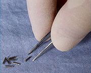
- Permanent brachytherapy, also known as seed implantation, involves placing small LDR radioactive seeds or pellets (about the size of a grain of rice) in the tumour or treatment site and leaving them there permanently to gradually decay. Over a period of weeks or months, the level of radiation emitted by the sources will decline to almost zero. The inactive seeds then remain in the treatment site with no lasting effect. Permanent brachytherapy is most commonly used in the treatment of prostate cancerProstate cancerProstate cancer is a form of cancer that develops in the prostate, a gland in the male reproductive system. Most prostate cancers are slow growing; however, there are cases of aggressive prostate cancers. The cancer cells may metastasize from the prostate to other parts of the body, particularly...
.
Clinical applications
Brachytherapy is commonly used to treat cancers of the cervixCervix
The cervix is the lower, narrow portion of the uterus where it joins with the top end of the vagina. It is cylindrical or conical in shape and protrudes through the upper anterior vaginal wall...
, prostate
Prostate
The prostate is a compound tubuloalveolar exocrine gland of the male reproductive system in most mammals....
, breast
Breast
The breast is the upper ventral region of the torso of a primate, in left and right sides, which in a female contains the mammary gland that secretes milk used to feed infants.Both men and women develop breasts from the same embryological tissues...
, and skin
Skin
-Dermis:The dermis is the layer of skin beneath the epidermis that consists of connective tissue and cushions the body from stress and strain. The dermis is tightly connected to the epidermis by a basement membrane. It also harbors many Mechanoreceptors that provide the sense of touch and heat...
.
Brachytherapy can also be used in the treatment of tumours of the brain
Human brain
The human brain has the same general structure as the brains of other mammals, but is over three times larger than the brain of a typical mammal with an equivalent body size. Estimates for the number of neurons in the human brain range from 80 to 120 billion...
, eye
Human eye
The human eye is an organ which reacts to light for several purposes. As a conscious sense organ, the eye allows vision. Rod and cone cells in the retina allow conscious light perception and vision including color differentiation and the perception of depth...
, head and neck region (lip, floor of mouth, tongue, nasopharynx and oropharynx), respiratory tract (trachea
Vertebrate trachea
In tetrapod anatomy the trachea, or windpipe, is a tube that connects the pharynx or larynx to the lungs, allowing the passage of air. It is lined with pseudostratified ciliated columnar epithelium cells with goblet cells that produce mucus...
and bronchi), digestive tract (oesophagus, gall bladder, bile-ducts
Bile duct
A bile duct is any of a number of long tube-like structures that carry bile.Bile, required for the digestion of food, is excreted by the liver into passages that carry bile toward the hepatic duct, which joins with the cystic duct to form the common bile duct, which opens into the intestine.The...
, rectum
Rectum
The rectum is the final straight portion of the large intestine in some mammals, and the gut in others, terminating in the anus. The human rectum is about 12 cm long...
, anus
Anus
The anus is an opening at the opposite end of an animal's digestive tract from the mouth. Its function is to control the expulsion of feces, unwanted semi-solid matter produced during digestion, which, depending on the type of animal, may be one or more of: matter which the animal cannot digest,...
), urinary tract (bladder
Bladder
Bladder usually refers to an anatomical hollow organBladder may also refer to:-Biology:* Urinary bladder in humans** Urinary bladder ** Bladder control; see Urinary incontinence** Artificial urinary bladder, in humans...
, urethra
Urethra
In anatomy, the urethra is a tube that connects the urinary bladder to the genitals for the removal of fluids out of the body. In males, the urethra travels through the penis, and carries semen as well as urine...
, penis
Penis
The penis is a biological feature of male animals including both vertebrates and invertebrates...
), female reproductive tract (uterus
Uterus
The uterus or womb is a major female hormone-responsive reproductive sex organ of most mammals including humans. One end, the cervix, opens into the vagina, while the other is connected to one or both fallopian tubes, depending on the species...
, vagina
Vagina
The vagina is a fibromuscular tubular tract leading from the uterus to the exterior of the body in female placental mammals and marsupials, or to the cloaca in female birds, monotremes, and some reptiles. Female insects and other invertebrates also have a vagina, which is the terminal part of the...
, vulva
Vulva
The vulva consists of the external genital organs of the female mammal. This article deals with the vulva of the human being, although the structures are similar for other mammals....
), and soft tissues.
As the radiation sources can be precisely positioned at the tumour treatment site, brachytherapy enables a high dose of radiation to be applied to a small area. Furthermore, because the radiation sources are placed in or next to the target tumour, the sources maintain their position in relation to the tumour when the patient moves or if there is any movement of the tumour within the body. Therefore, the radiation sources remain accurately targeted. This enables clinicians to achieve a high level of dose conformity – i.e. ensuring the whole of the tumour receives an optimal level of radiation. It also reduces the risk of damage to healthy tissue, organs or structures around the tumour, thus enhancing the chance of cure and preservation of organ function.
The use of HDR brachytherapy enables overall treatment times to be reduced compared with EBRT.
Patients receiving brachytherapy generally have to make fewer visits for radiotherapy compared with EBRT, and overall radiotherapy treatment plans can be completed in less time.
Many brachytherapy procedures are performed on an outpatient basis. This convenience may be particularly relevant for patients who have to work, older patients, or patients who live some distance from treatment centres, to ensure that they have access to radiotherapy treatment and adhere to treatment plans. Shorter treatment times and outpatient procedures can also help improve the efficiency of radiotherapy clinics.
Brachytherapy can be used with the aim of curing the cancer in cases of small or locally advanced tumours, provided the cancer has not metastasized (spread to other parts of the body). In appropriately selected cases, brachytherapy for primary tumours often represents a comparable approach to surgery, achieving the same probability of cure and with similar side effects.
However, in locally advanced tumours, surgery may not routinely provide the best chance of cure and is often not technically feasible to perform. In these cases radiotherapy, including brachytherapy, offers the only chance of cure.
In more advanced disease stages, brachytherapy can be used as palliative treatment for symptom relief from pain and bleeding.
In cases where the tumour is not easily accessible or is too large to ensure an optimal distribution of irradiation to the treatment area, brachytherapy can be combined with other treatments, such as EBRT and/or surgery. Combination therapy of brachytherapy exclusively with chemotherapy is rare.
Cervical cancer
Brachytherapy is commonly used in the treatment of early or locally confined cervical cancerCervical cancer
Cervical cancer is malignant neoplasm of the cervix uteri or cervical area. One of the most common symptoms is abnormal vaginal bleeding, but in some cases there may be no obvious symptoms until the cancer is in its advanced stages...
and is a standard of care in many countries.
Cervical cancer can be treated with either LDR, PDR or HDR brachytherapy.
Used in combination with EBRT, brachytherapy can provide better outcomes than EBRT alone.
The precision of brachytherapy enables a high dose of targeted radiation to be delivered to the cervix, while minimising radiation exposure to adjacent tissues and organs.
The chances of staying free of disease (disease-free survival) and of staying alive (overall survival) are similar for LDR, PDR and HDR treatments.
However, a key advantage of HDR treatment is that each dose can be delivered on an outpatient basis with a short administration time providing greater convenience for many patients.
Prostate cancer
Brachytherapy to treat prostate cancerProstate cancer
Prostate cancer is a form of cancer that develops in the prostate, a gland in the male reproductive system. Most prostate cancers are slow growing; however, there are cases of aggressive prostate cancers. The cancer cells may metastasize from the prostate to other parts of the body, particularly...
can be given either as permanent LDR seed implantation or as temporary HDR brachytherapy.
Permanent seed implantation is suitable for patients with a localised tumour and good prognosis
and has been shown to be a highly effective treatment to prevent the cancer from returning. The survival rate is similar to that found with EBRT or surgery (radical prostatectomy), but with fewer side effects such as impotence and incontinence
Urinary incontinence
Urinary incontinence is any involuntary leakage of urine. It is a common and distressing problem, which may have a profound impact on quality of life. Urinary incontinence almost always results from an underlying treatable medical condition but is under-reported to medical practitioners...
. The procedure can be completed quickly and patients are usually able to go home on the same day of treatment and return to normal activities after 1 to 2 days.
Permanent seed implantation is often a less invasive treatment option compared to the surgical removal of the prostate.
Temporary HDR brachytherapy is a newer approach to treating prostate cancer, but is currently less common than seed implantation. It is predominately used as to provide an extra dose in addition to EBRT (known as ‘”boost” therapy) as it offers an alternative method to deliver a high dose of radiation therapy that conforms to the shape of the tumour within the prostate, while sparing radiation exposure to surrounding tissues.
HDR brachytherapy as a boost for prostate cancer also means that the EBRT course can be shorter than when EBRT is used alone.
Breast cancer
Radiation therapy is standard of care for women who have undergone lumpectomyLumpectomy
Lumpectomy is a common surgical procedure designed to remove a discrete lump, usually a benign tumor or breast cancer, from an affected man or woman's breast...
or mastectomy
Mastectomy
Mastectomy is the medical term for the surgical removal of one or both breasts, partially or completely. Mastectomy is usually done to treat breast cancer; in some cases, women and some men believed to be at high risk of breast cancer have the operation prophylactically, that is, to prevent cancer...
surgery, and is an integral component of breast-conserving therapy.
Brachytherapy can be used after surgery, before chemotherapy or palliatively in the case of advanced disease. Brachytherapy to treat breast cancer
Breast cancer
Breast cancer is cancer originating from breast tissue, most commonly from the inner lining of milk ducts or the lobules that supply the ducts with milk. Cancers originating from ducts are known as ductal carcinomas; those originating from lobules are known as lobular carcinomas...
is usually performed with HDR temporary brachytherapy. Post surgery, breast brachytherapy can be used as a “boost” following irradiation of the whole breast using EBRT.
More recently, brachytherapy alone is applied in a technique called APBI (accelerated partial breast irradiation), involving delivery of radiation to only the immediate region surrounding the original tumour.
The main benefit of breast brachytherapy compared to EBRT is that a high dose of radiation can be precisely applied to the tumour while sparing radiation to healthy breast tissues and underlying structures such as the ribs and lungs. APBI can typically be completed over the course of a week. The option of brachytherapy may be particularly important in ensuring that working women, the elderly or women without easy access to a treatment centre, are able to benefit from breast-conserving therapy due to the short treatment course compared with EBRT (which often requires more visits over the course of 1–2 months). Brachytherapy has demonstrated excellent local control of breast cancer at follow-up of up to 6 years post treatment. A study is underway to compare patient outcomes of APBI in comparison to EBRT at up to 10 years after treatment.
There are two methods that can be used to deliver breast brachytherapy:
- Interstitial breast brachytherapy using multiple catheters
- Intracavitary breast brachytherapy using a balloon catheter
Interstitial breast brachytherapy involves the temporary placement of several flexible plastic catheters in the breast tissue. These are carefully positioned to allow optimal targeting of radiation to the treatment area while sparing the surrounding breast tissue. The catheters are connected to an afterloader, which delivers the planned radiation dose to the treatment area. Interstitial breast brachytherapy can be used as “boost” after EBRT, or as APBI.
Intracavitary breast brachytherapy (also known as “balloon brachytherapy”) involves the placement of a single catheter into the breast cavity left after the removal of the tumour (lumpectomy). The catheter can be placed at the time of the lumpectomy or postoperatively. Via the catheter, a balloon is then inflated in the cavity. The catheter is then connected to an afterloader, which delivers the radiation dose through the catheter and into the balloon. Currently, intracavitary breast brachytherapy is only routinely used for APBI.
There are also devices that combine the features of interstitial and intracavitary breast brachytherapy (e.g. SAVI). These devices use multiple catheters but are inserted through a single-entry point in the breast. Studies suggest the use of multiple catheters enables physicians to target the radiation more precisely.
Skin cancer
HDR brachytherapy for nonmelanomatous skin cancerSkin cancer
Skin neoplasms are skin growths with differing causes and varying degrees of malignancy. The three most common malignant skin cancers are basal cell cancer, squamous cell cancer, and melanoma, each of which is named after the type of skin cell from which it arises...
, such as basal cell carcinoma
Basal cell carcinoma
Basal-cell carcinoma is the most common type of skin cancer. It rarely metastasizes or kills. However, because it can cause significant destruction and disfigurement by invading surrounding tissues, it is still considered malignant. Statistically, approximately 3 out of 10 Caucasians may develop a...
and squamous cell carcinoma
Squamous cell carcinoma
Squamous cell carcinoma , occasionally rendered as "squamous-cell carcinoma", is a histologically distinct form of cancer. It arises from the uncontrolled multiplication of malignant cells deriving from epithelium, or showing particular cytological or tissue architectural characteristics of...
, provides an alternative treatment option to surgery. This is especially relevant for cancers on the nose, ears, eyelids or lips, where surgery may cause disfigurement or require extensive reconstruction. Various applicators can be used to ensure close contact between the radiation source(s) and the skin, which conform to the curvature of the skin and help ensure precision delivery of the optimal irradiation dose.
Brachytherapy for skin cancer provides good cosmetic results and clinical efficacy; studies with up to 5 years follow-up have shown that brachytherapy is highly effective in terms local control, and is comparable to EBRT. Treatment times are typically short, providing convenience for patients.
It has been suggested that brachytherapy may become a standard of treatment for skin cancer in the near future.
Coronary/Vascular
Brachytherapy can be used in the treatment of coronary in-stent restenosis, in which a catheter is placed inside blood vessels, through which sources are inserted and removed.In treating In-stent restenosis (ISR) Drug Eluting stents (DES)have been found to be superior to Intracoronary Brachytherapy (ICBT).
However, there is continued interest in vascular brachytherapy for persistent restenosis in failed stents and vein grafts.
The therapy has also been investigated for use in the treatment of peripheral vasculature stenosis and considered for the treatment of atrial fibrillation
Atrial fibrillation
Atrial fibrillation is the most common cardiac arrhythmia . It is a common cause of irregular heart beat, identified clinically by taking a pulse. Chaotic electrical activity in the two upper chambers of the heart result in the muscle fibrillating , instead of achieving coordinated contraction...
.
Side effects
The likelihood and nature of potential acute, sub-acute or long-term side-effects associated with brachytherapy depends on the location of the tumour being treated and the type of brachytherapy being used.Acute
Acute side effects associated with brachytherapy include localised bruising, swelling, bleeding, discharge or discomfort within the implanted region. These usually resolve within a few days following completion of treatment.Patients may also feel fatigued for a short period following treatment.
Brachytherapy treatment for cervical or prostate cancer can cause acute and transient urinary symptoms such as urinary retention, urinary incontinence or painful urination (dysuria).
Transient increased bowel frequency, diarrhoea, constipation or minor rectal bleeding, may also occur. Acute and subacute side effects usually resolve over a matter of days or a few weeks. In the case of permanent (seed) brachytherapy for prostate cancer, there is a small chance that some seeds may migrate out of the treatment region into the bladder or urethra and be passed in the urine.
Brachytherapy for skin cancer may result in a shedding of the outer layers of skin (desquamation) around the area of treatment in the weeks following therapy, which typically heals in 5–8 weeks. If the cancer is located on the lip, ulceration may occur as a result of brachytherapy, but usually resolves after 4–6 weeks.
Most of the acute side effects associated with brachytherapy can be treated with medication or through dietary changes, and usually disappear over time (typically a matter of weeks), once the treatment is completed. The acute side effects of HDR brachytherapy are broadly similar to EBRT.
Long-term
In a small number of people, brachytherapy may cause long-term side effects due to damage or disruption of adjacent tissues or organs. Long-term side effects are usually mild or moderate in nature. For example, urinary and digestive problems may persist as a result of brachytherapy for cervical or prostate cancer, and may require ongoing management.Brachytherapy for prostate cancer may cause erectile dysfunction in approximately 15-30% of patients. However, the risk of erectile dysfunction is related to age (older men are at a greater risk than younger men) and also the level of erectile function prior to receiving brachytherapy. In patients who do experience erectile dysfunction, the majority of cases can successfully be treated with drugs such as Viagra. Importantly, the risk of erectile dysfunction after brachytherapy is less than after radical prostatectomy.
Brachytherapy for breast or skin cancer may cause scar tissue to form around the treatment area. In the case of breast brachytherapy, fat necrosis may occur as a result of fatty acids entering the breast tissues. This can cause the breast tissue to become swollen and tender. Fat necrosis is a benign condition and typically occurs 4–12 months after treatment and affects about 2% of patients.
Safety around others
Patients often ask if they need to have special safety precautions around family and friends after receiving brachytherapy. If temporary brachytherapy is used, no radioactive sources remain in the body after treatment. Therefore, there is no radiation risk to friends or family from being in close proximity with them.If permanent brachytherapy is used, low dose radioactive sources (seeds) are left in the body after treatment - the radiation levels are very low and decrease over time. In addition, the irradiation only affects tissues within a few millimeters of the radioactive sources (i.e. the tumour being treated). As a precaution, some people receiving permanent brachytherapy may be advised to not hold any small children or be too close to pregnant women for a short time after treatment. Radiation oncologists or nurses can provide specific instructions to patients and advise for how long they need to be careful.
Procedure
Initial planning
In order to accurately plan the brachytherapy procedure, a thorough clinical examination is performed to understand the characteristics of the tumour. In addition, a range of imaging modalities can be used to visualise the shape and size of the tumour and its relation to surrounding tissues and organs. These include x-ray radiography, ultrasound, computed axial tomography (CT or CAT) scans and magnetic resonance imaging (MRI). The data from many of these sources can be used to create a 3D visualisation of the tumour and the surrounding tissues.Using this information, a plan of the optimal distribution of the radiation sources can be developed. This includes consideration of how the source carriers (applicators), which are used to deliver the radiation to the treatment site, should be placed and positioned. Applicators are non-radioactive and are typically needles or plastic catheters. The specific type of applicator used will depend on the type of cancer being treated and the characteristics of the target tumour.
This initial planning helps to ensure that ‘cold spots’ (too little irradiation) and ‘hot spots’ (too much irradiation) are avoided during treatment, as these can respectively result in treatment failure and side-effects.
Insertion and imaging of the applicator(s)
Before radioactive sources can be delivered to the tumour site, the applicators have to be inserted and correctly positioned in line with the initial planning.Imaging techniques, such as x-ray, fluoroscopy and ultrasound are typically used to help guide the placement of the applicators to their correct positions and to further refine the treatment plan. CAT scans and MRI can also be used. Once the applicators are inserted, they are held in place against the skin using sutures or adhesive tape to prevent them from moving. Once the applicators are confirmed as being in the correct position, further imaging can be performed to guide detailed treatment planning.
Creation of a virtual patient
The images of the patient with the applicators in situ are imported into treatment planning software and the patient is brought into a dedicated shielded room for treatment. The treatment planning software enables multiple 2D images of the treatment site to be translated into a 3D ‘virtual patient’, within which the position of the applicators can be defined. The spatial relationships between the applicators, the treatment site and the surrounding healthy tissues within this ‘virtual patient’ are a copy of the relationships in the actual patient.Optimizing the irradiation plan
To identify the optimal spatial and temporal distribution of radiation sources within the applicators of the implanted tissue or cavity, the treatment planning software allows virtual radiation sources to be placed within the virtual patient. The software shows a graphical representation of the distribution of the irradiation. This serves as a guide for the brachytherapy team to refine the distribution of the sources and provide a treatment plan that is optimally tailored to the anatomy of each patient before actual delivery of the irradiation begins. This approach is sometimes called ‘dose-painting’.Treatment delivery
The radiation sources used for brachytherapy are always enclosed within a non-radioactive capsule. The sources can be delivered manually, but are more commonly delivered through a technique known as ‘afterloading’.Manual delivery of brachytherapy is limited to a few LDR applications, due to risk of radiation exposure to clinical staff.
In contrast, afterloading involves the accurate positioning of non-radioactive applicators in the treatment site, which are subsequently loaded with the radiation sources. In manual afterloading, the source is delivered into the applicator by the operator.
Remote afterloading systems provide protection from radiation exposure to healthcare professionals by securing the radiation source in a shielded safe. Once the applicators are correctly positioned in the patient, they are connected to an ‘afterloader’ machine (containing the radioactive sources) through a series of connecting guide tubes. The treatment plan is sent to the afterloader, which then controls the delivery of the sources along the guide tubes into the pre-specified positions within the applicator. This process is only engaged once staff are removed from the treatment room. The sources remain in place for a pre-specified length of time, again following the treatment plan, following which they are returned along the tubes to the afterloader.
On completion of delivery of the radioactive sources, the applicators are carefully removed from the body. Patients typically recover quickly from the brachytherapy procedure, enabling it to often be performed on an outpatient basis.
Radiation sources
Commonly used radiation sources (radionuclides) for brachytherapy| Radionuclide | Type | Half-life | Energy |
|---|---|---|---|
| Caesium-137 Caesium-137 Caesium-137 is a radioactive isotope of caesium which is formed as a fission product by nuclear fission.It has a half-life of about 30.17 years, and decays by beta emission to a metastable nuclear isomer of barium-137: barium-137m . Caesium-137 is a radioactive isotope of caesium which is formed... (137Cs) |
γ-ray | 30.17 years | 0.662 MeV |
| Cobalt-60 Cobalt-60 Cobalt-60, , is a synthetic radioactive isotope of cobalt. Due to its half-life of 5.27 years, is not found in nature. It is produced artificially by neutron activation of . decays by beta decay to the stable isotope nickel-60... (60Co) |
γ-rays | 5.26 years | 1.17, 1.33 MeV |
| Iridium-192 (192Ir) | β--particles | 73.8 days | 0.38 MeV (mean) |
| Iodine-125 Iodine-125 Iodine-125 is a radioisotope of iodine which has uses in biological assays, nuclear medicine imaging and in radiation therapy as brachytherapy to treat prostate cancer and brain tumors. It is the second longest-lived radioisotope of iodine, after iodine-129.Its half-life is around 59 days and it... (125I) |
γ-rays | 59.6 days | 27.4, 31.4 and 35.5 keV |
| Palladium-103 (103Pd) | γ-ray | 17.0 days | 21 keV (mean) |
| Ruthenium-106 (106Ru) | β--particles | 1.02 years | 3.54 MeV |
Electronic brachytherapy
Electronic brachytherapy involves placement of miniature low energy x-ray tube sources into a pre-positioned applicator within body/tumour cavities to rapidly deliver high doses to target tissues while maintaining low doses to distant non-target tissues.See also
- External beam radiotherapyExternal beam radiotherapyExternal beam radiotherapy or teletherapy is the most common form of radiotherapy. The patient sits or lies on a couch and an external source of radiation is pointed at a particular part of the body...
- Prostate brachytherapyProstate brachytherapyBrachytherapy is a type of radiotherapy, or radiation treatment, offered to certain cancer patients. There are two types of brachytherapy – high dose-rate and low dose-rate...
- Targeted Intra-operative radiotherapy TARGITTARGITTARGIT is a technique of giving radiotherapy to the tissues surrounding a cancer after its surgical removal. The technique was designed in 1998 at the University College London...
External links
- European Society for Therapeutic Radiology and Oncology (ESTRO)
- American Society for Therapeutic Radiology and Oncology (ASTRO)
- American Brachytherapy Society (ABS)
- About Brachytherapy - a resource for patients and healthcare professionals
- Brachytherapy – an international disciplinary journal
- Journal of Contemporary Brachytherapy
- Cancerbackup – Brachytherapy
- About breast brachytherapy
- Brachytherapy for breast cancer
- Prostate brachytherapy advisory group
- Understanding Brachytherapy: resources, FAQs, glossary
- Nucletron Information on Brachytherapy service provision