
Two-photon excitation microscopy
Overview
Fluorescence
Fluorescence is the emission of light by a substance that has absorbed light or other electromagnetic radiation of a different wavelength. It is a form of luminescence. In most cases, emitted light has a longer wavelength, and therefore lower energy, than the absorbed radiation...
imaging technique that allows imaging of living tissue
Tissue (biology)
Tissue is a cellular organizational level intermediate between cells and a complete organism. A tissue is an ensemble of cells, not necessarily identical, but from the same origin, that together carry out a specific function. These are called tissues because of their identical functioning...
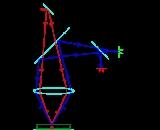
up to a very high depth, that is up to about one millimeter. Being a special variant of the multiphoton fluorescence microscope
Multiphoton fluorescence microscope
-Description:The MFM uses pulsed long-wavelength light to excite fluorophores within the specimen being observed. The fluorophore absorbs the energy from two long-wavelength photons which must arrive simultaneously in order to excite an electron into a higher energy state, from which it can decay,...
, it uses red-shifted
Redshift
In physics , redshift happens when light seen coming from an object is proportionally increased in wavelength, or shifted to the red end of the spectrum...
excitation light which can also excite fluorescent dyes. However for each excitation, two photons of the infrared
Infrared
Infrared light is electromagnetic radiation with a wavelength longer than that of visible light, measured from the nominal edge of visible red light at 0.74 micrometres , and extending conventionally to 300 µm...
light are absorbed. Using infrared light minimizes scattering in the tissue. Due to the multiphoton absorption the background signal is strongly suppressed.