

Ossification of the mandible
Encyclopedia
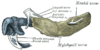
These cartilages form the cartilaginous bar of the mandibular arch (see p. 66), and are two in number, a right and a left.
Their proximal or cranial ends are connected with the ear capsules, and their distal extremities are joined to one another at the symphysis
Symphysis
A symphysis is a fibrocartilaginous fusion between two bones. It is a type of cartilaginous joint, specifically a secondary cartilaginous joint.1.A symphysis is an amphiarthrosis, a slightly movable joint.2.A growing together of parts or structures...
by mesodermal tissue.
They run forward immediately below the condyles and then, bending downward, lie in a groove near the lower border of the bone; in front of the canine tooth
Canine tooth
In mammalian oral anatomy, the canine teeth, also called cuspids, dogteeth, fangs, or eye teeth, are relatively long, pointed teeth...
they incline upward to the symphysis
Symphysis
A symphysis is a fibrocartilaginous fusion between two bones. It is a type of cartilaginous joint, specifically a secondary cartilaginous joint.1.A symphysis is an amphiarthrosis, a slightly movable joint.2.A growing together of parts or structures...
.
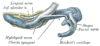
From the proximal end of each cartilage the malleus
Malleus
The malleus or hammer is a hammer-shaped small bone or ossicle of the middle ear which connects with the incus and is attached to the inner surface of the eardrum...
and incus
Incus
The incus or anvil is the anvil-shaped small bone or ossicle in themiddle ear. It connects the malleus to the stapes. It was first described by Alessandro Achillini of Bologna.The incus transmits sound vibrations from the malleus to the stapes....
, two of the bones of the middle ear, are developed; the next succeeding portion, as far as the lingula, is replaced by fibrous tissue, which persists to form the sphenomandibular ligament
Sphenomandibular ligament
The sphenomandibular ligament is a flat, thin band which is attached above to the spina angularis of the sphenoid bone, and, becoming broader as it descends, is fixed to the lingula of the mandibular foramen...
.
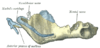
Between the lingula and the canine tooth the cartilage disappears, while the portion of it below and behind the incisor teeth becomes ossified and incorporated with this part of the mandible.
Ossification takes place in the membrane covering the outer surface of the ventral end of Meckel's cartilage (Figs. 178 to 181), and each half of the bone is formed from a single center which appears, near the mental foramen, about the sixth week of fetal life.
By the tenth week the portion of Meckel's cartilage which lies below and behind the incisor teeth is surrounded and invaded by the membrane bone.
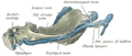
Somewhat later, accessory nuclei of cartilage make their appearance:
- a wedge-shaped nucleus in the condyloid processCondyloid processThe condyloid process is part of the mandible and is thicker than the coronoid, and consists of two portions: the condyle, and the constricted portion which supports it, the neck.-Condyle :...
and extending downward through the ramus; - a small strip along the anterior border of the coronoid processCoronoid processThe Coronoid process can refer to:* The coronoid process of the mandible, part of the ramus mandibulae of the mandible...
; - smaller nuclei in the front part of both alveolar walls and along the front of the lower border of the bone.
These accessory nuclei possess no separate ossific centers, but are invaded by the surrounding membrane bone and undergo absorption.
The inner alveolar border, usually described as arising from a separate ossific center (splenial
Splenial
The splenial is a small bone in the lower jaw of reptiles, amphibians and birds, usually located on the lingual side between the angular and suprangular....
center), is formed in the human mandible by an ingrowth from the main mass of the bone.
At birth the bone consists of two parts, united by a fibrous symphysis, in which ossification takes place during the first year.
The foregoing description of the ossification of the mandible is based on the researches of Low 44 and Fawcett, 45 and differs somewhat from that usually given.