
Coronary circulation
Encyclopedia
Coronary circulation is the circulation of blood in the blood vessel
s of the heart
muscle (the myocardium). The vessels that deliver oxygen-rich blood to the myocardium are known as coronary arteries
. The vessels that remove the deoxygenated blood from the heart muscle are known as cardiac veins.
The coronary arteries that run on the surface of the heart are called epicardial coronary arteries. These arteries, when healthy, are capable of autoregulation to maintain coronary blood flow at levels appropriate to the needs of the heart muscle. These relatively narrow vessels are commonly affected by atherosclerosis
and can become blocked, causing angina or a heart attack
. (See also: circulatory system
.) The coronary arteries that run deep within the myocardium are referred to as subendocardial.
The coronary arteries are classified as "end circulation", since they represent the only source of blood supply to the myocardium: there is very little redundant blood supply, which is why blockage of these vessels can be so critical.
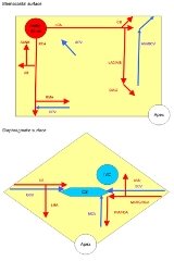
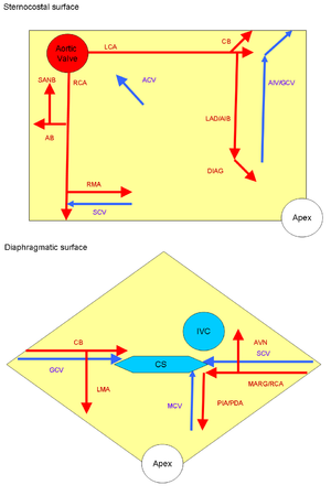 Both of these arteries originate from the left side of the heart at the beginning (root) of the aorta
, immediately above the aortic valve
. As discussed below, the left coronary artery originates from the left aortic sinus
, while the right coronary artery originates from the right aortic sinus.
Occasionally, a coronary artery will exist as a double structure (i.e. there are two arteries, parallel to each other, where ordinarily there would be one).
(PDA) (a.k.a. posterior interventricular artery) determines the coronary dominance.
Approximately 70% of the general population are right-dominant, 20% are co-dominant, and 10% are left-dominant.
A precise anatomic definition of dominance would be the artery which gives off supply to the AV node i.e. the AV nodal artery. Most of the times this is the right coronary artery.
s attach the mitral valve
(the valve between the left atrium
and the left ventricle
) and the tricuspid valve
(the valve between the right atrium
and the right ventricle
) to the wall of the heart. If the papillary muscles are not functioning properly, the mitral valve may leak during contraction of the left ventricle. This causes some of the blood to travel "in reverse", from the left ventricle to the left atrium, instead of forward to the aorta and the rest of the body. This leaking of blood to the left atrium is known as mitral regurgitation
. Similarly, the leaking of blood from the right ventricle through the tricuspid valve and into the right atrium can also occur, and this is described as tricuspid insufficiency
or tricuspid regurgitation.
The anterolateral papillary muscle more frequently receives two blood supplies: left anterior descending (LAD) artery and the left circumflex artery
(LCX).
It is therefore more frequently resistant to coronary ischemia
(insufficiency of oxygen-rich blood). On the other hand, the posteromedial papillary muscle is usually supplied only by the PDA. This makes the posteromedial papillary muscle significantly more susceptible to ischemia. The clinical significance of this is that a myocardial infarction
involving the PDA is more likely to cause mitral regurgitation.
myocardium (systole
), the subendocardial coronary vessels (the vessels that enter the myocardium) are compressed due to the high intraventricular pressures. However, the epicardial coronary vessels (the vessels that run along the outer surface of the heart) remain patent. Because of this, blood flow in the subendocardium stops. As a result most myocardial perfusion occurs during heart relaxation (diastole
) when the subendocardial coronary vessels are patent and under low pressure. This contributes to the filling difficulties of the coronary arteries.
Compression remains the same. Failure of oxygen delivery caused by a decrease in blood flow in front of increased oxygen demand of the heart results in tissue ischemia, a condition of oxygen debt. Brief ischemia is associated with intense chest pain, known as angina. Severe ischemia can cause the heart muscle to die from hypoxia, such as during a myocardial infarction
. Chronic moderate ischemia causes contraction of the heart to weaken, known as myocardial hibernation.
In addition to metabolism, the coronary circulation possesses unique pharmacologic characteristics. Prominent among these is its reactivity to adrenergic stimulation. The majority of vasculature in the body constricts to norepinephrine
, a sympathetic neurotransmitter the body uses to increase blood pressure. In the coronary circulation, norepinephrine
elicits vasodilation, due to the predominance of beta-adrenergic receptors in the coronary circulation. Agonists of alpha-receptors, such as phenylephrine, elicit very little constriction in the coronary circulation.
, the second artery is still able to supply oxygenated blood to the myocardium. However this can only occur if the atheroma progresses slowly, giving the anastomoses a chance to proliferate. Under the most common configuration of coronary arteries, there are three areas of anastomoses. Small branches of the LAD (left anterior descending/anterior interventricular) branch of the left coronary join with branches of the posterior interventricular branch of the right coronary in the interventricular groove. More superiorly, there is an anastomosis between the circumflex artery (a branch of the left coronary artery) and the right coronary artery in the atrioventricular groove. There is also an anastomoses between the septal branches of the two coronary arteries in the interventricular septum. The photograph shows area of heart supplied by the right and the left coronary arteries.(Right = yellow, left = red)
http://commons.wikimedia.org/wiki/File:Coronary_Arteries.tif
Blood vessel
The blood vessels are the part of the circulatory system that transports blood throughout the body. There are three major types of blood vessels: the arteries, which carry the blood away from the heart; the capillaries, which enable the actual exchange of water and chemicals between the blood and...
s of the heart
Heart
The heart is a myogenic muscular organ found in all animals with a circulatory system , that is responsible for pumping blood throughout the blood vessels by repeated, rhythmic contractions...
muscle (the myocardium). The vessels that deliver oxygen-rich blood to the myocardium are known as coronary arteries
Artery
Arteries are blood vessels that carry blood away from the heart. This blood is normally oxygenated, exceptions made for the pulmonary and umbilical arteries....
. The vessels that remove the deoxygenated blood from the heart muscle are known as cardiac veins.
The coronary arteries that run on the surface of the heart are called epicardial coronary arteries. These arteries, when healthy, are capable of autoregulation to maintain coronary blood flow at levels appropriate to the needs of the heart muscle. These relatively narrow vessels are commonly affected by atherosclerosis
Atherosclerosis
Atherosclerosis is a condition in which an artery wall thickens as a result of the accumulation of fatty materials such as cholesterol...
and can become blocked, causing angina or a heart attack
Myocardial infarction
Myocardial infarction or acute myocardial infarction , commonly known as a heart attack, results from the interruption of blood supply to a part of the heart, causing heart cells to die...
. (See also: circulatory system
Circulatory system
The circulatory system is an organ system that passes nutrients , gases, hormones, blood cells, etc...
.) The coronary arteries that run deep within the myocardium are referred to as subendocardial.
The coronary arteries are classified as "end circulation", since they represent the only source of blood supply to the myocardium: there is very little redundant blood supply, which is why blockage of these vessels can be so critical.
Coronary anatomy
Aorta
The aorta is the largest artery in the body, originating from the left ventricle of the heart and extending down to the abdomen, where it branches off into two smaller arteries...
, immediately above the aortic valve
Aortic valve
The aortic valve is one of the valves of the heart. It is normally tricuspid , although in 1% of the population it is found to be congenitally bicuspid . It lies between the left ventricle and the aorta....
. As discussed below, the left coronary artery originates from the left aortic sinus
Aortic sinus
An aortic sinus is one of the anatomic dilations of the ascending aorta, which occurs just above the aortic valve.There are generally three aortic sinuses, the left posterior, the right posterior and the anterior....
, while the right coronary artery originates from the right aortic sinus.
Variations
Four percent of people have a third, the posterior coronary artery. In rare cases, a person will have one coronary artery that runs around the root of the aorta.Occasionally, a coronary artery will exist as a double structure (i.e. there are two arteries, parallel to each other, where ordinarily there would be one).
Coronary artery dominance
The artery that supplies the posterior descending arteryPosterior descending artery
The posterior interventricular artery is an artery running in the posterior interventricular sulcus to the apex of the heart where it meets with the anterior interventricular artery....
(PDA) (a.k.a. posterior interventricular artery) determines the coronary dominance.
- If the posterior descending arteryPosterior descending arteryThe posterior interventricular artery is an artery running in the posterior interventricular sulcus to the apex of the heart where it meets with the anterior interventricular artery....
(PDA) (a.k.a. posterior interventricular artery) is supplied by the right coronary arteryRight coronary arteryThe right coronary artery originates above the right cusp of the aortic valve. It travels down the right atrioventricular groove, towards the crux of the heart.At the origin of the RCA is the conus artery....
(RCA), then the coronary circulation can be classified as "right-dominant". - If the posterior descending arteryPosterior descending arteryThe posterior interventricular artery is an artery running in the posterior interventricular sulcus to the apex of the heart where it meets with the anterior interventricular artery....
(PDA) is supplied by the circumflex artery (CX), a branch of the left artery, then the coronary circulation can be classified as "left-dominant". - If the posterior descending arteryPosterior descending arteryThe posterior interventricular artery is an artery running in the posterior interventricular sulcus to the apex of the heart where it meets with the anterior interventricular artery....
(PDA) is supplied by both the right coronary artery (RCA) and the circumflex artery, then the coronary circulation can be classified as "co-dominant".
Approximately 70% of the general population are right-dominant, 20% are co-dominant, and 10% are left-dominant.
A precise anatomic definition of dominance would be the artery which gives off supply to the AV node i.e. the AV nodal artery. Most of the times this is the right coronary artery.
Blood supply of the papillary muscles
The papillary musclePapillary muscle
In anatomy, the papillary muscles are muscles located in the ventricles of the heart. They attach to the cusps of the atrioventricular valves via the chordae tendinae and contract to prevent inversion or prolapse of these valves.- Action :There are five total papillary muscles in the heart, three...
s attach the mitral valve
Mitral valve
The mitral valve is a dual-flap valve in the heart that lies between the left atrium and the left ventricle...
(the valve between the left atrium
Left atrium
The left atrium is one of the four chambers in the human heart. It receives oxygenated blood from the pulmonary veins, and pumps it into the left ventricle, via the mitral valve.-Foramen ovale:...
and the left ventricle
Left ventricle
The left ventricle is one of four chambers in the human heart. It receives oxygenated blood from the left atrium via the mitral valve, and pumps it into the aorta via the aortic valve.-Shape:...
) and the tricuspid valve
Tricuspid valve
The tricuspid valve, or right atrioventricular valve, is on the right dorsal side of the mammalian heart, between the right atrium and the right ventricle. The normal tricuspid valve usually has three leaflets and three papillary muscles. They are connected to the papillary muscles by the chordae...
(the valve between the right atrium
Right atrium
The right atrium is one of four chambers in the hearts of mammals and archosaurs...
and the right ventricle
Right ventricle
The right ventricle is one of four chambers in the human heart. It receives deoxygenated blood from the right atrium via the tricuspid valve, and pumps it into the pulmonary artery via the pulmonary valve and pulmonary trunk....
) to the wall of the heart. If the papillary muscles are not functioning properly, the mitral valve may leak during contraction of the left ventricle. This causes some of the blood to travel "in reverse", from the left ventricle to the left atrium, instead of forward to the aorta and the rest of the body. This leaking of blood to the left atrium is known as mitral regurgitation
Mitral regurgitation
Mitral regurgitation , mitral insufficiency or mitral incompetence is a disorder of the heart in which the mitral valve does not close properly when the heart pumps out blood. It is the abnormal leaking of blood from the left ventricle, through the mitral valve, and into the left atrium, when...
. Similarly, the leaking of blood from the right ventricle through the tricuspid valve and into the right atrium can also occur, and this is described as tricuspid insufficiency
Tricuspid insufficiency
Tricuspid insufficiency , a valvular heart disease also called tricuspid regurgitation , refers to the failure of the heart's tricuspid valve to close properly during systole. As a result, with each heart beat some blood passes from the right ventricle to the right atrium, the opposite of the...
or tricuspid regurgitation.
The anterolateral papillary muscle more frequently receives two blood supplies: left anterior descending (LAD) artery and the left circumflex artery
Left circumflex artery
The "LCX", or left circumflex artery is an artery of the heart.-Course:...
(LCX).
It is therefore more frequently resistant to coronary ischemia
Ischemia
In medicine, ischemia is a restriction in blood supply, generally due to factors in the blood vessels, with resultant damage or dysfunction of tissue. It may also be spelled ischaemia or ischæmia...
(insufficiency of oxygen-rich blood). On the other hand, the posteromedial papillary muscle is usually supplied only by the PDA. This makes the posteromedial papillary muscle significantly more susceptible to ischemia. The clinical significance of this is that a myocardial infarction
Myocardial infarction
Myocardial infarction or acute myocardial infarction , commonly known as a heart attack, results from the interruption of blood supply to a part of the heart, causing heart cells to die...
involving the PDA is more likely to cause mitral regurgitation.
Coronary flow
During contraction of the ventricularVentricle (heart)
In the heart, a ventricle is one of two large chambers that collect and expel blood received from an atrium towards the peripheral beds within the body and lungs. The Atria primes the Pump...
myocardium (systole
Systole (medicine)
Systole is the contraction of the heart. Used alone, it usually means the contraction of the left ventricle.In all mammals, the heart has 4 chambers. The left and right ventricles pump together. The atria and ventricles pump in sequence...
), the subendocardial coronary vessels (the vessels that enter the myocardium) are compressed due to the high intraventricular pressures. However, the epicardial coronary vessels (the vessels that run along the outer surface of the heart) remain patent. Because of this, blood flow in the subendocardium stops. As a result most myocardial perfusion occurs during heart relaxation (diastole
Diastole
Diastole is the period of time when the heart fills with blood after systole . Ventricular diastole is the period during which the ventricles are relaxing, while atrial diastole is the period during which the atria are relaxing...
) when the subendocardial coronary vessels are patent and under low pressure. This contributes to the filling difficulties of the coronary arteries.
Compression remains the same. Failure of oxygen delivery caused by a decrease in blood flow in front of increased oxygen demand of the heart results in tissue ischemia, a condition of oxygen debt. Brief ischemia is associated with intense chest pain, known as angina. Severe ischemia can cause the heart muscle to die from hypoxia, such as during a myocardial infarction
Myocardial infarction
Myocardial infarction or acute myocardial infarction , commonly known as a heart attack, results from the interruption of blood supply to a part of the heart, causing heart cells to die...
. Chronic moderate ischemia causes contraction of the heart to weaken, known as myocardial hibernation.
In addition to metabolism, the coronary circulation possesses unique pharmacologic characteristics. Prominent among these is its reactivity to adrenergic stimulation. The majority of vasculature in the body constricts to norepinephrine
Norepinephrine
Norepinephrine is the US name for noradrenaline , a catecholamine with multiple roles including as a hormone and a neurotransmitter...
, a sympathetic neurotransmitter the body uses to increase blood pressure. In the coronary circulation, norepinephrine
Norepinephrine
Norepinephrine is the US name for noradrenaline , a catecholamine with multiple roles including as a hormone and a neurotransmitter...
elicits vasodilation, due to the predominance of beta-adrenergic receptors in the coronary circulation. Agonists of alpha-receptors, such as phenylephrine, elicit very little constriction in the coronary circulation.
Anastomoses
There is some anastomoses between branches of the two coronary arteries. However the coronary arteries are functionally end arteries and so these meetings are referred to as anatomical anastamoses, which lack function, as opposed to functional or physiological anastomoses like that in the palm of the hand. This is as blockage of one coronary artery generally results in death of the heart tissue due to lack of sufficient blood supply from the other branch. When two arteries or their branches join, the area of the myocardium receives dual blood supply. These junctions are called anastomoses. If one coronary artery is obstructed by an atheromaAtheroma
In pathology, an atheroma is an accumulation and swelling in artery walls that is made up of macrophage cells, or debris, that contain lipids , calcium and a variable amount of fibrous connective tissue...
, the second artery is still able to supply oxygenated blood to the myocardium. However this can only occur if the atheroma progresses slowly, giving the anastomoses a chance to proliferate. Under the most common configuration of coronary arteries, there are three areas of anastomoses. Small branches of the LAD (left anterior descending/anterior interventricular) branch of the left coronary join with branches of the posterior interventricular branch of the right coronary in the interventricular groove. More superiorly, there is an anastomosis between the circumflex artery (a branch of the left coronary artery) and the right coronary artery in the atrioventricular groove. There is also an anastomoses between the septal branches of the two coronary arteries in the interventricular septum. The photograph shows area of heart supplied by the right and the left coronary arteries.(Right = yellow, left = red)
http://commons.wikimedia.org/wiki/File:Coronary_Arteries.tif