
Nuchal lines
Encyclopedia
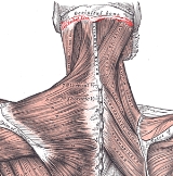
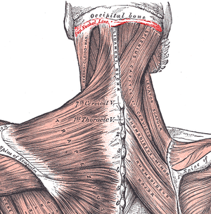
The nuchal lines are four curved lines on the external surface of the occipital bone
:
Occipital bone
The occipital bone, a saucer-shaped membrane bone situated at the back and lower part of the cranium, is trapezoidal in shape and curved on itself...
:
- The upper, often faintly marked, is named the highest nuchal line, but is sometimes referred to as the Mempin Line, and to it the galea aponeuroticaGalea aponeuroticaThe galea aponeurotica is a tough layer of dense fibrous tissue which covers the upper part of the cranium; behind, it is attached, in the interval between its union with the Occipitales, to the external occipital protuberance and highest nuchal lines of the occipital bone; in front, it forms a...
is attached.
- Below the highest nuchal line is the superior nuchal line. To it is attached the Occipitalis muscleOccipitalis muscleThe Occipitalis, thin and quadrilateral in form, arises by tendinous fibers from the lateral two-thirds of the superior nuchal line of the occipital bone, and from the mastoid part of the temporal. It ends in the galea aponeurotica....
, and Splenius capitis muscleSplenius capitis muscleThe splenius capitis is a broad, straplike muscle in the back of the neck. It pulls on the base of the skull from vertebrae in the neck and upper thorax.-Origin, insertion, and Innervation:...
, and the trapezius muscleTrapezius muscleIn human anatomy, the trapezius is a large superficial muscle that extends longitudinally from the occipital bone to the lower thoracic vertebrae and laterally to the spine of the scapula...
.
- From the external occipital protuberance a ridge or crest, the median nuchal line, often faintly marked, descends to the foramen magnum, and affords attachment to the ligamentum nuchæ.
- Running from the middle of this line is the inferior nuchal line. Attached are the Obliquus capitis superior muscleObliquus capitis superior muscleThe Obliquus capitis superior muscle arises from the lateral mass of the atlas bone. It passes superiorly and posteriorly to insert into the lateral half of the inferior nuchal line. It is innervated by the suboccipital nerve, the dorsal ramus of the first spinal nerve.It acts at the...
, Rectus capitis posterior major muscleRectus capitis posterior major muscleThe Rectus capitis posterior major arises by a pointed tendon from the spinous process of the axis, and, becoming broader as it ascends, is inserted into the lateral part of the inferior nuchal line of the occipital bone and the surface of the bone immediately below the line.In 2011, Scali et al.,...
, and Rectus capitis posterior minor muscleRectus capitis posterior minor muscleThe Rectus capitis posterior minor arises by a narrow pointed tendon from the tubercle on the posterior arch of the atlas, and, widening as it ascends, is inserted into the medial part of the inferior nuchal line of the occipital bone and the surface between it and the foramen magnum, and also...
.
Additional images