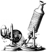
|
|
|
|
Microscope
|
| |
|
| |
A microscope = small + (skopein) = to look at) is an instrument for viewing objects that are too small to be seen by the naked or unaided eye. The science of investigating small objects using such an instrument is called microscopy. The term microscopic means minute or very small, not visible with the eye unless aided by a microscope. HistoryThe microscopes used in schools and homes trace their history back almost 1200 years with Abbas Ibn Firnas's corrective lenses, and it was Ibn al-Haytham's Book of Optics—which was written when he was under house arrest from 1011 to 1021—that laid the foundation for optical technology.
The first useful microscope was made around 1595 in Middleburg, Holland.

Discussion
Ask a question about 'Microscope'
Start a new discussion about 'Microscope'
Answer questions from other users
|
Recent Posts

Timeline
Encyclopedia
A microscope = small + (skopein) = to look at) is an instrument for viewing objects that are too small to be seen by the naked or unaided eye. The science of investigating small objects using such an instrument is called microscopy. The term microscopic means minute or very small, not visible with the eye unless aided by a microscope.
HistoryThe microscopes used in schools and homes trace their history back almost 1200 years with Abbas Ibn Firnas's corrective lenses, and it was Ibn al-Haytham's Book of Optics—which was written when he was under house arrest from 1011 to 1021—that laid the foundation for optical technology.
The first useful microscope was made around 1595 in Middleburg, Holland. Three different eyeglass makers have been given credit for the invention: Hans Lippershey (who also developed the first real telescope); Hans Janssen; and his son, Zacharias. The coining of the name "microscope" has been credited to Giovanni Faber, who gave that name to Galileo Galilei's compound microscope in 1625,. (Galileo had called it the "occhiolino" or "little eye".)
The most common type of microscope—and the first to be invented—is the optical microscope. This is an optical instrument containing one or more lenses that produce an enlarged image of an object placed in the focal plane of the lens(es). There are, however, many other microscope designs.
Types "Microscopes" can largely be separated into three classes, optical theory microscopes, electron microscopes and scanning probe microscopes.
Optical theory microscopes are microscopes which function through the optical theory of lenses in order to magnify the image generated by the passage of a wave through the sample. The waves used are either electromagnetic in optical microscopes or electron beams in electron microscopes. The types are the Compound Light, Stereo, and the electron microscope.
Optical microscopes Optical microscopes, through their use of visible wavelengths of light, are the simplest and hence most widely used type of microscope.
Optical microscopes use refractive lenses, typically of glass and occasionally of plastic, to focus light into the eye or another light detector. Typical magnification of a light microscope is up to 1500x with a theoretical resolution limit of around 0.2 micrometres or 200 nanometers. Specialised techniques (e.g., scanning confocal microscopy) may exceed this magnification but the resolution is an insurmountable diffraction limit.
Various wavelengths of light are sometimes used for special purposes, for example, in the study of biological tissue. Ultraviolet light is used to illuminate the object being viewed in order to excite a fluorescent dye which then emits visible light. Infrared light is used to study thick slices of biological tissue because infrared light's low diffraction coefficient permits viewing deeper into tissue.
Other microscopes which use electromagnetic wavelengths not visible to the human eye are often called optical microscopes. The most common of these, due to its high resolution yet no requirement for a vacuum like electron microscopes, is the x-ray microscope.
Electron microscopes Electron microscopes, which use beams of electrons instead of light, are designed for very high magnification usage. Electrons, which can be accelerated to produce a much smaller wavelength than visible light, allow a much higher resolution. The main limitation of the electron beam is that it must pass through a vacuum as air molecules would otherwise scatter the beam.
Instead of relying on refraction, lenses for electron microscopes are specially designed electromagnets which generate magnetic fields that are approximately parallel to the direction that electrons travel. The electrons are typically detected by a phosphor screen, photographic film or a charge-coupled device (CCD).
Two major variants of electron microscopes exist:
- Scanning electron microscope: looks at the surface of bulk objects by scanning the surface with a fine electron beam and measuring reflection. May also be used for spectroscopy.
- Transmission electron microscope: passes electrons completely through the sample, analogous to basic optical microscopy. This requires careful sample preparation, since electrons are scattered so strongly by most materials.This is a scientific device that allows people to see objects that could normally not be seen by the naked or unaided eye.
Scanning probe microscopeIn scanning probe microscopy (SPM), a physical probe is used either in close contact to the sample or nearly touching it. By rastering the probe across the sample, and by measuring the interactions between the sharp tip of the probe and the sample, a micrograph is generated. The exact nature of the interactions between the probe and the sample determines exactly what kind of SPM is being used. Because this kind of microscopy relies on the interactions between the tip and the sample, it generally only measures information about the surface of the sample.
A variation of the SPM is the SECM (Scanning ElectroChemical Microscope). A SECM images a sample in a similar manner as a SPM but the sample is in an electrolyte solution with the SECM using electrochemically active tip.
Other microscopesScanning acoustic microscopes use sound waves to measure variations in acoustic impedance. Similar to Sonar in principle, they are used for such jobs as detecting defects in the subsurfaces of materials including those found in integrated circuits.
See also
in: A Study Guide to the Science of Botany at Wikibooks
External links
|
| |
|
|