
High resolution electron energy loss spectroscopy
Encyclopedia
High Resolution Electron Energy Loss Spectroscopy (HREELS) is a tool used in surface science
. The inelastic scattering
of electron
s from surfaces is utilized to study electronic excitations or vibrational modes of the surface or of molecules adsorbed to a surface. Hence in contrast to other Electron Energy Loss Spectroscopies (EELS) HREELS deals with small energy losses in the range of 10-3 eV to 1 eV.
It plays an important role in the investigation of surface structure, catalysis
, dispersion
of surface phonon
s and the monitoring of epitaxial growth
.
 In general Electron Energy Loss Spectroscopy bases on the energy losses of electrons when inelastically scattered on matter. An incident beam of electrons with a known energy (Ei) is scattered on a sample. The scattering of these electrons can excite the electronic structure of the sample. If this is the case the scattered electron loses the specific energy (ΔE) needed to cause the excitation. Those scattering processes are called inelastic. It may be easiest to imagine that the energy loss is for example due to an excitation of an electron from an atomic K-shell to the M-shell. This energy for this excitation is taken away from the electrons kinetic energy.
Then the energies of the scattered electrons (Es) are measured and the energy loss can be calculated. From the measured data an intensity versus energy loss diagram is established. In the case of scattering on phonons the so called energy loss can also be a gain of energy (see: Raman spectroscopy
).
These energy losses allow, using comparison to other experiments, or theory, to draw conclusion about surface properties of the sample.
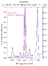
Excitations of the surface structure are usually very low energetic ranging from 10-3 eV to 10 eV. In spectra electrons with only small energy losses, like also Raman scattering, the interesting features are all located very close together and especially near to the very strong elastic scattering peak. Hence the used spectrometers require a high resolution. Therefore this regime of EELS is called High Resolution EELS.
In this context resolution shall be defined as the energy difference in which two features in a spectrum are just distinguishable divided by the mean energy of those features:
In the case of EELS the first thing to think of in order to achieve high resolution is using incident electrons of a very precisely defined energy and a high quality analyzer.
Further high resolution is only possible when the energies of the incident electrons are not far bigger than the energy losses. For HREELS the energy of the incident electrons is therefore mostly significantly smaller than 102 eV.
Considering that 102 eV electrons have a mean free path of around 1 nm (corresponds to a few monolayers), which is decreasing with lower energies, this automatically implies that HREELS is a surface sensitive technique.
This is the reason why HREELS must be measured in reflection and has to be implemented in ultra high vacuum (UHV
). In contrast to e.g. Core Level EELS which operates at very high energies and can therefore also be found in Transmission Electron Microscopes
(TEM).
In HREELS not only the electron energy loss can be measured, often the angular distribution, of electrons of a certain energy loss, in reference to the specular direction gives interesting insight to the structures on the surface.
Surface science
Surface science is the study of physical and chemical phenomena that occur at the interface of two phases, including solid–liquid interfaces, solid–gas interfaces, solid–vacuum interfaces, and liquid-gas interfaces. It includes the fields of surface chemistry and surface physics. Some related...
. The inelastic scattering
Inelastic scattering
In particle physics and chemistry, inelastic scattering is a fundamental scattering process in which the kinetic energy of an incident particle is not conserved . In an inelastic scattering process, some of the energy of the incident particle is lost or gained...
of electron
Electron
The electron is a subatomic particle with a negative elementary electric charge. It has no known components or substructure; in other words, it is generally thought to be an elementary particle. An electron has a mass that is approximately 1/1836 that of the proton...
s from surfaces is utilized to study electronic excitations or vibrational modes of the surface or of molecules adsorbed to a surface. Hence in contrast to other Electron Energy Loss Spectroscopies (EELS) HREELS deals with small energy losses in the range of 10-3 eV to 1 eV.
It plays an important role in the investigation of surface structure, catalysis
Catalysis
Catalysis is the change in rate of a chemical reaction due to the participation of a substance called a catalyst. Unlike other reagents that participate in the chemical reaction, a catalyst is not consumed by the reaction itself. A catalyst may participate in multiple chemical transformations....
, dispersion
Dispersion relation
In physics and electrical engineering, dispersion most often refers to frequency-dependent effects in wave propagation. Note, however, that there are several other uses of the word "dispersion" in the physical sciences....
of surface phonon
Phonon
In physics, a phonon is a collective excitation in a periodic, elastic arrangement of atoms or molecules in condensed matter, such as solids and some liquids...
s and the monitoring of epitaxial growth
Epitaxy
Epitaxy refers to the deposition of a crystalline overlayer on a crystalline substrate, where the overlayer is in registry with the substrate. In other words, there must be one or more preferred orientations of the overlayer with respect to the substrate for this to be termed epitaxial growth. The...
.
Overview of HREELS
Then the energies of the scattered electrons (Es) are measured and the energy loss can be calculated. From the measured data an intensity versus energy loss diagram is established. In the case of scattering on phonons the so called energy loss can also be a gain of energy (see: Raman spectroscopy
Raman scattering
Raman scattering or the Raman effect is the inelastic scattering of a photon. It was discovered by Sir Chandrasekhara Venkata Raman and Kariamanickam Srinivasa Krishnan in liquids, and by Grigory Landsberg and Leonid Mandelstam in crystals....
).
These energy losses allow, using comparison to other experiments, or theory, to draw conclusion about surface properties of the sample.
Excitations of the surface structure are usually very low energetic ranging from 10-3 eV to 10 eV. In spectra electrons with only small energy losses, like also Raman scattering, the interesting features are all located very close together and especially near to the very strong elastic scattering peak. Hence the used spectrometers require a high resolution. Therefore this regime of EELS is called High Resolution EELS.
In this context resolution shall be defined as the energy difference in which two features in a spectrum are just distinguishable divided by the mean energy of those features:
In the case of EELS the first thing to think of in order to achieve high resolution is using incident electrons of a very precisely defined energy and a high quality analyzer.
Further high resolution is only possible when the energies of the incident electrons are not far bigger than the energy losses. For HREELS the energy of the incident electrons is therefore mostly significantly smaller than 102 eV.
Considering that 102 eV electrons have a mean free path of around 1 nm (corresponds to a few monolayers), which is decreasing with lower energies, this automatically implies that HREELS is a surface sensitive technique.
This is the reason why HREELS must be measured in reflection and has to be implemented in ultra high vacuum (UHV
UHV
UHV may refer to:* University of Houston–Victoria* Ultra high vacuum* Ultra high voltage power line...
). In contrast to e.g. Core Level EELS which operates at very high energies and can therefore also be found in Transmission Electron Microscopes
Transmission electron microscopy
Transmission electron microscopy is a microscopy technique whereby a beam of electrons is transmitted through an ultra thin specimen, interacting with the specimen as it passes through...
(TEM).
In HREELS not only the electron energy loss can be measured, often the angular distribution, of electrons of a certain energy loss, in reference to the specular direction gives interesting insight to the structures on the surface.
Physics of HREELS
As mentioned above HREELS involves an inelastic scattering process on a surface. For those processes the conservation of energy as well as the conservation of momentum’s projection parallel to the surface hold: |
 |} E are energies k and q are wave vectors and G denotes a reciprocal lattice vector. One should mention at this point that for non perfect surfaces G is not in any case a well defined quantum number, what has to be considered when using the second relation. Variables subscripted with i denote values of incident electrons those subscripted with s values of scattered electrons. “||” denotes parallel to the surface. For the description of the inelastic scattering processes due to the excitation of vibrational modes of adsorbates different approaches exist. The simplest approach distinguishes between regimes of small and large scattering angles: Dipole scatteringThe so called dipole scattering can be applied when the scattered beam is very near to the specular direction. In this case a macroscopic theory can be applied to explain the results. It can be approached using the so called dielectrical theory of which a quantum mechanical treatment was first presented by E. Evans and D.L. Mills in the early 1970s. Or by a more unformular model which exactly only holds for perfect conductors:A unit cell at the surface does not have homogenous surrounding hence it is supposed to have an electrical dipole moment. When a molecule is adsorbed to the surface there can be an additional dipole moment and the total dipole moment P is present. This dipole moment causes a long range electronic potential in the vacuum above the surface. On this potential the incident electron can scatter inelastically what means it excites vibrations in the dipole structure. The dipole moment can than be written as  . When the adsorbate sticks to a metal surface imaginary dipoles occur as shown in the figure on the right. Hence for an adsorbed dipole normal to the surface the dipole moment “seen” from the vacuum doubles. Whereas the dipole moment of a parallel to the surface adsorbed dipole vanishes. Hence an incident electron can excite the adsorbed dipole only when it is adsorbed normal to the surface and the vibrational mode can be detected in the energy loss spectrum. If the dipole is adsorbed parallel then no energy losses will be detected and the vibrational modes of the dipole are missing in the energy loss spectrum. . When the adsorbate sticks to a metal surface imaginary dipoles occur as shown in the figure on the right. Hence for an adsorbed dipole normal to the surface the dipole moment “seen” from the vacuum doubles. Whereas the dipole moment of a parallel to the surface adsorbed dipole vanishes. Hence an incident electron can excite the adsorbed dipole only when it is adsorbed normal to the surface and the vibrational mode can be detected in the energy loss spectrum. If the dipole is adsorbed parallel then no energy losses will be detected and the vibrational modes of the dipole are missing in the energy loss spectrum.The dielectric model holds also when the material on which the molecule adsorbs is not a metal. The picture shown above is then the limit for  where where  denotes the relative dielectrical constant. denotes the relative dielectrical constant.When measuring the intensity of the electron energy loss peaks and comparing to other experimental results or to theoretical models it can also be told whether a molecule is adsorbed normal to the surface or tilted by an angle. As the incident electron in this model has to be scattered in the region above the surface it does not come to a direct impact at the surface and as the amount of momentum transferred is therefore small the scattering of is very much into the specular direction. Impact scatteringImpact scattering is the regime which deals with electrons that are scattered further away from the specular direction. In those cases no macroscopic theory exists and a microscopic theoryMicroscopic theory A microscopic theory is one that contains an explanation at the atomic or subatomic level in contrast to a higher level or classical macroscopic or phenomenological theory. eg in superconductivity BCS theory is a microscopic theory.... like, quantum mechanical dispersion theory, has to be applied. Symmetry considerations than also results in certain selection rules (They assume that the energy lost in the inelastic scattering process is negligible):
All those selection rules make it possible to identify the normal coordinates of the adsorbed molecules. Intermediate negative ion resonanceIntermediate negative ion resonance: The electron forms a compound state with an adsorbed molecule during the scattering process. But the lifetime of those states are so short that this type of scattering is barely observed.All of those regimes can at once be described with the help of the single microscopic theory the selection rule find their origins that in symmetry considerations. Selection rules for dipole scattering from the perspective of vibrational eigenmodesA microscopic theory makes it for example possible to approach the selection rule for dipole scattering in a more exact way. The scattering cross section is only none vanishing in the case of a nonzero matrix element . .Where i denotes the initial and f the final vibrational mode of the adsorbed molecule and pz the z component of its dipole moment. As the dipole moment is something like charge times length pz has the same symmetry properties as z, which is totally symmetric. Hence the product of i and f has to be a totally symmetric function, too, otherwise the matrix element would vanish. Hence excitations from the totally symmetrical ground state of a molecule are only possible to a totally symmetric vibrational state. This is the surface selection rule for dipole scattering. Note that it says nothing about how big the intensity for scattering is and that not the displacement of the atoms of the adsorbate but its total dipole moment is the operator in the matrix element. This is important as a vibration of the atoms parallel to the surface can cause and vibration of the dipole moment normal to the surface, too. So the result in the section "Dipole scattering" was not exactly correct. When trying to gain information from selection rules it has to be carefully considered whether really a pure dipole or impact scattering region is investigated. Further symmetry breaking due to strong bindings to the surface must be considered. Another problem is that in cases of bigger molecules often many vibrational modes are degenerate, which could again be resolved due to strong molecule surface interactions. Those interactions can also generate completely new dipole moments which the molecule normally does not have. But when investigating carefully it is due to analysis of normal dipole modes mostly possible to get a very good picture of how the molecule adheres to the surface. High Resolution Electron Energy Loss SpectrometerAs the electrons used for HREELS are low energetic they do not only have a very short free mean path length in the sample materials but also under normal atmospheric conditions. Therefore one has to setup the spectrometer in UHV.The spectrometer is in general a computer simulated design which tries to optimize the resolution while keeping an acceptable electron flux.
Next some general problems of HREEL Spectrometers are noted. Due to the electron flux the apertures can become negatively charged which makes them effectively smaller for the passing electrons. This has to be considered when doing the design of the setup as it is anyway difficult to keep different potentials, of repeller, lenses, screening elements, and the reflector, constant. Unstable potentials on lenses or CHA deflectors would cause fluctuations in the measured signal. Similar problems are caused by external electric of magnetic fields, either they cause fluctuations in the signal, or add a constant offset. That is why the sample is normally shielded by equipotential, metal electrodes to keep the region of the sample field free so that neither the probe electrons nor the sample is affected by external electric fields. Further a cylinder of a material with a high magnetic permeability, e.g. Mu-metal Mu-metal Mu-metal is a nickel-iron alloy that is notable for its high magnetic permeability. The high permeability makes mu-metal very effective at screening static or low-frequency magnetic fields, which cannot be attenuated by other methods. The name came from the Greek letter mu which represents... , built around the whole spectrometer to keep magnetic fields or field inhomogeneties at the experiment down to 10 mG or 1mG/cm. Because of the same reason the whole experiment, except the lenses which are normally made of coated copper, is designed in stainless antimagnetic steel and insulating parts are avoided wherever possible. External links
The source of this article is wikipedia, the free encyclopedia. The text of this article is licensed under the GFDL.
|
 for the monochromator. This means, the electrons leaving the monochromator with e.g. 10 eV have an energy accurate to 10-1 eV. The beam’s flux is then in the orders of 10-8 A to 10-10 A. The radii of the CHA are in the order of several 10 mm. And the deflector electrodes have a saw tooth profile to backscatter electrons which are reflected from the walls in order to reduce the background of electrons with the wrong Ei. The electrons are then focused by a lens system onto the sample. These lenses are, in contrary to those of the emitter system very flexible, as it is important is to get a good focus on the sample. To enable measurements of angular distributions all those elements are mounted on a rotate able table with the axis cantered at the sample.Its negative charge causes the electron beam to broaden. What can be prevented by charging the top and bottom plates of the CHA deflectors negative. What again causes a change in the deflection angle and has to be considered when designing the experiment.
 higher resolution as in the monochromator is wanted. Hence the radial dimensions of this CHA are mostly bigger by like a factor 2. Due to aberrations of the lens systems the beam has also broadened. To sustain a high enough electron flux to the analyzer the apertures are also about a factor 2 bigger. To make the analysis more accurate, especially to reduce the background of in the deflector scattered electrons often two analyzers are used, or additional apertures are added behind the analyzers as scattered electrons of the wrong energy normally leave the CHAs under large angles. In this way energy losses of 10-2 eV to 10 eV can be detected with accuracies of about 10-2 eV.