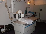
All Topics  
X-ray machine

 
X Ray Machine

   Email Print
   Bookmark   Link


X-ray machine


An X-ray machine is a device used by radiographers to acquire an x-ray
X-ray

X-radiation is a form of electromagnetic radiation. X-rays have a wavelength in the range of 10 to 0.01 nanometers, corresponding to frequency in the range 30 Hertz to 30 Hertz and energies in the range 120 Electron volt to 120 keV....
 image. They are used in various fields, notably medicine
Medicine

Medicine is the art and science of healing. It encompasses a range of health care practices evolved to maintain and restore health by the prevention and treatment of illness....
 and security
Security

Security is the degree of protection against danger, loss, and criminals. Individuals or actions that encroach upon the condition of protection are responsible for a "breach of security."...
.

M. Anderson and Ryan W. Preuninger formulated mathematical equations for X-rays. Physicist Johann Hittorf observed tubes with energy rays extending from a negative electrode. William Crookes
William Crookes

Sir William Crookes, Order of Merit , Fellow of the Royal Society was an England chemist and physicist who attended the Royal College of Chemistry, in London, and worked on spectroscopy....
 investigated the effects of energy discharges on rare gases.


Discussion
Ask a question about 'X-ray machine'
Start a new discussion about 'X-ray machine'
Answer questions from other users
Full Discussion Forum


Recent Posts


Encyclopedia


An X-ray machine is a device used by radiographers to acquire an x-ray
X-ray

X-radiation is a form of electromagnetic radiation. X-rays have a wavelength in the range of 10 to 0.01 nanometers, corresponding to frequency in the range 30 Hertz to 30 Hertz and energies in the range 120 Electron volt to 120 keV....
 image. They are used in various fields, notably medicine
Medicine

Medicine is the art and science of healing. It encompasses a range of health care practices evolved to maintain and restore health by the prevention and treatment of illness....
 and security
Security

Security is the degree of protection against danger, loss, and criminals. Individuals or actions that encroach upon the condition of protection are responsible for a "breach of security."...
.

History

Shay M. Anderson and Ryan W. Preuninger formulated mathematical equations for X-rays. Physicist Johann Hittorf observed tubes with energy rays extending from a negative electrode. William Crookes
William Crookes

Sir William Crookes, Order of Merit , Fellow of the Royal Society was an England chemist and physicist who attended the Royal College of Chemistry, in London, and worked on spectroscopy....
 investigated the effects of energy discharges on rare gases. Heinrich Hertz began experimenting and demonstrated that cathode rays could penetrate very thin metal foil (such as aluminium). In 1887, Nikola Tesla
Nikola Tesla

Nikola Tesla was an inventor and a mechanical engineer and electrical engineer. Tesla was born in the village of Smiljan near the town of Gospic, in Croatia ....
 began to investigate X-rays and produced the bremsstrahlung
Bremsstrahlung

Bremsstrahlung , is electromagnetic radiation produced by the deceleration of a charged particle, such as an electron, when deflected by another charged particle, such as an atomic nucleus....
 process. In 1895, Wilhelm Röntgen began observing and further documenting X-rays while experimenting with vacuum tubes.

One of the first X-ray photographs was made of the hand of Röntgen's wife. The image displayed both her wedding ring and bones. On January 18, 1896 an X-ray machine was formally displayed by H.L. Smith. Upon discovery in 1895, X-Rays were advertised as the new scientific wonder and seized upon by entertainers. Circus patrons viewed their own skeletons and were given pictures of their own bony hands wearing silhouetted jewelry. While many people were fascinated by this discovery, others feared that it would allow strangers to look through doors and invade people's privacy.

In the 1940s and 50s, (real time) X-ray machines were used in stores to help sell footwear. These were known as fluoroscopes. However, as the harmful effects of X-ray
X-ray

X-radiation is a form of electromagnetic radiation. X-rays have a wavelength in the range of 10 to 0.01 nanometers, corresponding to frequency in the range 30 Hertz to 30 Hertz and energies in the range 120 Electron volt to 120 keV....
 radiation
Radiation

In physics, radiation describes any process in which energy emitted by one body travels through a medium or through space, ultimately to be absorbed by another body....
 were properly considered, they finally fell out of use. Shoe-fitting use of the device was first banned by the state of Pennsylvania
Pennsylvania

The Commonwealth of Pennsylvania , often colloquially referred to as PA by natives and Northeasterners, is a U.S. state located in the Northeastern United States and Mid-Atlantic States regions of the United States....
 in 1957. (They were more a clever marketing tool to attract customers, rather than a fitting aid.)

Overview

An X-ray imaging system consists of a X-ray source or generator (X-ray tube
X-ray tube

An X-ray tube is a vacuum tube that produces X-rays. They are part of X-ray machines. X-rays are part of the electromagnetic spectrum, an ionizing radiation with wavelength just shorter than ultraviolet light....
), and an image detection system which can either be comprised of film (analog technology) or a digital capture system (such as a picture archiving and communication system
Picture archiving and communication system

In medical imaging, picture archiving and communication systems are computers or networks dedicated to the storage, retrieval, distribution and presentation of images....
).

X-ray sources

In the typical X-ray source of less than 450 kV, X-ray
X-ray

X-radiation is a form of electromagnetic radiation. X-rays have a wavelength in the range of 10 to 0.01 nanometers, corresponding to frequency in the range 30 Hertz to 30 Hertz and energies in the range 120 Electron volt to 120 keV....
 photons are produced by an electron beam striking a target. The electrons that make up the beam are emitted from a heated cathode filament. The electrons are then focused and accelerated towards an angled anode target. The point where the electron beam strikes the target is called the focal spot. Most of the kinetic energy
Kinetic energy

The kinetic energy of an object is the extra energy which it possesses due to its motion. It is defined as the mechanical work needed to accelerate a body of a given mass from rest to its current velocity....
 contained in the electron beam is converted to heat, but around 1% of the energy is converted into X-ray photons, the excess heat is dissipated via a heat sink. At the focal spot, X-ray photons are emitted in all directions from the target surface, the highest intensity being around 60deg to 90deg from the beam due to the angle of the anode target to the approaching X-ray photons. There is a small round window in the X-ray tube directly above the angled target. This window allows the X-ray to exit the tube with little attenuation while maintaining a vacuum seal required for the X-ray tube operation.

X-ray machines work by applying controlled voltage
Voltage

Electrical tension is the potential difference between two points of an electrical or electronic circuit, expressed in volts. It is the measurement of the potential for an electric field to cause an electric current in an electrical conductor....
 and current
Electric current

Electric current is the flow of electric charge. The electric charge may be either electrons or ions.The International System of Units unit of electric current intensity is the ampere....
 to the X-ray tube
X-ray tube

An X-ray tube is a vacuum tube that produces X-rays. They are part of X-ray machines. X-rays are part of the electromagnetic spectrum, an ionizing radiation with wavelength just shorter than ultraviolet light....
, which results in a beam of X-ray
X-ray

X-radiation is a form of electromagnetic radiation. X-rays have a wavelength in the range of 10 to 0.01 nanometers, corresponding to frequency in the range 30 Hertz to 30 Hertz and energies in the range 120 Electron volt to 120 keV....
s. The beam is projected on matter
Matter

In common usage, matter is anything that has both mass and volume . A more rigorous definition is used in science: matter is what atoms and molecules are made of....
. Some of the X-ray beam will pass through the object, while some are absorbed. The resulting pattern of the radiation is then ultimately detected by a detection medium including rare earth
Rare earth element

According to IUPAC, rare earth elements or rare earth metals are a collection of seventeen chemical elements in the periodic table, namely scandium, yttrium, and the fifteen lanthanoids....
 screens (which surround photographic film), semiconductor detectors, or X-ray image intensifier
X-ray image intensifier

An X-ray image intensifier , sometimes referred to as a C-Arm or Fluoroscope in medical settings, is a highly complex piece of equipment which uses x-rays and produces a 'live' image feed which is displayed on a TV screen....
s.

Detection

In healthcare applications in particular, the x-ray detection system rarely consists of the detection medium. For example, a typical stationary radiographic x-ray machine also includes an ion chamber and grid. The ion chamber is basically a hollow plate located between the detection medium and the object being imaged. It determines the level of exposure by measuring the amount of x-rays that have passed through the electrically charged, gas-filled gap inside the plate. This allows for minimization of patient radiation exposure by both ensuring that an image is not underdeveloped to the point the exam needs to be repeated and ensuring that more radiation than needed is not applied. The grid is usually located between the ion chamber and object and consists of many aluminum slats stacked next to each other (resembling a polaroid lens). In this manner, the grid allows straight x-rays to pass through to the detection medium but absorbs reflected x-rays. This improves image quality by preventing scattered (non-diagnostic) x-rays from reaching the detection medium, but using a grid creates higher exam radiation doses overall.

Image
Image

An image is an artifact, usually two-dimensional , that has a similar appearance to some subject —usually a physical object or a person....
s taken with such devices are known as X-ray photographs or radiographs. The older term Röentgenogram continues to be used by radiologists. and

Applications

X-ray technology
Technology

Technology is a broad concept that deals with an animal species' usage and knowledge of tools and crafts, and how it affects an animal species' ability to control and adapt to its Natural environment....
 is used in health care
Health care

File:Ear surgery on a patient.jpgFile:Monoclonal antibodies3.jpgHealth care, or healthcare, refers to the treatment and management of illness, and the preservation of health through services offered by the Medicine, pharmaceutical, Dentistry, clinical laboratory sciences , nursing, and allied health professions....
 for visualising bone structures and other dense tissues such as tumours. Non-medicial applications include security
Security

Security is the degree of protection against danger, loss, and criminals. Individuals or actions that encroach upon the condition of protection are responsible for a "breach of security."...
 and material analysis.

Medicine

The two main fields in which x-ray machines are used in medicine are radiography
Radiography

Radiography is the use of X-rays to view unseen or hard-to-image objects. The main diagnostic purposes of X-rays are to see inside one's body, most commonly the bones which can be viewed at an optimum resolution ....
 and dentistry
Dentistry

Dentistry is the known evaluation, diagnosis, prevention, and treatment of diseases, disorders and conditions of the mouth, maxillofacial area and the adjacent and associated structures and their impact on the human body....
.

Radiography
Radiography

Radiography is the use of X-rays to view unseen or hard-to-image objects. The main diagnostic purposes of X-rays are to see inside one's body, most commonly the bones which can be viewed at an optimum resolution ....
 is used for fast, highly penetrating images, and is usually used in areas with a high bone content. Some forms of radiography include:
  • orthopantomogram
    Orthopantomogram

    An Orthopantomogram , also known as an "orthopantogram" or "panorex", is a panorama scanning dental radiography of the upper and lower jaw. It shows a 2-D view of a semi-circle from ear to ear....
     — a panoramic x-ray of the jaw
    Jaw

    The jaw is either of the two opposable structures forming, or near the entrance to the mouth.The term jaws is also broadly applied to the whole of the structures constituting the vault of the mouth and serving to open and close it and is part of the body plan of most animals....
     showing all the teeth at once
  • mammography
    Mammography

    Mammography is the process of using low-dose amplitude-X-rays to examine the human breast. The goal of mammography is the early detection of breast cancer, typically through detection of characteristic masses and/or microcalcifications....
     — x-rays of breast tissue
  • tomography
    Tomography

    Tomography is imaging by sections or sectioning. A device used in tomography is called a tomograph, while the image produced is a tomogram....
     — x-ray imaging in sections


Radiotherapy — the use of x-ray radiation to treat malignant cancer cell
Cancer Cell

Cancer Cell is the first Cell Press journal to focus on a specific disease field, with an editorial scope spanning from basic to clinical-oriented researches and a strong emphasis on translational research....
s, a non-imaging application

Fluoroscopy
Fluoroscopy

Fluoroscopy is an imaging technique commonly used by physicians to obtain real-time moving images of the internal structures of a patient through the use of a fluoroscope....
 is used in cases where real-time visualization is necessary (and is most commonly encountered in everyday life at airport security
Airport security

Airport security refers to the techniques and methods used in protecting airports and aircraft from crime.Large numbers of people pass through airports....
). Some medical applications of fluorography include:
  • angiography — used to examine blood vessel
    Blood vessel

    The blood vessels are the part of the circulatory system that transport blood throughout the body. There are three major types of blood vessels: the artery, which carry the blood away from the heart, the capillary, which enable the actual exchange of water and chemicals between the blood and the tissues; and the veins, which carry blood from...
    s in real time
  • barium enema
    Barium enema

    A lower gastrointestinal series, also called a barium enema, is a medical procedure used to examine and diagnose problems with the human colon ....
     — a procedure used to examine problems of the colon
    Colón

    Col?n is a Spanish surname, comparable to the Italian and Portuguese Colombo . It may refer to:* Crist?bal Col?n, the Spanish language name for the explorer Christopher Columbus...
     and lower gastrointestinal tract
    Gastrointestinal tract

    The digestive tract is the system of Organ s within multicellular animals that takes in food, digestion it to extract energy and nutrients, and expels the remaining waste....
  • barium swallow
    Barium swallow

    A barium swallow is a medical imaging procedure used to examine the upper GI tract, which includes the esophagus and, to a lesser extent, the stomach....
     — similar to a barium enema, but used to examine the upper gastroinstestional tract
  • biopsy
    Biopsy

    A biopsy is a medical test involving the removal of Cell_s or Biological tissues for examination. It is the removal of tissue from a living subject to determine the presence or extent of a disease....
     — the removal of tissue for examination


X-rays are highly penetrating, ionizing radiation
Ionizing radiation

Ionizing radiation consists of subatomic particle radiation or electromagnetic radiation that are energetic enough to detach electrons from atoms or molecules, ionize them....
, therefore X-ray machines are used to take pictures of dense tissues such as bones and teeth. This is because bones absorb the radiation more than the less dense soft tissue
Soft tissue

In medicine, the term soft tissue refers to Tissue that connect, support, or surround other structures and Organ s of the body.Soft tissue includes tendons, ligaments, fascia, Fibrous connective tissue, fat, and synovial membranes , and muscles, nerves and blood vessels ....
. X-rays from a source pass through the body and onto a photographic cassette. Areas where radiation is absorbed show up as lighter shades of grey (closer to white). This can be used to diagnose broken or fractured bones. In fluoroscopy, imaging of the digestive tract is done with the help of a radiocontrast agent
Radiocontrast

Radiocontrast agents are a type of medical contrast medium used to improve the visibility of internal bodily structures in an X-ray based imaging techniques such as Computed tomography or Radiography ....
 such as barium sulfate
Barium sulfate

Barium sulfate is a white crystalline solid with the chemical formula BaSO4. It is Solubility in water and other traditional solvents but is soluble in concentrated sulfuric acid....
, which is opaque to X-rays.

Security

Flughafenkontrolle
X-ray machines are used to screen objects non-invasively. Luggage at airport
Airport security

Airport security refers to the techniques and methods used in protecting airports and aircraft from crime.Large numbers of people pass through airports....
s and student baggage at many school
School

File:Primary Student of Pakistan.JPGA school , is an institution designed to allow and encourage students to education, under the supervision of teachers....
s are examined for possible weapons, including bombs. These machines are very low dose and safe to be around. The main parts of an X-ray Baggage Inspection System are the generator used to generate x-rays, the detector to detect radiation after passing through the baggage, signal processor unit (usually a PC) to process the incoming signal from the detector, and a conveyor system for moving baggage into the system.

Operation
When baggage is placed on the conveyor, it is moved into the machine by the operator. There is an infrared
Infrared

Infrared radiation is electromagnetic radiation whose wavelength is longer than that of visible light , but shorter than that of terahertz radiation and microwaves ....
 transmitter and receiver assembly to detect the baggage when it enters the tunnel. This assembly gives the signal to switch on the generator and signal processing system. The signal processing system processes incoming signals from the detector and reproduce an image based upon the type of material and material density inside the baggage. This image is then sent to the display unit.

Colour classification
The colour of the image displayed depends upon the material and material density . Organic material such as paper, clothes and most explosives are dispayed in orange. Mixed materials such as aluminium are displayed in green. Inorganic materials such as copper are displayed in blue and non-penetrable items are displayed in black (some machines display this as a yellowish green or red). The darkness of the colour depends upon the density or thickness of the material.

The material density determination is achieved by two-layer detector. The layers of the detector pixels are separated with a strip of metal. The metal absorbs soft gamma, letting the shorter, more penetrating wavelengths through to the bottom layer of detectors, turning the detector to a crude two-band spectrometer. Signal from the top layer is displayed as orange, signal from the bottom layer as blue.

Advances in X-ray technology

A film of carbon nanotube
Carbon nanotube

Carbon nanotubes are allotropes of carbon with a nanostructure that can have a length-to-diameter ratio of up to 28,000,000:1, which is significantly larger than any other material....
s (as a cathode) that emits electrons at room temperature when exposed to an electrical field has been fashioned into an X-ray device. An array of these emitters can be placed around a target item to be scanned and the images from each emitter can be assembled by computer software to provide a 3-dimensional image of the target in a fraction of the time it takes using a conventional X-ray device. The carbon nanotube emitters also use less energy than conventional X-ray tubes leading to lower operational costs.

See also

  • Fluoroscope
  • Backscatter X-ray
    Backscatter X-ray

    In contrast to the traditional X-ray machine, which detects hard and soft materials by the variation in transmission through the target, backscatter X-ray is a newer imaging system which detects the radiation which comes back from the target....
     e.g., for security scanning passengers (rather than baggage)
  • X-ray crystallography
    X-ray crystallography

    X-ray crystallography is a method of determining the arrangement of atoms within a crystal, in which a beam of X-rays strikes a crystal and scatters into many different directions....
  • Radiography
    Radiography

    Radiography is the use of X-rays to view unseen or hard-to-image objects. The main diagnostic purposes of X-rays are to see inside one's body, most commonly the bones which can be viewed at an optimum resolution ....
  • X-ray fluorescence
    X-ray fluorescence

    X-ray fluorescence is the emission of characteristic "secondary" X-rays from a material that has been excited by bombarding with high-energy X-rays or gamma rays....
  • X-ray astronomy
    X-ray astronomy

    X-ray astronomy is an observational branch of astronomy, which deals with the study of X-ray emission from celestial objects. X-ray radiation is absorbed by the Earth's Earth's atmosphere, so instruments to observe X-rays must be taken to high altitude, in the past with balloons and sounding rockets....
     Just detectors.


External links