

Vertico SMI
Encyclopedia
Vertico-SMI is currently the fastest light microscope for the 3D analysis of complete cells in the nanometer range. It is based on two technologies developed in 1996, SMI and SPDM. The effective optical resolution of this optical nanoscope has reached the vicinity of 5 nm in 2D and 40 nm in 3D and is therefore substantially better than the physical limit of 200 nm predicted by Abbe‘s law. Abbe postulated in 1873 the theoretical limit of resolution of optical microscopy.
The Vertico-SMI microscope was developed by Christoph Cremer
, Professor of Applied Optics and Information Processing at Heidelberg University and is based on the combination of light optical techniques of localization microscopy (SPDM, Spectral Precision Distance Microscopy) and structured illumination (SMI, Spatially Modulated Illumination).
Since March 2008 many standard fluorescent dyes like GFP
and Alexa fluorescent dyes can be used with this so-called SPDMphymod (physically modifiable fluorophores) localization microscopy, for which only one single laser wavelength of suitable intensity is sufficient for nanoimaging.
but also of living cells with an optical resolution below 10 nanometers (1 nanometer = 1 nm = 1 × 10⁻⁹ m).
A particularity of this technology compared with focusing techniques such as 4Pi microscopy, is the wide field exposures which allow entire cells to be depicted at the nano scale. Such a 3D exposure of a whole cell with a typical object size of 20 µm × 20 µm require only 2 minutes. Wide field exposures signify that the entire object is illuminated and detected simultaneously.
-Engineering. These are processes which modify the Point Spread Function (PSF) of a microscope
in a suitable manner to either increase the optical resolution, to maximize the precision of distance
measurements of fluorescent objects that are small relative to the wavelength
of the illuminating light, or to extract other structural parameters in the nanometer range.
The SMI microscope currently being developed at the Kirchhoff Institute for Physics at Heidelberg University achieves this in the following manner: The illumination intensity within the object range is not uniform, unlike conventional wide field fluorescence microscopes, but is spatially modulated in a precise manner by the use of two opposing interfering laser beams along the axis. The principle of the spatially-modulated wave field was developed in 1993 by Bailey et al. The SMI microscopy approach used in the Heidelberg application moves the object in high-precision steps through the wave field, or the wave field itself is moved relative to the object by phase shift. This results in an improved axial size and distance resolution.
) is being registered. This is possible when molecules within such a region all carry different spectral markers (e.g. different colors or other usable differences in the light emission of different particles).
The structural resolution achievable using SPDM can be expressed in terms of the smallest measurable distance between two in their spatial position determined punctiform particle of different spectral characteristics ("topological resolution“). Modeling has shown that under suitable conditions regarding the precision of localization, particle density etc., the "topological resolution" corresponds to a "space frequency" which in terms of the classical definition is equivalent to a much improved optical resolution.
SPDM is a localization microscopy which achieves an effective optical resolution several times better than the conventional optical resolution (approx. 200-250 nm), represented by the half-width of the main maximum of the effective point image function. By applying suitable laser optical precision processes, position and distances significantly smaller than the half-width of the point spread function (conventionally 200-250 nm) can be measured with nanometer accuracy between targets with different spectral signatures. An important area of application is genome research (study of the functional organization of the genome
). Another important area of use is research into the structure of membranes.
One of the most important basics of the localization microscopy in general is the first experimental work for the localization of fluorescent objects in the nanoscale (3D) in 1996 and theoretical and experimental proof for a localization accuracy using visible light in the range of 1 nm – the basis for localization microscopy better than 1/100 of the wave length.
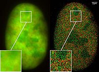 Only in the past two years have molecules been used in nanoscopic studies which emit the same spectral light frequency (but with different spectral signatures based on the flashing characteristics) but which can be switched on and off by means of light as is necessary for spectral precision distance microscopy. By combining many thousands of images of the same cell, it was possible using laser optical precision measurements to record localization images with significantly improved optical resolution. The application of these novel nanoscopy processes appeared until recently very difficult because it was assumed that only specially manufactured molecules could be switched on and off in a suitable manner by using light.
In March 2008 Christoph Cremer
’s lab discovered that this was also possible for many standard fluorescent dye like GFP
, Alexa dyes and fluorescein molecules, provided certain photo-physical conditions are present. Using this so-called SPDMphymod (physically modifiable fluorophores) technology a single laser wavelength of suitable intensity is sufficient for nanoimaging. In contrast other localization microscopies need two laser wavelengths when special photo-switchable/photo-activatable fluorescence molecules are used.
The GFP gene has been introduced and expressed in many procaryotic and eucaryotic cells and the Nobel Prize
in Chemistry
2008 was awarded to Martin Chalfie
, Osamu Shimomura
, and Roger Y. Tsien
for their discovery and development of the green fluorescent protein. The finding that these standard fluorescent molecules can be used extends the applicability of the SPMD method to numerous research fields in biophysics
, cell biology
and medicine
.
Standard fluorescent dyes already successfully used with the SPDMphymod technology: GFP, RFP, YFP, Alexa 488, Alexa 568, Alexa 647, Cy2, Cy3, Atto 488 and fluorescein.
The 3D images using Vertico-SMI are made possible by the combination of SMI and SPDM, whereby first the SMI and then the SPDM process is applied. The SMI process determines the center of particles and their spread in the direction of the microscope axis. While the center of particles/molecules can be determined with a 1-2 nm precision, the spread around this point can be determined down to an axial diameter of approx. 30-40 nm.
Subsequently, the lateral position of the individual particles/molecules is determined using SPDM, achieving a precision of a few nanometers. At present, SPDM achieves 16 frames/sec with an effective resolution of 10 nm in 2D (object plane); approximately 2000 such frames are combined with SMI data (currently ca. 10 sec acquisition time) to achieve a three dimensional image of highest resolution (effective optical 3D resolution ca. 40-50 nm). With a faster camera
, one can expect even higher rates (up to several hundred frames/sec, currently under development). Using suitable dyes, even higher effective optical 3D resolutions should be possible
By combining SPDMphymod with SMI (both invented in Christoph Cremer´s lab in 1996) a 3D dual colour reconstruction of the spatial arrangements of Her2/neu and Her3 clusters was achieved. The positions in all three directions of the protein clusters could be determined with an accuracy of about 25 nm.
The Vertico-SMI microscope was developed by Christoph Cremer
Christoph Cremer
Christoph Cremer is a German physicist and professor at the Ruprecht-Karls-University Heidelberg, who has successfully overcome the conventional limit of resolution that applies to light based investigations by a range of different methods Christoph Cremer (born in Freiburg im Breisgau, Germany)...
, Professor of Applied Optics and Information Processing at Heidelberg University and is based on the combination of light optical techniques of localization microscopy (SPDM, Spectral Precision Distance Microscopy) and structured illumination (SMI, Spatially Modulated Illumination).
Since March 2008 many standard fluorescent dyes like GFP
Green fluorescent protein
The green fluorescent protein is a protein composed of 238 amino acid residues that exhibits bright green fluorescence when exposed to blue light. Although many other marine organisms have similar green fluorescent proteins, GFP traditionally refers to the protein first isolated from the...
and Alexa fluorescent dyes can be used with this so-called SPDMphymod (physically modifiable fluorophores) localization microscopy, for which only one single laser wavelength of suitable intensity is sufficient for nanoimaging.
Configuration
SMI stands for a special type of laser optical illumination (Spatially Modulated Illumination) and Vertico reflects the vertical arrangement of the microscope axis which renders possible the analysis of fixed cellsCell (biology)
The cell is the basic structural and functional unit of all known living organisms. It is the smallest unit of life that is classified as a living thing, and is often called the building block of life. The Alberts text discusses how the "cellular building blocks" move to shape developing embryos....
but also of living cells with an optical resolution below 10 nanometers (1 nanometer = 1 nm = 1 × 10⁻⁹ m).
A particularity of this technology compared with focusing techniques such as 4Pi microscopy, is the wide field exposures which allow entire cells to be depicted at the nano scale. Such a 3D exposure of a whole cell with a typical object size of 20 µm × 20 µm require only 2 minutes. Wide field exposures signify that the entire object is illuminated and detected simultaneously.
SMI: Spatially Modulated Illumination
SMI microscopy is a light optical process of the so-called Point Spread FunctionPoint spread function
The point spread function describes the response of an imaging system to a point source or point object. A more general term for the PSF is a system's impulse response, the PSF being the impulse response of a focused optical system. The PSF in many contexts can be thought of as the extended blob...
-Engineering. These are processes which modify the Point Spread Function (PSF) of a microscope
Microscope
A microscope is an instrument used to see objects that are too small for the naked eye. The science of investigating small objects using such an instrument is called microscopy...
in a suitable manner to either increase the optical resolution, to maximize the precision of distance
Distance
Distance is a numerical description of how far apart objects are. In physics or everyday discussion, distance may refer to a physical length, or an estimation based on other criteria . In mathematics, a distance function or metric is a generalization of the concept of physical distance...
measurements of fluorescent objects that are small relative to the wavelength
Wavelength
In physics, the wavelength of a sinusoidal wave is the spatial period of the wave—the distance over which the wave's shape repeats.It is usually determined by considering the distance between consecutive corresponding points of the same phase, such as crests, troughs, or zero crossings, and is a...
of the illuminating light, or to extract other structural parameters in the nanometer range.
The SMI microscope currently being developed at the Kirchhoff Institute for Physics at Heidelberg University achieves this in the following manner: The illumination intensity within the object range is not uniform, unlike conventional wide field fluorescence microscopes, but is spatially modulated in a precise manner by the use of two opposing interfering laser beams along the axis. The principle of the spatially-modulated wave field was developed in 1993 by Bailey et al. The SMI microscopy approach used in the Heidelberg application moves the object in high-precision steps through the wave field, or the wave field itself is moved relative to the object by phase shift. This results in an improved axial size and distance resolution.
SPDM: Localization Microscopy
SPDM (Spectral Precision Distance Microscopy), the basic localization microscopy technology is a light optical process of fluorescence microscopy which allows position, distance and angle measurements on "optically isolated" particles (e.g. molecules) well below the theoretical limit of resolution for light microscopy. "Optically isolated" means that at a given point in time, only a single particle/molecule within a region of a size determined by conventional optical resolution (typically approx. 200-250 nm diameterDiameter
In geometry, a diameter of a circle is any straight line segment that passes through the center of the circle and whose endpoints are on the circle. The diameters are the longest chords of the circle...
) is being registered. This is possible when molecules within such a region all carry different spectral markers (e.g. different colors or other usable differences in the light emission of different particles).
The structural resolution achievable using SPDM can be expressed in terms of the smallest measurable distance between two in their spatial position determined punctiform particle of different spectral characteristics ("topological resolution“). Modeling has shown that under suitable conditions regarding the precision of localization, particle density etc., the "topological resolution" corresponds to a "space frequency" which in terms of the classical definition is equivalent to a much improved optical resolution.
SPDM is a localization microscopy which achieves an effective optical resolution several times better than the conventional optical resolution (approx. 200-250 nm), represented by the half-width of the main maximum of the effective point image function. By applying suitable laser optical precision processes, position and distances significantly smaller than the half-width of the point spread function (conventionally 200-250 nm) can be measured with nanometer accuracy between targets with different spectral signatures. An important area of application is genome research (study of the functional organization of the genome
Genome
In modern molecular biology and genetics, the genome is the entirety of an organism's hereditary information. It is encoded either in DNA or, for many types of virus, in RNA. The genome includes both the genes and the non-coding sequences of the DNA/RNA....
). Another important area of use is research into the structure of membranes.
One of the most important basics of the localization microscopy in general is the first experimental work for the localization of fluorescent objects in the nanoscale (3D) in 1996 and theoretical and experimental proof for a localization accuracy using visible light in the range of 1 nm – the basis for localization microscopy better than 1/100 of the wave length.
SPDMphymod: Standard fluorescent dyes in the blinking mode like GFP
In March 2008 Christoph Cremer
Christoph Cremer
Christoph Cremer is a German physicist and professor at the Ruprecht-Karls-University Heidelberg, who has successfully overcome the conventional limit of resolution that applies to light based investigations by a range of different methods Christoph Cremer (born in Freiburg im Breisgau, Germany)...
’s lab discovered that this was also possible for many standard fluorescent dye like GFP
Green fluorescent protein
The green fluorescent protein is a protein composed of 238 amino acid residues that exhibits bright green fluorescence when exposed to blue light. Although many other marine organisms have similar green fluorescent proteins, GFP traditionally refers to the protein first isolated from the...
, Alexa dyes and fluorescein molecules, provided certain photo-physical conditions are present. Using this so-called SPDMphymod (physically modifiable fluorophores) technology a single laser wavelength of suitable intensity is sufficient for nanoimaging. In contrast other localization microscopies need two laser wavelengths when special photo-switchable/photo-activatable fluorescence molecules are used.
The GFP gene has been introduced and expressed in many procaryotic and eucaryotic cells and the Nobel Prize
Nobel Prize
The Nobel Prizes are annual international awards bestowed by Scandinavian committees in recognition of cultural and scientific advances. The will of the Swedish chemist Alfred Nobel, the inventor of dynamite, established the prizes in 1895...
in Chemistry
Chemistry
Chemistry is the science of matter, especially its chemical reactions, but also its composition, structure and properties. Chemistry is concerned with atoms and their interactions with other atoms, and particularly with the properties of chemical bonds....
2008 was awarded to Martin Chalfie
Martin Chalfie
Martin Chalfie is an American scientist. He is the William R. Kenan, Jr. Professor of Biological Sciences at Columbia University, where he is also chair of the department of biological sciences. He shared the 2008 Nobel Prize in Chemistry along with Osamu Shimomura and Roger Y. Tsien "for the...
, Osamu Shimomura
Osamu Shimomura
is a Japanese organic chemist and marine biologist, and Professor Emeritus at Marine Biological Laboratory in Woods Hole, Massachusetts and Boston University Medical School...
, and Roger Y. Tsien
Roger Y. Tsien
Roger Yonchien Tsien is a Chinese American biochemist and a professor at the Department of Chemistry and Biochemistry, University of California, San Diego...
for their discovery and development of the green fluorescent protein. The finding that these standard fluorescent molecules can be used extends the applicability of the SPMD method to numerous research fields in biophysics
Biophysics
Biophysics is an interdisciplinary science that uses the methods of physical science to study biological systems. Studies included under the branches of biophysics span all levels of biological organization, from the molecular scale to whole organisms and ecosystems...
, cell biology
Cell biology
Cell biology is a scientific discipline that studies cells – their physiological properties, their structure, the organelles they contain, interactions with their environment, their life cycle, division and death. This is done both on a microscopic and molecular level...
and medicine
Medicine
Medicine is the science and art of healing. It encompasses a variety of health care practices evolved to maintain and restore health by the prevention and treatment of illness....
.
Standard fluorescent dyes already successfully used with the SPDMphymod technology: GFP, RFP, YFP, Alexa 488, Alexa 568, Alexa 647, Cy2, Cy3, Atto 488 and fluorescein.
LIMON: 3D Super Resolution Microscopy
LIMON (Light MicrOscopical nanosizing microscopy) has been invented in 2001 at the University of Heidelberg and combines localization microscopy and spatially modulated illumination to the 3D super resolution microscopy.The 3D images using Vertico-SMI are made possible by the combination of SMI and SPDM, whereby first the SMI and then the SPDM process is applied. The SMI process determines the center of particles and their spread in the direction of the microscope axis. While the center of particles/molecules can be determined with a 1-2 nm precision, the spread around this point can be determined down to an axial diameter of approx. 30-40 nm.
Subsequently, the lateral position of the individual particles/molecules is determined using SPDM, achieving a precision of a few nanometers. At present, SPDM achieves 16 frames/sec with an effective resolution of 10 nm in 2D (object plane); approximately 2000 such frames are combined with SMI data (currently ca. 10 sec acquisition time) to achieve a three dimensional image of highest resolution (effective optical 3D resolution ca. 40-50 nm). With a faster camera
Camera
A camera is a device that records and stores images. These images may be still photographs or moving images such as videos or movies. The term camera comes from the camera obscura , an early mechanism for projecting images...
, one can expect even higher rates (up to several hundred frames/sec, currently under development). Using suitable dyes, even higher effective optical 3D resolutions should be possible
By combining SPDMphymod with SMI (both invented in Christoph Cremer´s lab in 1996) a 3D dual colour reconstruction of the spatial arrangements of Her2/neu and Her3 clusters was achieved. The positions in all three directions of the protein clusters could be determined with an accuracy of about 25 nm.